
Ischnothyreus tergemintus Liu, Xu & Henrard, 2019
|
publication ID |
https://doi.org/10.11646/zootaxa.4701.3.2 |
|
publication LSID |
lsid:zoobank.org:pub:316AC45C-6247-4982-B272-3E923DF727BD |
|
persistent identifier |
https://treatment.plazi.org/id/03B79468-0457-FFC9-FF6F-F90E0379FD53 |
|
treatment provided by |
Plazi |
|
scientific name |
Ischnothyreus tergemintus Liu, Xu & Henrard |
| status |
sp. nov. |
Ischnothyreus tergemintus Liu, Xu & Henrard , sp. nov.
( Figures 1−5 View FIGURE 1 View FIGURE 2 View FIGURE 4 View FIGURE 5 , 16)
urn:lsid:zoobank.org:act:
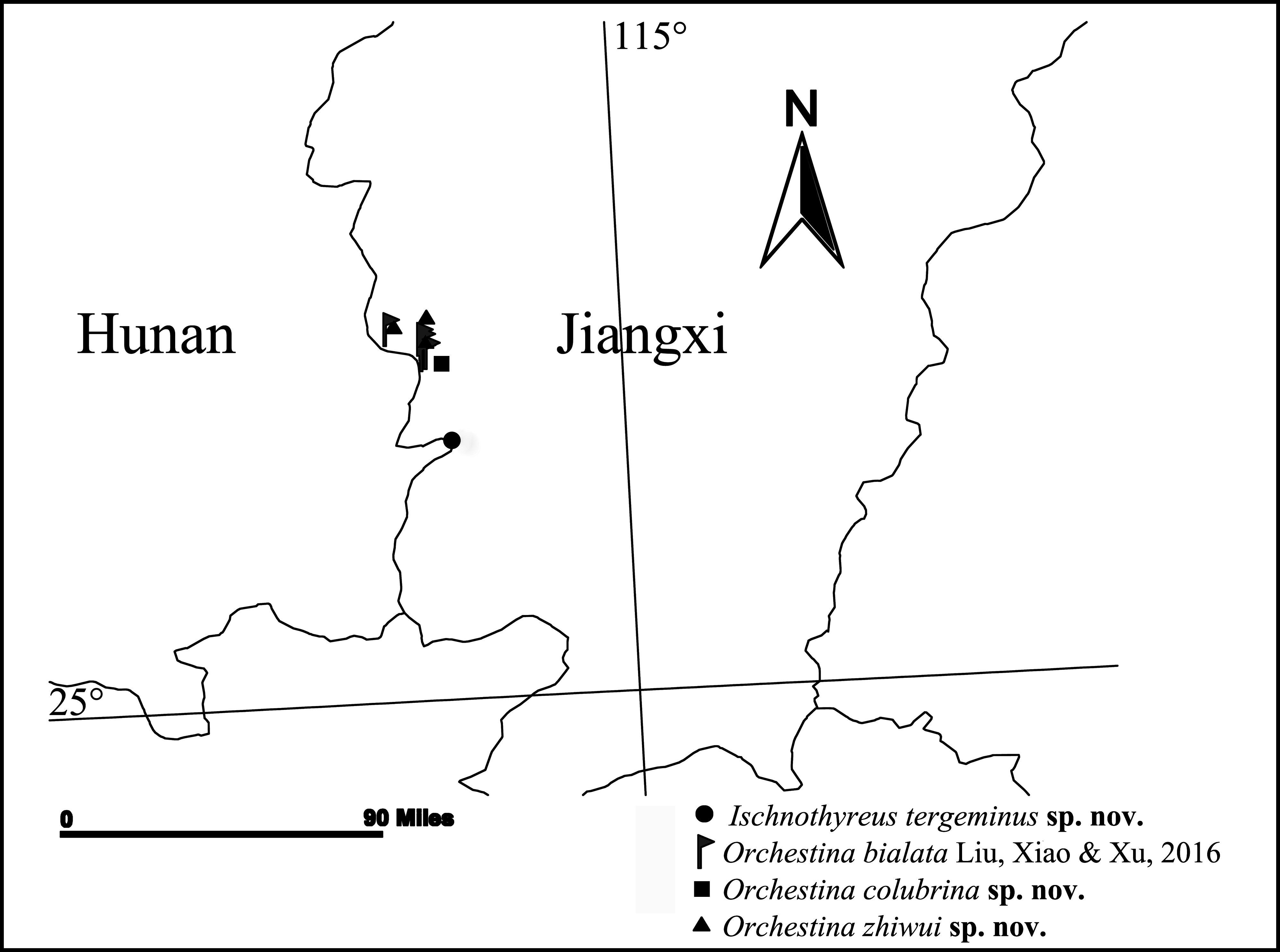
Type material: Holotype male: China: Jiangxi Province, Ji’an City, Jinggangshan County Level City , Xiangzhou Village , forest, 26.222°N, 114.265°E, 459m, 6.VIII.2015, Keke Liu, ZeyuanMeng, Sha Wu, Ce Xu, Shicong He and Yifan Zhao leg. ( OON 33 ) GoogleMaps . Paratypes: 2 males ( OON 34 ), same data as holotype GoogleMaps .
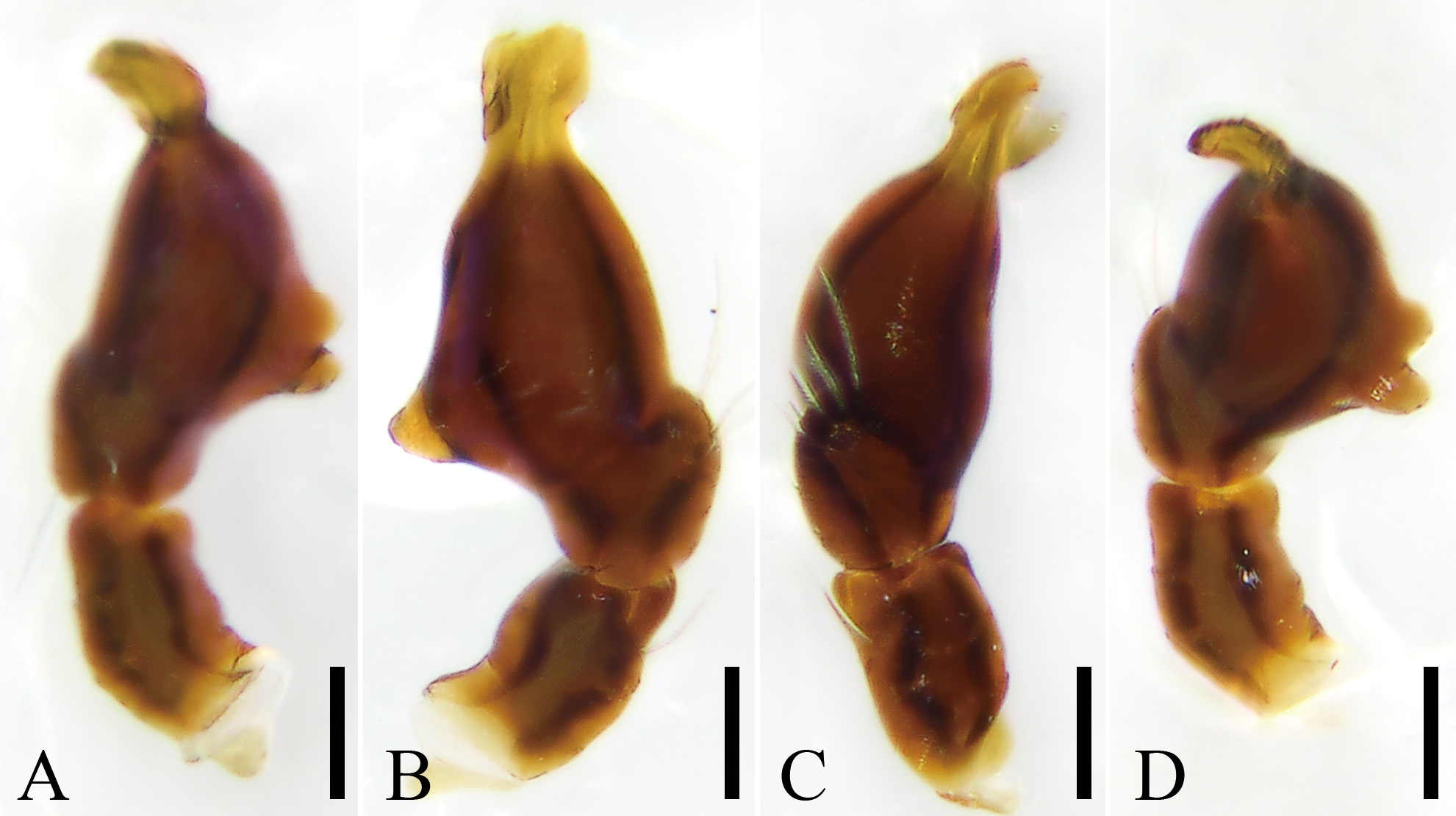
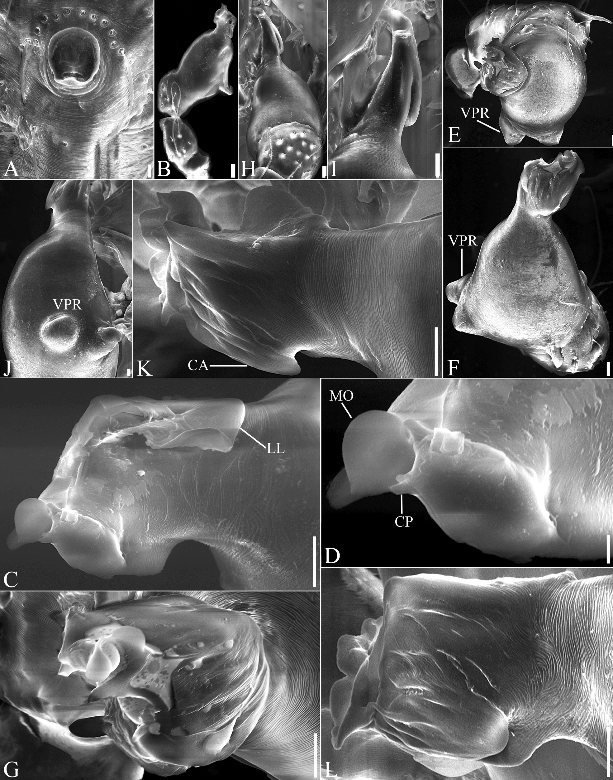
Etymology: The specific name derives from the Latin “tergeminus”, meaning “three pairs” and refers to the three pairs of corniculate projections (CP) triangularly inserted in the palpal tip.
Diagnosis: Male of Ischnothyreus tergeminus resembles those of I. falcatus Tong & Li, 2008 , I. flagellichelis Xu, 1989 and I. zhoujiayan Tong & Li, 2018 (especially the strongly modified seta on the chelicerae), but can be distinguished by Y-shaped frontal seta in median part of chelicerae (sickle-shaped in I. falcatus: Tong & Li (2008) : fig. 2C, E; flagelliform in I. flagellichelis: Xu (1989) : fig. 2; fork-shaped in I. zhoujiayan: Tong et al. (2018) : figs 1b, g, 2a, b, e−g) and distal tip of bulb bearing three pairs of corniculate projections (CP) apically ( Figs 2D View FIGURE 2 , 5D View FIGURE 5 ) (three membranous outgrowths in I. falcatus: Tong & Li (2008) : fig. 2H−J; hook-shaped in I. flagellichelis: Xu (1989) : figs 3−5; elongated with three apophysis in I. zhoujiayan: Tong et al. (2018) : fig. 2a−i).
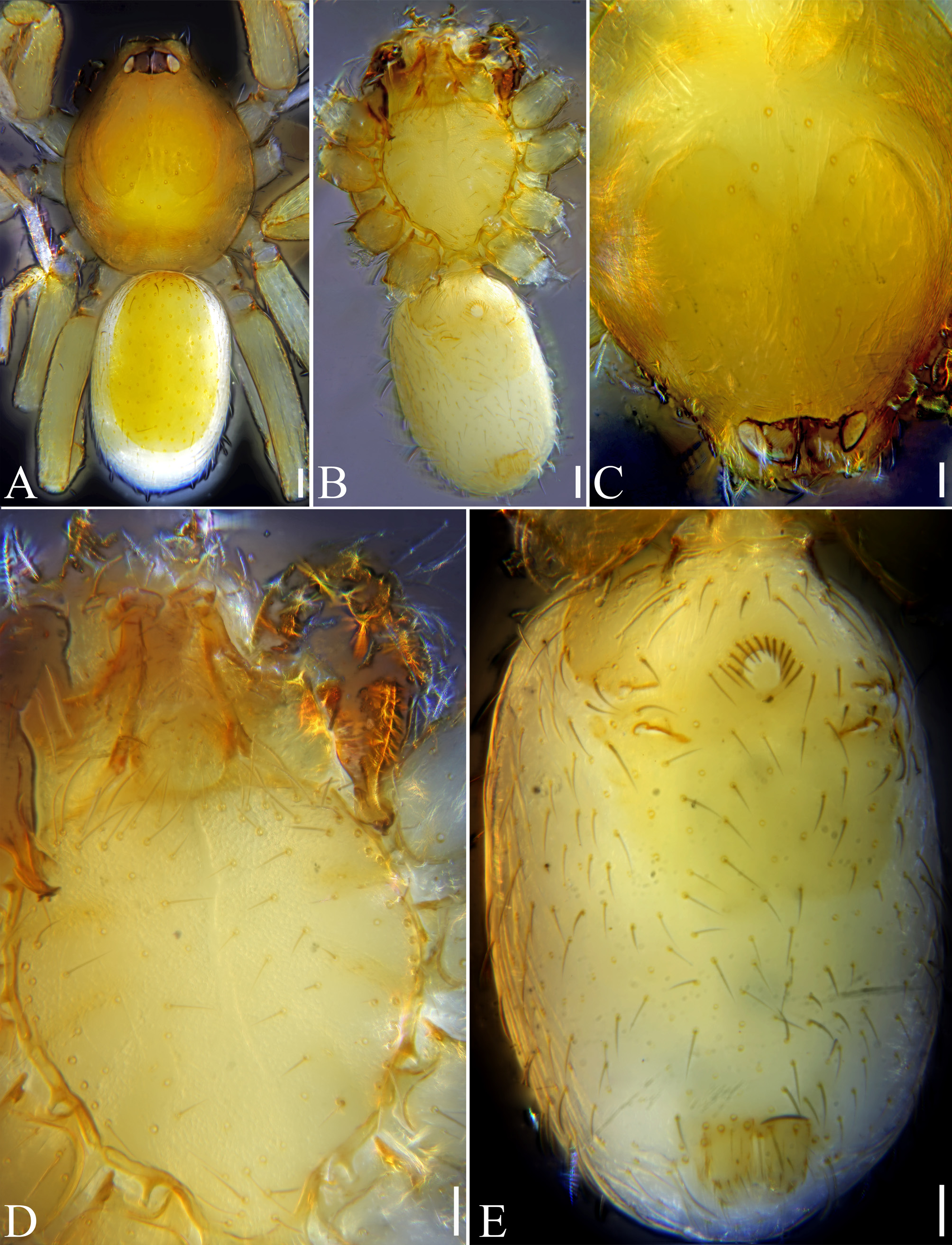
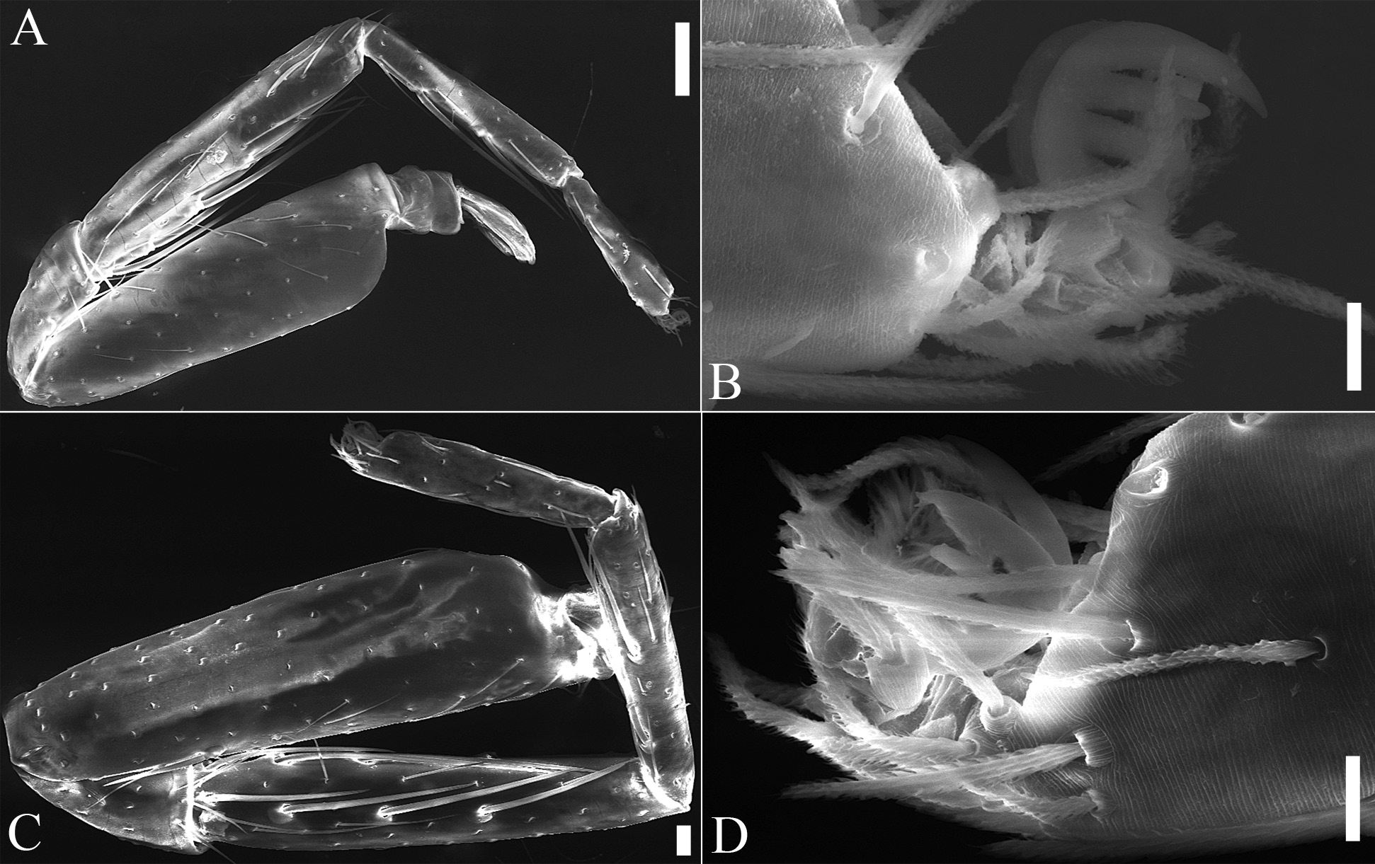
Description: Male holotype. Habitus as in Fig. 1A, B View FIGURE 1 . Total length 1.52, carapace ( Figs 1A, C View FIGURE 1 , 3A, B) length 0.79, width 0.64, smooth on top, lateral margin slightly reticulated, broadly oval in dorsal view, pars cephalica strongly elevated in frontal view, anteriorly narrowed to between 0.3 and 0.5 times its maximum width, with large pair light yellowish-green, oval patches, touching anteriorly, with three rows of setal alveoli surrounded by numerous, small scaly attachments behind eyes in dorsal view, median row located on axis of carapace. Eyes ( Figs 1A, C View FIGURE 1 , 3A−C): six, well developed and clustered, ALE and PME larger than PLE, ALE and PME roughly circular, PLE oval; ALE separated from edge of carapace by less than their radii; PER slightly procurved in dorsal view, ALE touching, PME touching, ALE–PLE touching, PLE–PME touching, ALE–PME separated from less than ALE’s radius; diameters: ALE 0.07, PME 0.07, PLE 0.06. Clypeus (Fig. 3A−C) straight in frontal view, curved down- wards in ventral view, with two groups of five setae on laterally rounded corners near ALE. Sternum ( Figs 1B, D View FIGURE 1 , 3H) slightly longer than wide, shield-shaped, pale yellow, surface smooth, without pits, anteriorly with abundant, needle-like setae, not fused to carapace, distance between coxae approximately equal, precoxal triangular extensions present. Mouthparts ( Figs 1B, D View FIGURE 1 , 3C–J): Chelicerae (Fig. 3C–G) light yellow; straight, subbasally with strong, sclerotized, Y-shaped apophyses (YA) divided in distal two thirds in frontal view, with anterior tip short and hooked, posterior tip strongly convolute in median part and gradually narrowing and strongly curving distally; with 6–8 needle-like setae near the base of YA, sparse setae on posterior margin, a row of 4–6 flat, smooth setae on anterior margin near the fang groove, and distally with heavily sclerotized process (HSP) at the base of fang. Labium ( Figs 1B, D View FIGURE 1 , 3H, I) rectangular, orange, fused to sternum, anterior margin with 14 strong setae, with two strong setae near anterolateral margin and 12 short, strong setae in median part. Endites ( Figs 1B, D View FIGURE 1 , 3G–I), yellowish brown, with slightly sclerotized rounded tip and numerous short setae on inner margin. Abdomen ( Fig. 1A, B, E View FIGURE 1 ) ovoid in lateral and oval, almost cylindrical in dorsal view, length 0.74, width 0.47, soft portions of dorsum white; FIGURE 3A–I. Ischnothyreus tergeminus sp. nov., SEM views of male paratype (OON 34). A. Carapace, frontal view. B. As previous, frontal view, slightly lateral. C. As previous, frontal view, detail of ALE, clypeus and chelicerae. D. Chelicerae, detail of YA situated in median part of strongly sclerotized apophyses, frontal view, slightly lateral. E. As previous, detail of right strongly sclerotized apophyses, frontal view. F. As previous, detail of left strongly sclerotized apophyses, frontal view. G. As previous, detail of HSP and strongly sclerotized apophyses, ventral view. H. Chelicerae, endites, labium and sternum, ventral view. I. Detail of endites and labium, ventral view.Abbreviations: HSP, heavily sclerotized process; VPR, ventral protuberance; YA, Y-shaped apophyses. Scale bars: A, B, H, 0.1 mm; C, E−G 10 µm; D, I, 20 µm. dorsal scutum slightly sclerotized, light yellowh-green, covering more than 3/4 of abdomen, not fused with epigastric scutum, abundant setae evenly distributed on dorsal part; pedicel tube short, ribbed, slightly sclerotized; spinnerets light yellow-green, cylindrical, sparse setae. Legs ( Figs 1A View FIGURE 1 , 4 View FIGURE 4 A–D): orange, tibial and metatarsal spines longer than each segmental width, femora I and II slightly thickened, coniform, tarsi distally widened, bearing uniseriate claws with three large teeth in lateral view of upper claw, scopula present between claws; spine formula: I p2*-0-4-2-0, r1*-0-4-2-0; II p1*-0-4-2-0, r1*-0-4-2-0; III and IV absent. Genitalia ( Figs 1B, E View FIGURE 1 , 2 View FIGURE 2 A−D, 4E, 5A–L): epigastric region with faint scutum, sperm pore large, circular, hollow with rebordered margins, situated at level of anterior spiracles, anteriorly surrounded by 16 strong needle-like setae. Palp ( Figs 2 View FIGURE 2 A−D, 5B–L) strongly sclero- tized, right and left palps symmetrical, brown, patellar plus tibia shorter than bulb, cymbium fused to bulb, setae sparse, bulb inverted drop-shaped in frontal view, tapering apically, with two clearly ventral protuberance (VPR) located on the ventromedian part, distal part with dorsal membranous outgrowth (MO), large prolateral lamellar lobe (LL), ventral coniform apophysis (CA), and apically with three pairs of triangularly inserted CP. Embolus not discernible, probably fused to the bulb.
Female: Unknown.
Distribution: Only known from Jiangxi, China ( Fig. 16 View FIGURE 16 ).
No known copyright restrictions apply. See Agosti, D., Egloff, W., 2009. Taxonomic information exchange and copyright: the Plazi approach. BMC Research Notes 2009, 2:53 for further explanation.
|
Kingdom |
|
|
Phylum |
|
|
Class |
|
|
Order |
|
|
Family |
|
|
Genus |