
Scydmaenus (Mascarensia) kasuganus Franz
|
publication ID |
https://doi.org/ 10.11646/zootaxa.5093.1.2 |
|
publication LSID |
lsid:zoobank.org:pub:8A1AC49B-D684-455A-9B76-9B2C014627C9 |
|
DOI |
https://doi.org/10.5281/zenodo.5903203 |
|
persistent identifier |
https://treatment.plazi.org/id/03BB6660-FFAD-FF87-FF5F-F9A6C2B2FAB8 |
|
treatment provided by |
Plazi |
|
scientific name |
Scydmaenus (Mascarensia) kasuganus Franz |
| status |
|
Scydmaenus (Mascarensia) kasuganus Franz View in CoL
Scydmaenus (Mascarensia) kasuganus Franz, 1976: 58 View in CoL . Redescribed by Hoshina, 2007: 2.
Scydmaenus (Mascarensia) honshuensis Franz, 1976: 57 . Lapsus for kasuganus.
( Figs 1–26 View FIGURES 1–3 View FIGURES 4–10 View FIGURES 11–15 View FIGURES 16–18 View FIGURES 19–27 )
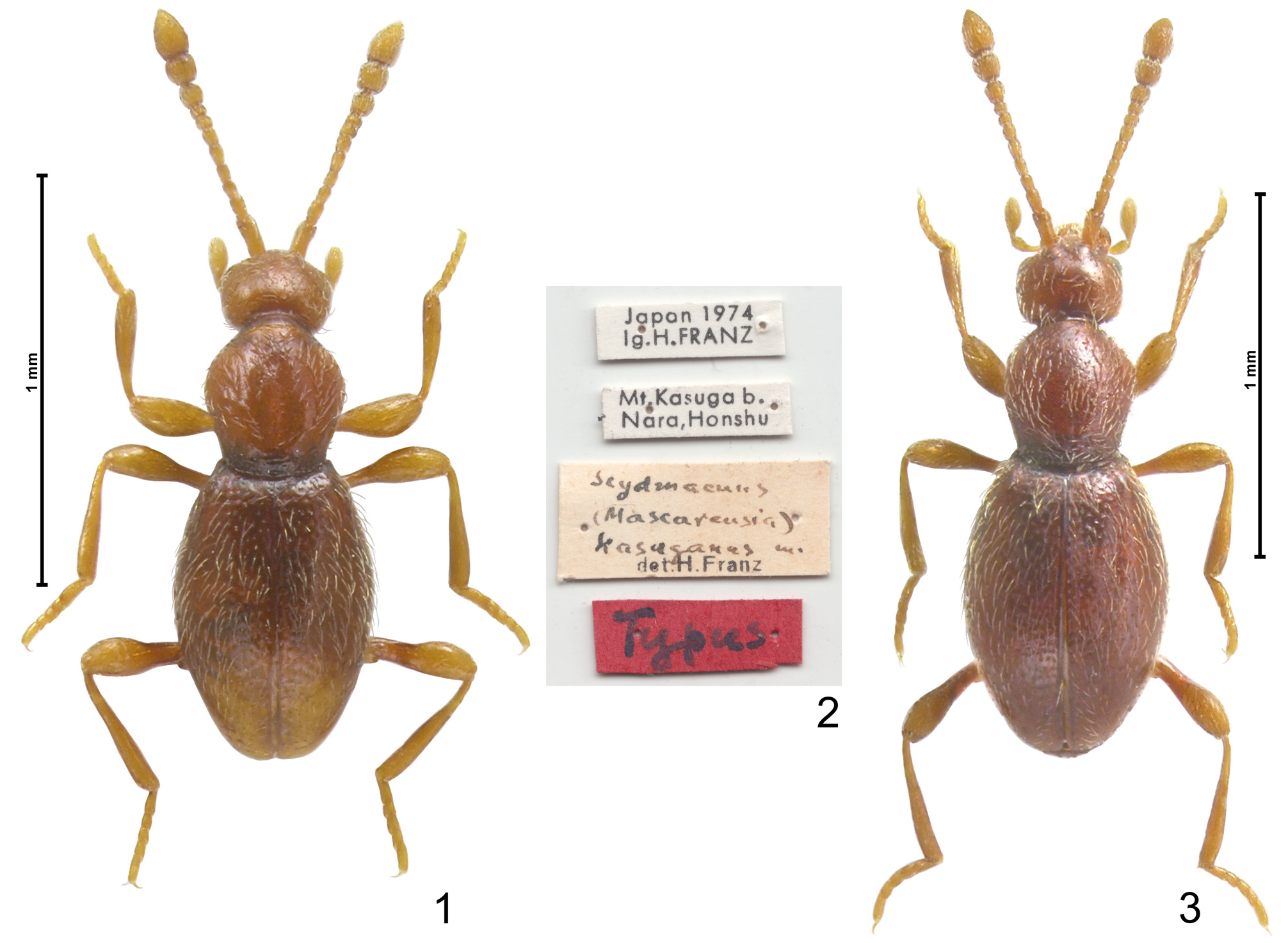
Type material examined. Holotype ( JAPAN: Nara Pref.): ♂ ( Fig. 1 View FIGURES 1–3 ), four labels ( Fig. 2 View FIGURES 1–3 ): „Japan 1974 / lg.H. FRANZ ” [white, printed], “ Mt.Kasuga b. / Nara,Honshu” [white, printed, with “ Ja 126” handwritten on reverse side], “ Scydmaenus / ( Mascarensia ) / kasuganus m. / det. H.Franz ” [creamy white, handwritten and printed], “Typus” [red, handwritten] ( NHMW).
Additional material examined (5 exx). Fukui Pref.: 1 ♂, Hakusan-jinja, 350 m, Shimo-Uchinami, Ono City , 27.04.2003, under bark of deciduous log, leg. P. Jałoszyński (cPJ) . Nara Pref.: 1 ♂, 1 ♀, Mt. Kasuga ad Nara, ca. 200 m, 17.05.2002, under bark of dead, standing Quercus tree, leg. P. Jałoszyński (cPJ); 2 ♂♂, Mt. Kasuga ad Nara, 05.05.2001, T. Tsuru leg. (cPJ) .
Revised diagnosis. Body length ~ 1.3‒1.4 mm; pronotum about as long as broad; punctures on head inconspicuous, on pronotum fine and dense, on elytra most distinct, conspicuous, deep and dense; pronotal base with only one (inner) pair of pits, which are minute and indistinct; pit on flattened proximal region of metafemur barely discernible; aedeagus with extremely narrow constriction delimiting large and distinctly elongate apical region and tubular proximal portion, in lateral view apical region curved dorsad.
Redescription. Body of male ( Figs 1, 3 View FIGURES 1–3 ) elongate and weakly convex, light brown with slightly lighter appendages, in some specimens elytra indistinctly darker than head and pronotum; vestiture of setae slightly lighter than cuticle; BL 1.28–1.40 mm.
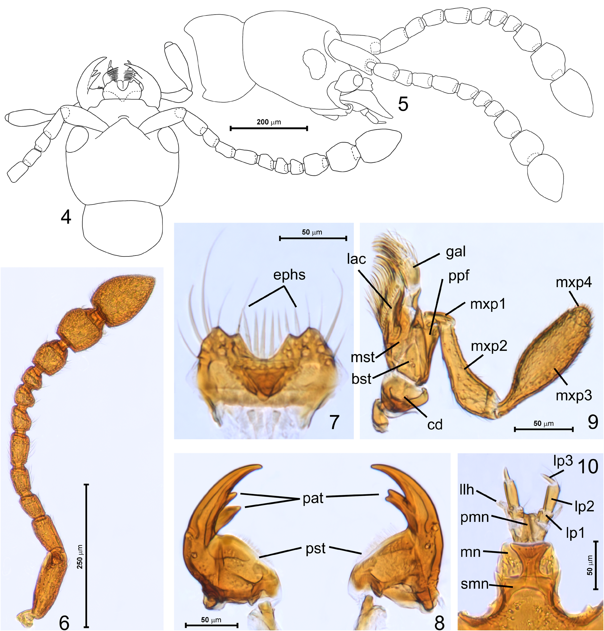
Head in dorsal view ( Fig. 4 View FIGURES 4–10 ) broadest at eyes, indistinctly transverse, HL 0.23–0.25 mm, HW 0.28; vertex and frons confluent, evenly convex; supraantennal tubercles feebly marked; tempora nearly three times as long as eye, eyes weakly convex and finely faceted, not projecting from head silhouette, in lateral view ( Fig. 5 View FIGURES 4–10 ) oval, posteriorly emarginate. Dorsal surface of head virtually impunctate and covered with sparse, short and suberect setae. Antennae ( Figs 4–6 View FIGURES 4–10 ) slender, as long as about half of body length, AnL 0.65–0.73 mm; scape and pedicel strongly elongate, antennomeres 3–5 each elongate (5 the longest) and symmetrical, 6–8 each distinctly asymmetrical, with mesal margin shorter than external one, antennomere 6 slightly elongate, 7 and 8 each about as long as broad, antennomeres 9–11 forming distinct and symmetrical club covered with setae denser than those on remaining antennomeres, 9 slightly elongate, 10 about as long as broad, 11 slightly broader than 10, much shorter than 9 and 10 combined, 1.4 × as long as broad.
Mouthparts ( Figs 7–10 View FIGURES 4–10 ) directed anterad. Labrum ( Fig. 7 View FIGURES 4–10 ) strongly transverse and bilobate, with broad and deep median U-shaped emargination flanked by short tooth, sides broadly rounded, dorsal surface with anteriorlydirected almost symmetrically and sparsely distributed long setae; epipharynx with 6–7 conspicuously long setiform sensilla ( Fig. 7 View FIGURES 4–10 ; ephs) projecting far beyond anterior labral margin and well-visible in dorsal view. Mandibles ( Fig. 8 View FIGURES 4–10 ) asymmetrical, falciform, long and in closed position overlapping, each with broad base and long setose prostheca ( Fig. 8 View FIGURES 4–10 ; pst) and slender distal region with sharp apical tooth; left mandible with two separate preapical teeth ( Fig. 8 View FIGURES 4–10 ; pat), right mandible with one preapical tooth with bifid apex. Maxilla ( Fig. 9 View FIGURES 4–10 ) with semicircular cardo ( Fig. 9 View FIGURES 4–10 ; ca), elongate, subtriangular basistipes ( Fig. 9 View FIGURES 4–10 ; bst), elongate and quadrangular mediostipes ( Fig. 9 View FIGURES 4–10 ; mst); maxillary palpomere inserted on elongate palpifer ( Fig. 9 View FIGURES 4–10 ; ppf) and composed of four palpomeres: palpomere 1 ( Fig. 9 View FIGURES 4–10 ; mxp1) minute, elongate, palpomere 2 ( Fig. 9 View FIGURES 4–10 ; mxp2) pipe-shaped, strongly elongate, palpomere 3 ( Fig. 9 View FIGURES 4–10 ; mxp3) the largest, narrow at base, broadening to nearly middle and then indistinctly narrowing toward truncate apex, palpomere 4 ( Fig. 9 View FIGURES 4–10 ; mxp4) dome-shaped, densely setose. Labium ( Fig. 10 View FIGURES 4–10 ) with submentum ( Fig. 10 View FIGURES 4–10 ; smn) not demarcated posteriorly, its anterior margin straight at middle and its anterolateral corners forming subtriangular projections; mentum ( Fig. 10 View FIGURES 4–10 ; mn) rectangular with slightly rounded sides, distinctly transverse, with weakly concave anterior margin; prementum ( Fig. 10 View FIGURES 4–10 ; pmn) largely membranous, with non-differentiated ligula and trimerous labial palps: palpomere 1 ( Fig. 10 View FIGURES 4–10 ; lp1) slightly shorter than long, palpomere 2 ( Fig. 10 View FIGURES 4–10 ; lp2) the longest, about 3 × as long as broad and nearly cylindrical, palpomere 3 ( Fig. 10 View FIGURES 4–10 ; lp3) minute, nearly aciculate; lateral lobes of hypopharynx ( Fig. 10 View FIGURES 4–10 ; llh) short and inconspicuous.
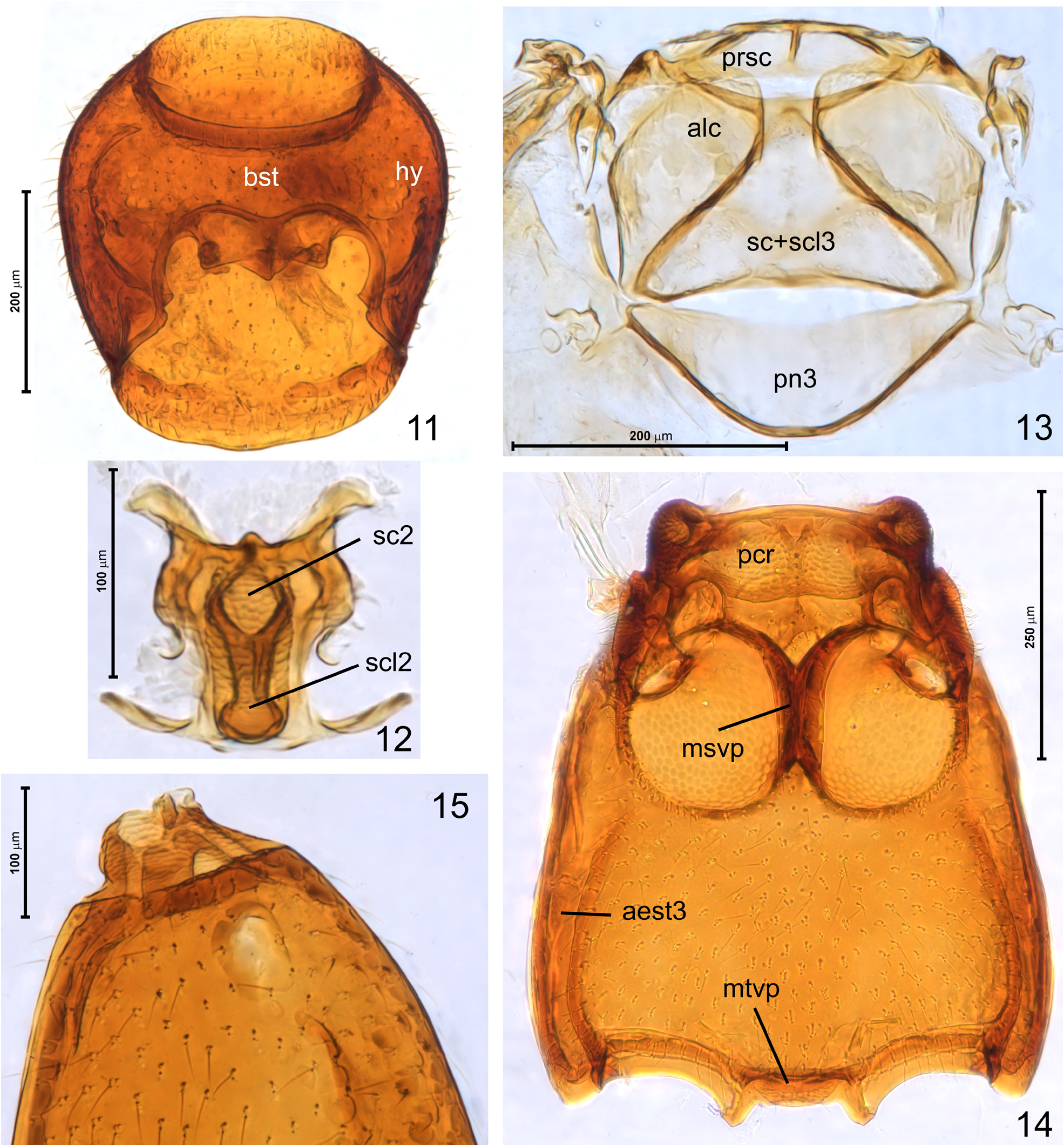
Pronotum in dorsal view broadest near anterior third; PL 0.35–0.38 mm, PW 0.35–0.38 mm; anterior margin and sides in anterior third rounded, sides in posterior half indistinctly rounded or nearly straight and strongly converging posterad; posterior margin arcuate, with median region slightly more profoundly projecting posterad; base with one pair of small, shallow and poorly marked pits. Pronotal disc distinctly punctate, punctures small and shallow but well-discernible, those on median area separated by spaces subequal to or slightly shorter than their diameters, punctures reducing in diameters and depths toward pronotal margins. Setae on disc moderately dense, short and suberect. Prosternum with its basisternal region ( Fig. 11 View FIGURES 11–15 ; bst) slightly longer than coxal region and laterally indistinguishably fused with convex hypomera ( Fig. 11 View FIGURES 11–15 ; hy).
Mesonotum ( Fig. 12 View FIGURES 11–15 ) with mesoscutum ( Fig. 12 View FIGURES 11–15 ; sc2) and mesoscutellum ( Fig. 12 View FIGURES 11–15 ; scl2) fused so that scutoscutellar suture is indiscernible, scutellar shield not exposed between elytral bases in intact specimens, with broadly rounded posterior margin.
Metanotum ( Fig. 13 View FIGURES 11–15 ) as in all winged Scydmaenus species , with strongly shortened alacristae ( Fig. 13 View FIGURES 11–15 ; alc) not reaching middle of scutum; metascutum and metascutellum fused ( Fig. 13 View FIGURES 11–15 ; sc+scl3). Prescutum ( Fig. 13 View FIGURES 11–15 ; prsc) relatively long, with well-visible median phragma; metapostnotum ( Fig. 13 View FIGURES 11–15 ; pn3) long and subtriangular, with broadly rounded posterior margin.
Mesoventrite ( Fig. 14 View FIGURES 11–15 ) strongly transverse, with massive prepectus, pair of weakly concave and asetose anteromedian impressions functioning as procoxal rests ( Fig. 14 View FIGURES 11–15 ; pcr) and mesocoxal projections weakly projecting laterad; mesoventral intercoxal process ( Fig. 14 View FIGURES 11–15 ; msvp) broad and nearly parallel-sided.
Metaventrite ( Fig. 4 View FIGURES 4–10 ) about as long as broad, slightly trapezoidal, broadening posterad up to posterior 1/6 and then narrowing, with distinctly rounded sides. Mesocoxal rests nearly circular, carinate posteriorly; metaventral intercoxal process ( Fig. 14 View FIGURES 11–15 ; mtvp) short and broad, with posterolateral subtriangular projections. Metaventrite unmodified. Metanepisterna ( Fig. 14 View FIGURES 11–15 ; aest3) broad and largely separated from metaventrite, but fused to it near posterolateral margins of mesocoxal rests.
Elytra ( Figs 1, 3 View FIGURES 1–3 , 15 View FIGURES 11–15 ) together oval, broadest slightly in front of middle, EL 0.70–0.78 mm, EW 0.48–0.56 mm, EI 1.38–1.47. Humeral calli prominent; basal impressions and basal foveae lacking; basal articulating lobe delimited from disc by anteriorly concave step-wise ridge. Elytra covered with punctures larger and deeper than those on pronotal disc, slightly unevenly distributed, so that some punctures touch each other, and some are separated by spaces subequal to their diameters; punctures reducing in diameters and depths toward sides and apices of elytra. Hind wings present, functional.
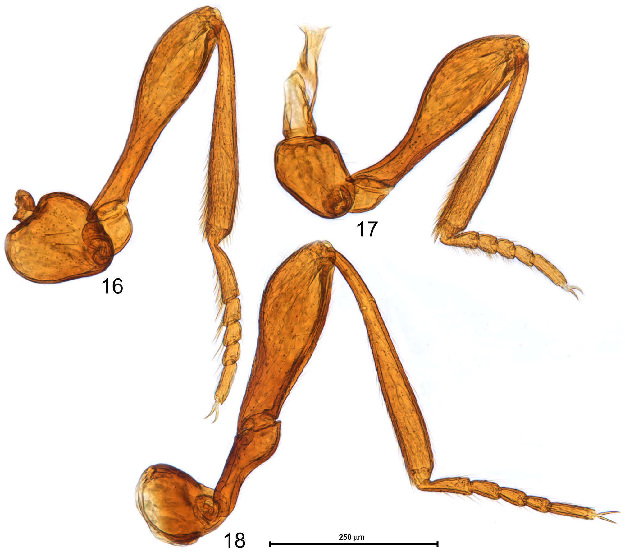
Legs ( Figs 16–18 View FIGURES 16–18 ) moderately long and slender, lacking male secondary sexual modifications. Fore ( Fig. 16 View FIGURES 16–18 ) and middle ( Fig. 17 View FIGURES 16–18 ) leg with subtriangular trochanter, clavate femur with slender, nearly cylindrical proximal half and nearly straight tibia; hind leg ( Fig. 18 View FIGURES 16–18 ) with long trochanter conspicuously broadened in distal region, femur with broad and strongly flattened proximal half bearing scale-like microsculpture and a trace of barely discernible pit on its external surface, tibia slightly curved near base. All tarsi slender and subcylindrical, protarsus not modified, but ventral surface of tarsomeres 1–4 bearing slightly longer setae than those on remaining legs.
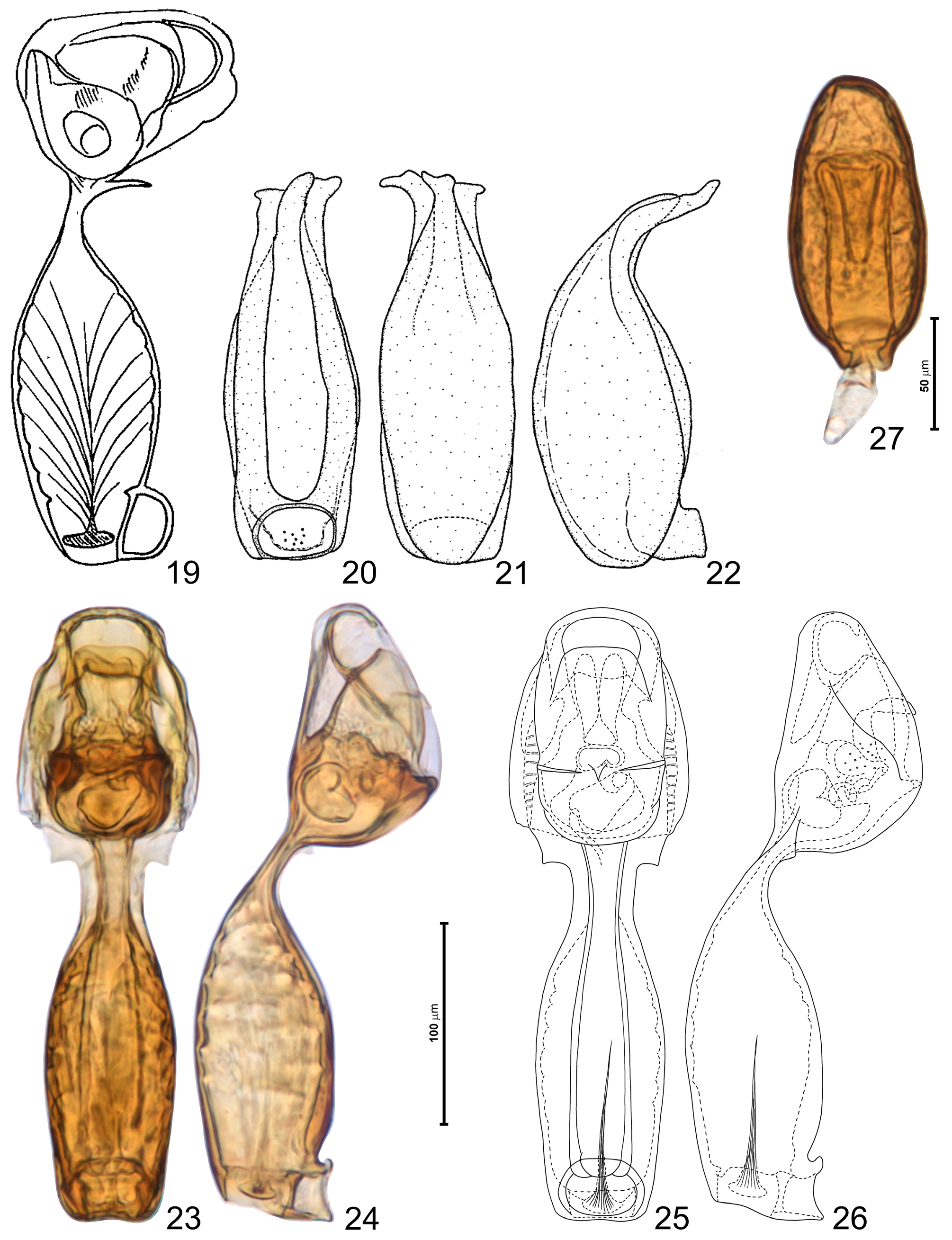
Aedeagus ( Figs 23–26 View FIGURES 19–27 ) strongly elongate, AeL 0.30 mm; median lobe divided by stalk-like region into tubular proximal portion and much broader but shorter distal capsular portion; in dorsal view basal region broadest near its middle, narrowest site as wide as half width of basal region and about 1/3 as wide as width of distal region; in lateral view stalk-like portion extremely narrow and curved dorsad. Distal region in dorsal view broadest near its base, with a pair of basal translucent subtriangular lobes on sides of stalk-like portion. Dorsally open ostium complex, with symmetrical external structures; endophallus with asymmetrical median capsular structure situated near base of broadened distal aedeagal region. Traces of parameres fused with median lobe can be seen on dorsal surface of tubular proximal region of median lobe.
Female. Externally indistinguishable from male. Only one female was available for measurements: BL 1.43 mm; HL 0.25 mm, HW 0.29 mm, AnL 0.73 mm; PL 0.40 mm, PW 0.39 mm; EL 0.78 mm, EW 0.55 mm, EI 1.41. Spermatheca ( Fig. 27 View FIGURES 19–27 ) darkly sclerotized, strongly elongate and symmetrical, length 0.13 mm.
Distribution. Honshu (prefectures Nara and newly recorded from Fukui-ken), Japan.
Remarks. I collected this species from under bark of dead deciduous trees, the one in Nara most likely Quercus sessilifolia Blume , a large, dead standing tree in a shaded forest on WS slope of Mt. Kasuga.
| NHMW |
Naturhistorisches Museum, Wien |
| T |
Tavera, Department of Geology and Geophysics |
No known copyright restrictions apply. See Agosti, D., Egloff, W., 2009. Taxonomic information exchange and copyright: the Plazi approach. BMC Research Notes 2009, 2:53 for further explanation.
|
Kingdom |
|
|
Phylum |
|
|
Class |
|
|
Order |
|
|
Family |
|
|
SubFamily |
Scydmaeninae |
|
Genus |
|
|
SubGenus |
Mascarensia |
Scydmaenus (Mascarensia) kasuganus Franz
| Jałoszyński, Paweł 2022 |
Scydmaenus (Mascarensia) kasuganus
| Hoshina, H. 2007: 2 |
| Franz, H. 1976: 58 |
Scydmaenus (Mascarensia) honshuensis
| Franz, H. 1976: 57 |