
Pseudocandona becca, Smith & Kamiya, 2015
|
publication ID |
https://doi.org/ 10.5852/ejt.2015.136 |
|
publication LSID |
lsid:zoobank.org:pub:530F395F-97A9-46F1-957C-8E57B9C3ACD9 |
|
DOI |
https://doi.org/10.5281/zenodo.3794649 |
|
persistent identifier |
https://treatment.plazi.org/id/F3A75D11-C834-4673-817D-E962EA9F9347 |
|
taxon LSID |
lsid:zoobank.org:act:F3A75D11-C834-4673-817D-E962EA9F9347 |
|
treatment provided by |
Carolina |
|
scientific name |
Pseudocandona becca |
| status |
sp. nov. |
Pseudocandona becca sp. nov.
urn:lsid:zoobank.org:act:F3A75D11-C834-4673-817D-E962EA9F9347
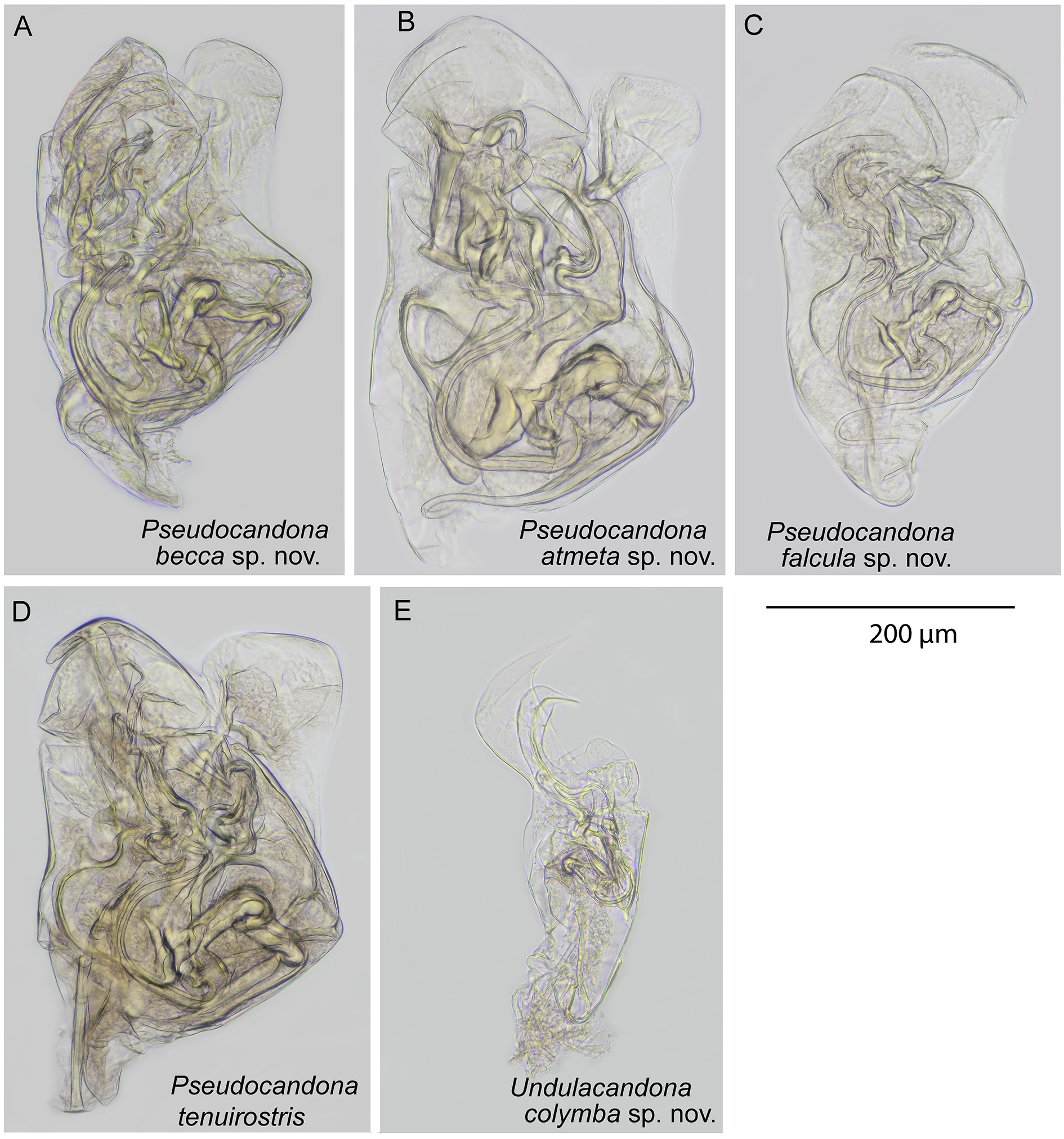
Figs 2 View Fig A–E, 3–5, 6A
Pseudocandona View in CoL sp. – Smith et al. 2014: appendix S1.
Diagnosis
Posterior margin more inflated than anterior margin, hinge straight, slightly sloping towards anterior. Calcified inner lamella with distinctive convexity in antero-ventral region. Dorsal view with slight anterior beak. Surface of valves without pits. Second endopodal segment of male antenna sub-divided and with long male bristles (t2 and t3). Female antennal claw G2 approximately two-thirds length of G3. Mandible with 3+1+beta setae on second segment of palp, and with long gamma seta and short alpha and beta setae; beta slightly shorter than alpha. Walking leg with long d1 seta, and very short e, f and g setae. Seventh limb with five segments, and short, curved, reflexed h1 seta and long h2 and h3 setae. Outer and inner lobes (a and b respectively) of hemipenes sub-equal in length. Outer lobe (a) subquadrate distally, inner lobe (b) sub-triangular distally. Medial lobe (h) shorter than other lobes, with distinctive small, triangular to quadrate protrusion on outer edge.
Etymology
From the Latin beccus, meaning “beak” or “bill”, and referring to the small but characteristic, beakshaped projection on the medial lobe (h) of the hemipenes (marked with a black triangle on Fig. 5C View Fig ).
Type material
Holotype
♂ ( LBM 1430006264 View Materials ), dissected with appendages sealed in a glass slide and valves stored dry in a micropalaeontological cavity slide. Collected from the type locality on 5 Dec. 2010.
Allotype
♀ ( LBM 1430006265 View Materials ), dissected with appendages sealed in a glass slide and valves stored dry in a micropalaeontological cavity slide. Collected from the type locality on 5 Dec. 2010.
Paratypes
2 ♀♀ ( LBM 1430006266, LBM 1430006267), dissected with appendages sealed in a glass slide and valves stored dry in a micropalaeontological cavity slide. 1 ♂ ( LBM 1430006268), whole, stored dry in a micropalaeontological cavity slide. 1♀ ( LBM 1430006269), valves, stored dry in a micropalaeontological cavity slide. All collected from the type locality on 5 Dec. 2010.
Type locality
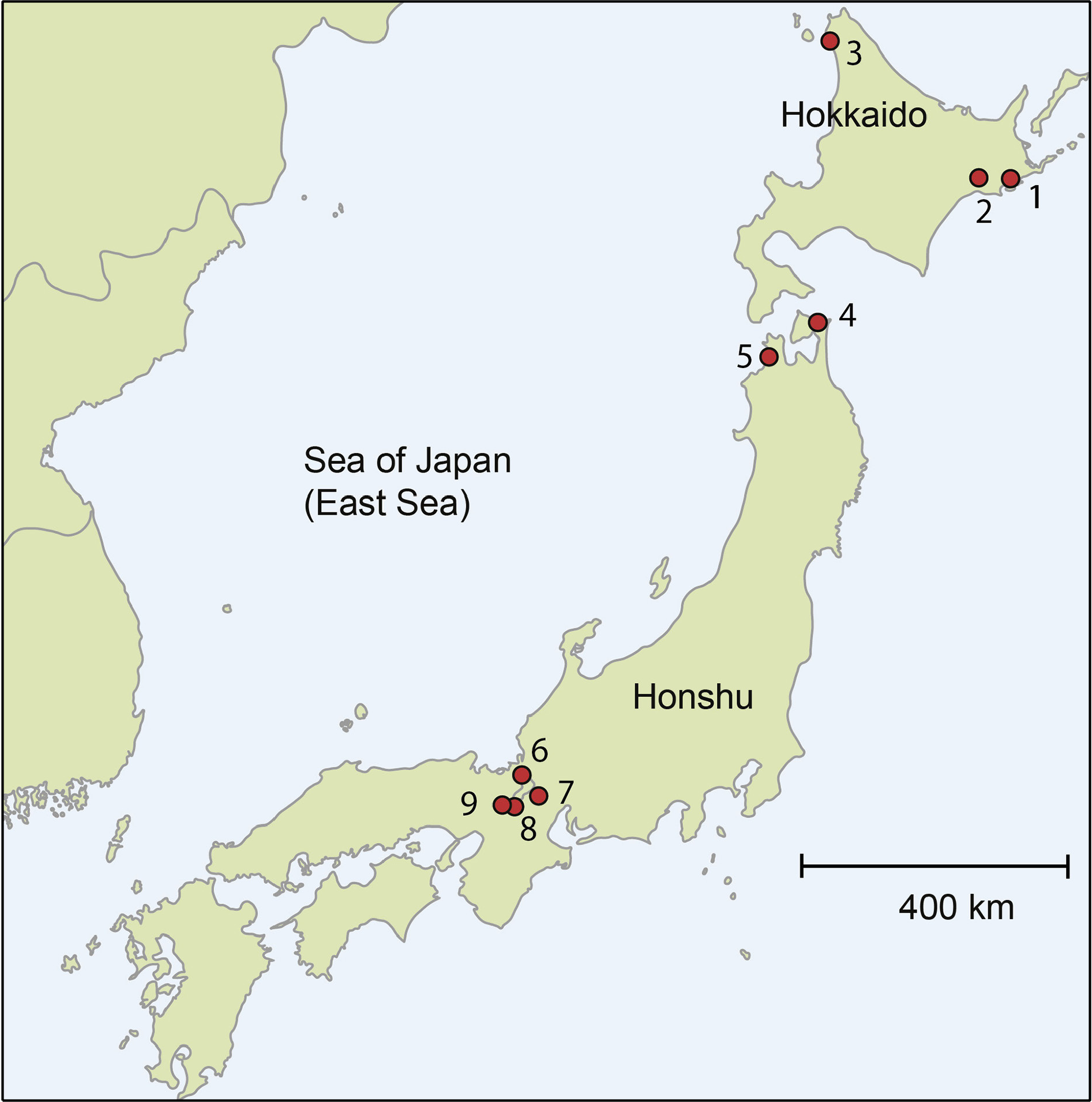
Sunny Beach in Nishihama, Makino, Lake Biwa, Shiga Prefecture, Japan, 35º27’26.58” N, 136º03’49.5” E. Locality 6 on Fig. 1 View Fig .
Other material examined
11 ♀♀, 6 ♂♂, collected from the type locality on 5 Dec. 2010.
Description
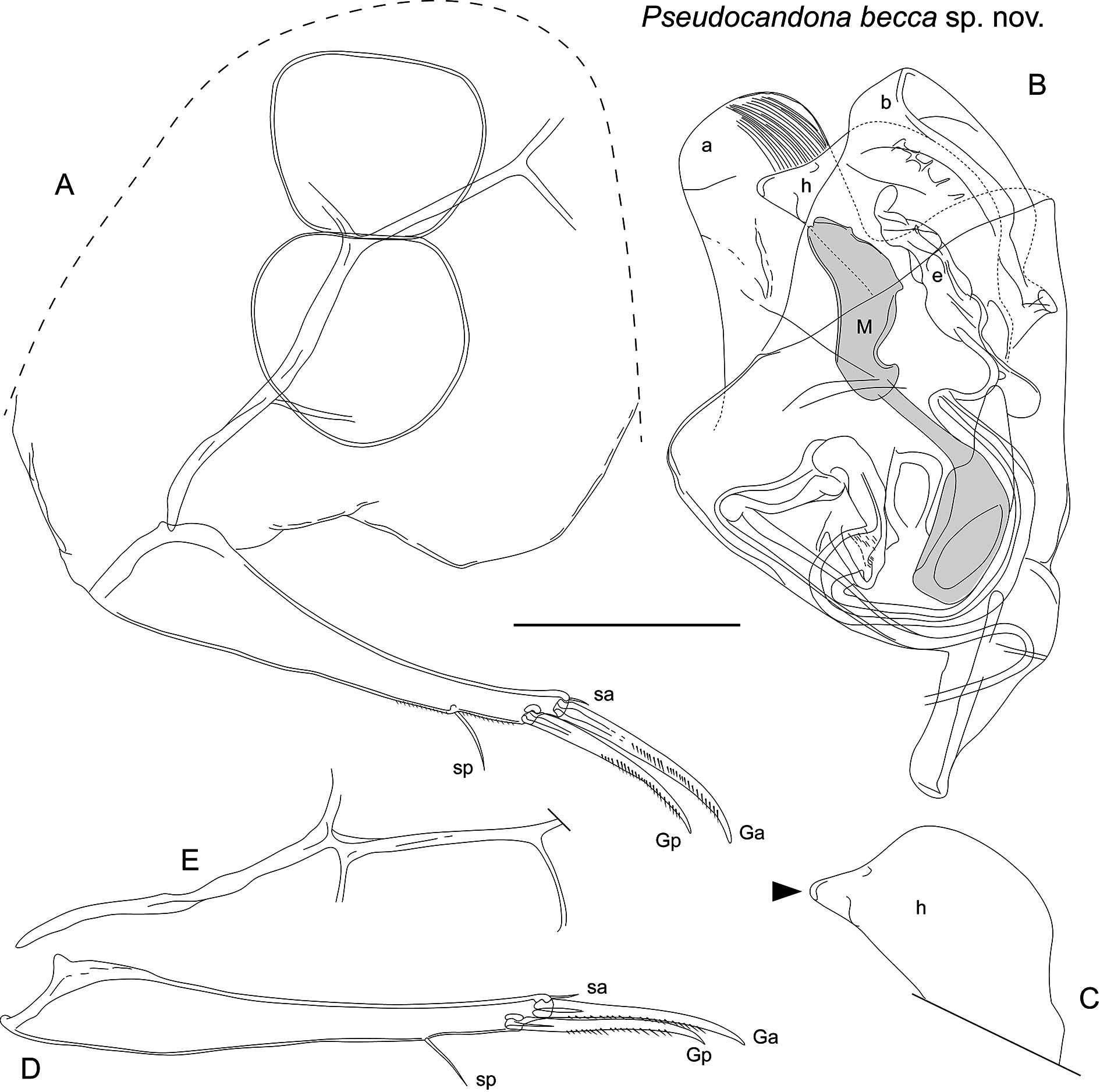
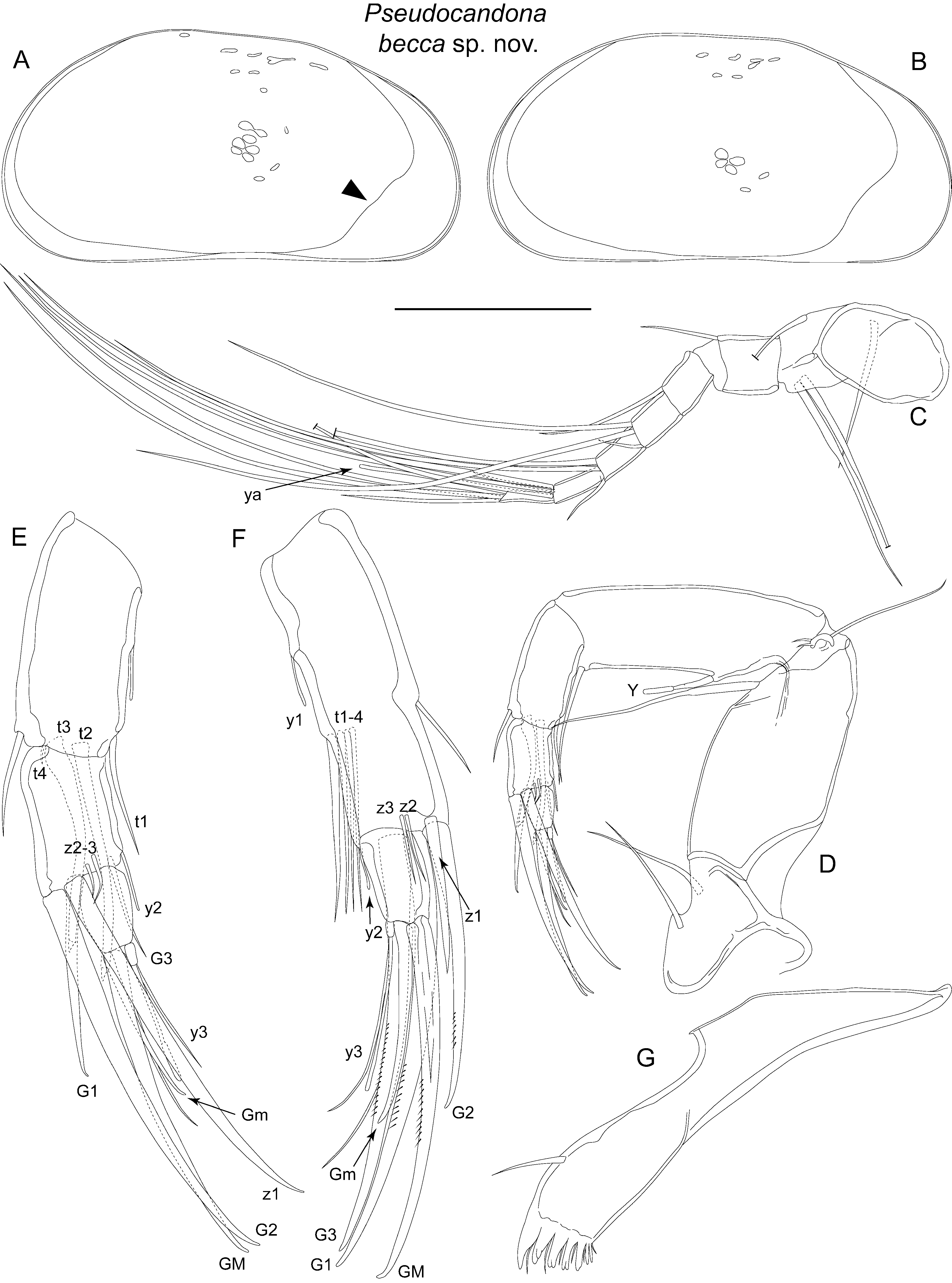
Carapace ( Figs 2 View Fig A–E, 3A–B) length 1135–1181 µm, height 547–594 µm, female carapace slightly less high than male. Posterior margin more inflated than anterior margin, both margins unevenly curved, with maximum curvature below mid-height. Ventral margin slightly concave, dorsal margin straight, sloping down towards anterior. Dorsal view compressed, with anterior end slightly beak-shaped, posterior end slightly pointed. Calcified inner lamella wider anteriorly than posteriorly. Anterior calcified inner lamella not evenly curved but with convexity in the antero-ventral region, forming distinct wider section (marked with triangle on Fig 3A View Fig ). Carapace delicate, with smooth surface. Colour translucent white.
Antennule with seven articulated segments ( Fig. 3C View Fig ). First segment large, supporting two setae on dorsal edge and two long setae on ventral-apical corner. Second and third segments quadrate, with one dorsal-apical seta each. Fourth and fifth segments each with two long dorsal-apical setae and one short ventral-apical seta. Sixth segment with three long and one short apical setae. Final segment with two long and one short setae, and aesthetasc ya.
Male antenna with second endopodite segment sub-divided ( Fig. 3 View Fig D–E). Setae t2 and t3 represented by long male bristles, both similar in morphology, each terminating with small, triangular process. Seta t4 tiny, protruding from near base of t3. Setae z2 and z3 very short, z1 represented by well-developed claw, of similar size to claw G2. Claw G1 about half length of claw G2. Claw Gm on final segment half length of claw GM.
Female antennal ( Fig. 3F View Fig ) claw G2 about two-thirds length of G1, z1 stout and claw-like, about half length of G2, setae z2 and z3 very short. Claw Gm just over half length of claw GM. Exopodite of both sexes with longest seta relatively short, not reaching to end of first endopodal segment ( Fig. 3D View Fig ).
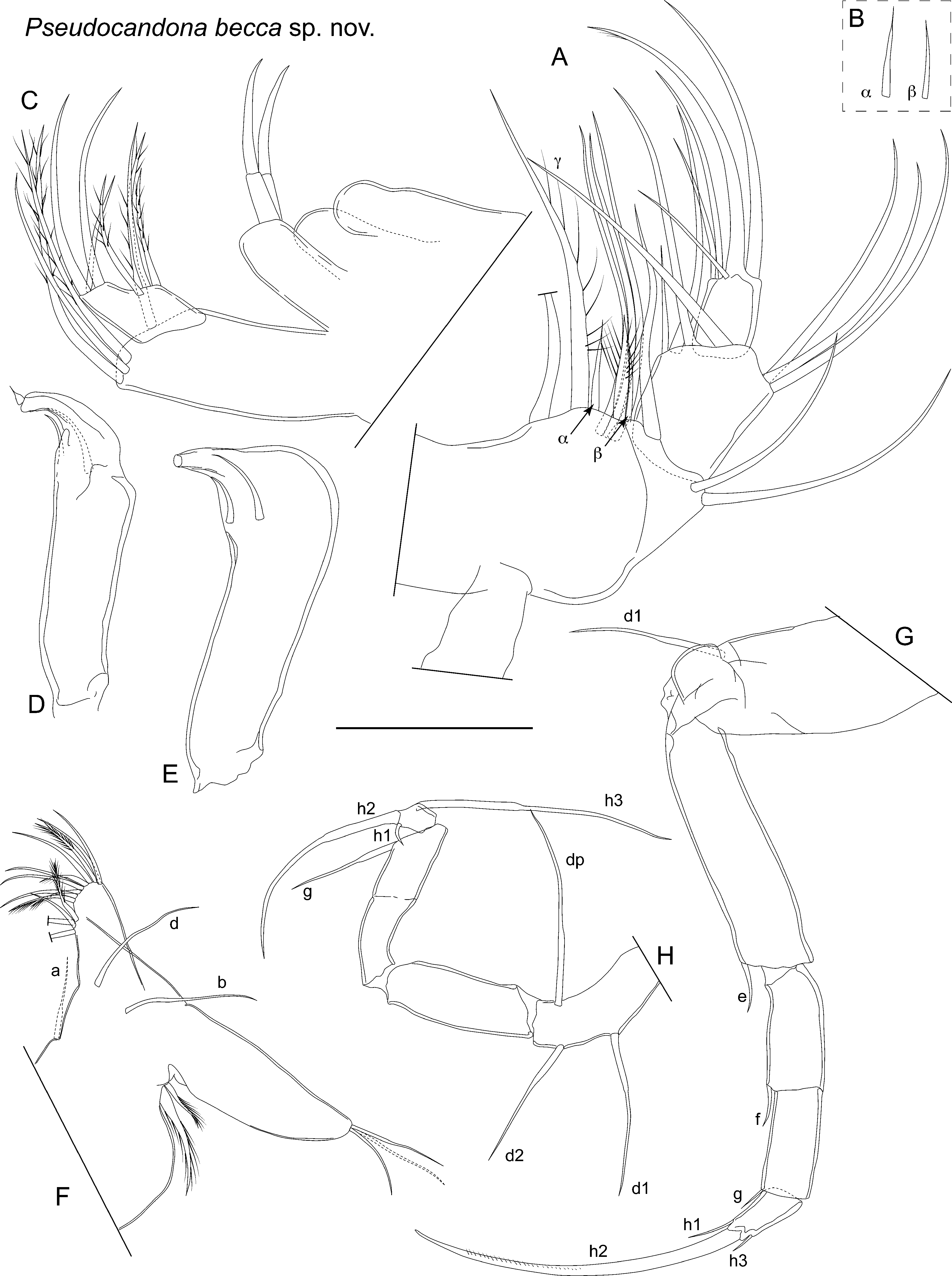
Mandibular palp ( Fig. 4A View Fig ) with four segments. Alpha seta of first segment short ( Fig. 4 View Fig A–B). Inner edge of second segment with 3+1+beta arrangement of setae; beta seta slightly shorter than alpha ( Fig. 4B View Fig ). Outer edge of second segment with two apical setae. Third segment with three long sub-apical setae on outer edge, and three long and one short setae arranged along apical edge; outer-most gamma seta, long and without obvious setules. Final segment with robust seta on outer edge, thick claw-like seta in mid-apical position and three shorter setae on inner apical edge. Six long setae on branchial plate. Mandibular coxa ( Fig. 3G View Fig ) with five well-developed teeth plus two much smaller, spine-like teeth.
Maxillula ( Fig. 4C View Fig ) palp first segment with three setae on apical outer margin, and one apical seta offset towards inner edge. Second segment with stepped apical margin, with outer part more distal than inner part. Outer part of apical margin with two long and one mid-length setae, and inner part with three midlength setae. Branchial plate with morphology typical of subfamily, supporting 18–19 normals rays; number of reflexed rays not observed.
Fifth limb palps of male asymmetrical ( Fig. 4 View Fig D–E). Right palp widens distally to large bulbous hookshaped end, with two sub-apical setae towards inner edge. Left palp distally narrower than right, with finger-like distal hook, and with two sub-apical setae. Fifth limb of female ( Fig. 4 F View Fig ) with long d and b setae, and one a seta.
Sixth limb ( Fig. 4 G View Fig ) five-segmented, with first segment bearing long d1 seta. Setae e and f of second and third segments respectively, both less than half length of next respective segment. Fourth segment with short g seta. Fifth segment with short h1 and h3 seta and well-developed claw h2.
Seventh limb ( Fig. 4 H View Fig ) with five segments, but division between third and fourth segments weak. First segment with long d1, d2 and dp setae. Second and third segments with no setae. Fourth segment with long g seta. Final segment with two long setae (h2 and h3) and one short, reflexed, hooked setae (h1).
Caudal ramus ( Fig. 5 A, D View Fig ) with inflated proximal end, tapering distally, slightly curved. Claw Gp approximately 90% length of claw Ga. Seta sa very short. Seta sp short, about 30% length of claw Gp.
Male sexual organ ( Figs 5 B View Fig , 6 A View Fig ) outer lobe (a) tongue-like, elongate with rounded end and striations towards distal edge. Inner lobe (b) wide, slightly sub-triangular, protruding just beyong outer lobe. Medial lobe (h) shorter than both outer and inner lobes, unevenly rounded with triangular to sub-quadrate process on outer edge ( Fig. 5 C View Fig ). M-process with rounded proximal part tapering to thin, straight central part, and then expanding to large, elongate, roughly triangular distal part; inner edge with indentation towards base of distal part. Bursa copulatrix (e) elongate and irregular in shape, with distal-most part lobe-like and bent off-axis.
Female genital lobe ( Fig. 5 A View Fig ) protruding, sub-triangular to rounded.
Remarks
Smith et al. (2014) reported that this species, as Pseudocandona sp., has sperm ranging from 424 to 475 µm in length.
The combination of the calcified inner lamella with a distinctive convexity in the antero-ventral region of the carapace, the reflexed h1 seta on the seventh limb, and especially the morphology of the hemipenes clearly separate this species from its congeners.
The lateral view of the carapace is similar to that of Pseudocandona renoensis ( Gutentag & Benson, 1962) and Pseudocandona delormei ( Karanovic, 2006) . Pseudocandona renoensis was described from Pleistocene deposits in Kanas, USA ( Gutentag & Benson 1962), but later living specimens were reported from Canada ( Delorme 1970). The lateral view of the female carapace of P. renoensis is similar to that of the female of Pseudocandona becca sp. nov., but the male is higher and more rounded posteriorly than the male of Pseudocandona becca sp. nov. Delorme’s (1970) figure of P. renoensis shows a similar distinctive convexity in the antero-ventral region of the calcified inner lamella of the carapace to that of Pseudocandona becca sp. nov., but this feature is missing in Gutentag & Benson’s (1962) original description of P. renoensis .
Pseudocandona delormei was erected by Karanovic (2006) based on photographs of Pseudocandona hartwigi (G.W. Müller, 1900) and Pseudocandona sarsi (Hartwig, 1899) in Delorme (1970) . Delorme’s specimens of Pseudocandona sarsi were designated by Karanovic (2006) as the type material of P. delormei , even though she was unable to trace this material. Karanovic’s (2006) opinion that the specimens of Pseudocandona hartwigi and Pseudocandona sarsi figured by Delorme (1970) are the same species is probably incorrect, as the lateral views of the valves of both species are different in Delorme’s figures. Pseudocandona hartwigi figured by Delorme (1970) has an distinctive convexity in the antero-ventral region of the calcified inner lamella, similar to that of Pseudocandona becca sp. nov. (absent in Pseudocandona sarsi and other reports of Pseudocandona hartwigi ), but the carapace in lateral view is noticeably higher than that of Pseudocandona becca sp. nov. and the hemipenes are also differently shaped.
Ecology and distribution
So far, this species is only known from the type locality. The species was found by digging a small, shallow hole in the sand at the water’s edge (psammon environment) of the beach at Makino in the north-western part of Lake Biwa.

| LBM |
Lake Biwa Museum |
| F |
Field Museum of Natural History, Botany Department |
| G |
Conservatoire et Jardin botaniques de la Ville de Gen�ve |
| H |
University of Helsinki |
| A |
Harvard University - Arnold Arboretum |
| B |
Botanischer Garten und Botanisches Museum Berlin-Dahlem, Zentraleinrichtung der Freien Universitaet |
| C |
University of Copenhagen |
No known copyright restrictions apply. See Agosti, D., Egloff, W., 2009. Taxonomic information exchange and copyright: the Plazi approach. BMC Research Notes 2009, 2:53 for further explanation.
|
Kingdom |
|
|
Phylum |
|
|
Class |
|
|
Order |
|
|
Family |
|
|
Genus |
Pseudocandona becca
| Smith, Robin James & Kamiya, Takahiro 2015 |
Pseudocandona
| Kaufmann 1900 |