
Tmetopis chinensis Kiritshenko, 1947
|
publication ID |
https://doi.org/ 10.11646/zootaxa.4303.2.3 |
|
publication LSID |
lsid:zoobank.org:pub:F5747B94-6DB7-4680-BD74-EDC201129B7C |
|
DOI |
https://doi.org/10.5281/zenodo.6025417 |
|
persistent identifier |
https://treatment.plazi.org/id/03BC87B9-FFB0-0917-3F95-FEB2FA89F9FC |
|
treatment provided by |
Plazi |
|
scientific name |
Tmetopis chinensis Kiritshenko, 1947 |
| status |
|
Tmetopis chinensis Kiritshenko, 1947
( Figs. 1 View FIGURES 1 – 2 –28)
Tmetopis chinensis Kiritshenko, 1947: 73 , 75. Holotype: ♂, China; ZMAS!
Prionaca jiangxiensis Lin & Zhang, 1989: 332, 334. Holotype: ♂, China: Jiangxi, Waknzai (sic) [= Wanzai]; IZAS! New subjective synonym.
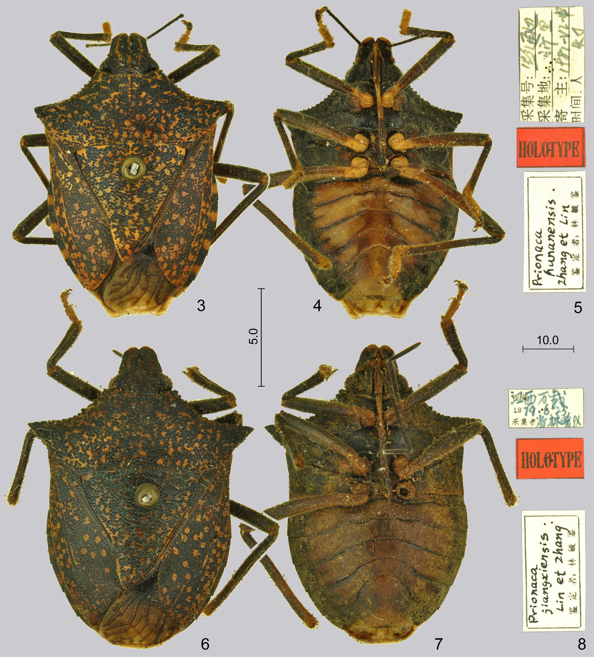
Prionaca hunanensis Lin & Zhang, 1989: 333 , 334. Holotype: ♂, China: Hunan, Tongdao; IZAS! New subjective synonym.
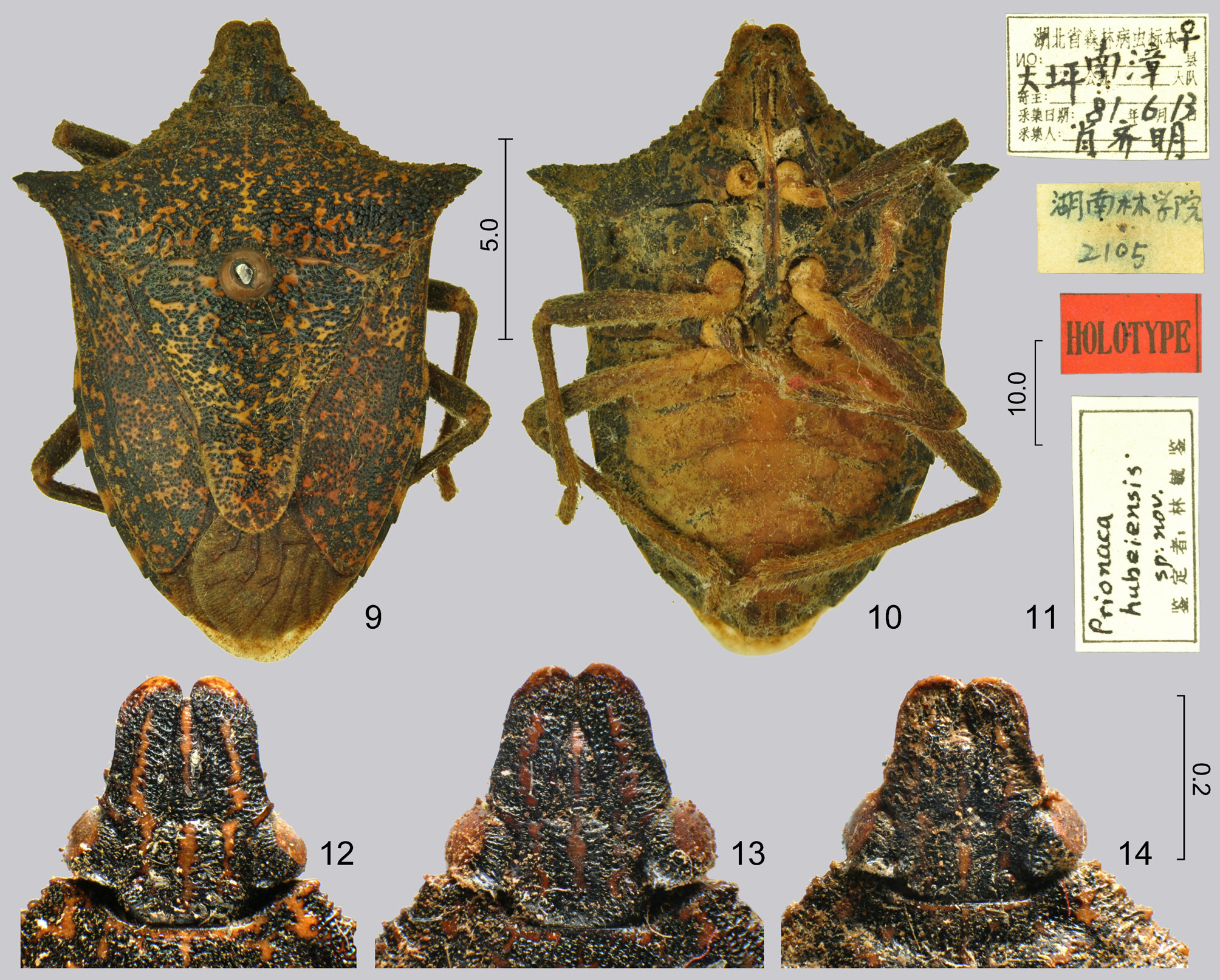
Prionaca hubeiensis Zhang, Lin & Zhao, 1990: 8, 9. Holotype: ♀, China: Hube [i]: Nanchang [= Nanzhang]; IZAS! New subjective synonym.
Tmetopis chinensis: Stichel (1961: 743) (catalogue, distribution), Stichel (1962: 224) (catalogue, distribution), Hua (2000: 180) (listed, distribution), Rider et al. (2002: 145) (type locality, distribution), Rider (2006: 312) (catalogue, distribution).
Prionaca sp.: Zhang & Zhao (1988: 32) (records, host plant).
Prionaca jiangxiensis: Zhang (1994: 34) (listed, distribution), Hua (2000: 179) (listed, distribution), Rider et al. (2002: 144) (listed, distribution), Rider (2006: 336) (catalogue, distribution).
Prionaca hunanensis: Zhang et al. (1992: 245) (redescription, habitus, host plant, record, distribution), Lin et al. 1993: 180 (record, host plant), Hua (2000: 179) (listed, distribution), Rider et al. (2002: 144) (listed, distribution), Rider (2006: 336) (catalogue, distribution).
Prionaca hubeiensis: Zhang et al. (1992: 245) (redescription, habitus, distribution), Lei & Zhou (1998: 41) (listed, distribution), Hua (2000: 179) (listed, distribution), Rider et al. (2002: 144) (listed, distribution), Rider (2006: 336) (catalogue, distribution).
Diagnosis. Being the single known member of Tmetopis, this species can be recognized based on the characters given for the genus.
Redescription. Colour. Ground colour of dorsum appears dark brown due to closely adjacent and confluent black punctures, irregular callose interspaces of punctures form a yellow to orange marmorate pattern on head, pronotum, scutellum, and sclerotized portions of fore wings; dorsum of head with three more or less distinct but irregularly interrupted longitudinal callose vittae, one median along whole length of head, and a pair on mandibular plates from level of apex of clypeus to ocelli; callose patches usually form a distinct median vitta on pronotum and basal portion of scutellum; membrane smoky, veins brownish; connexivum black, segments III–VII each with a large yellow patch occupying whole width of its segment, situated somewhat closer to the posterior margin of the segment than to the anterior one; venter of head and thorax dark blackish brown; abdominal venter ochraceous, broadly margined by blackish brown laterally, but ventral surface usually concealed by a thick, continuous wax layer; antennae brownish black; labium dark ochraceous, black apically; legs blackish brown, coxae, trochanters, and bases of femora (extreme base on fore legs, broader portions on mid and hind legs) ochraceous, tarsi dark ochraceous.
Head ( Figs. 12–14 View FIGURES 9 – 14 , 27–28) about 1.05 times as broad as its median length (measured to an imaginary line connecting apices of mandibular plates), mandibular plates closely approaching ( Fig. 12 View FIGURES 9 – 14 ) or adjacent ( Figs. 13–14 View FIGURES 9 – 14 ) anteriad of apex of clypeus; anteocular projection variously developed, directed laterad or posterolaterad, occasionally might be lacking. Pronotum 2.9–3.0 times as broad as its median length, humeral process somewhat variable, usually relatively longer and narrower in males, more robust in females.
External male genitalia (Figs. 15–23). Genital capsule (Figs. 15–17) broadly oval, infolding of ventral rim (Fig. 15: vif) deeply recessed and form a pair of large, broad excavations ventrolaterally, the wall of which is connected to the external wall of the genital capsule with cuticular struts laterally (Fig. 15: cus); posterior rim deeply emarginate at midline; posteroventral part of genital capsule broadly and deeply impressed medially below posterior rim, this region is connected with the median part of the cuplike sclerite by cuticular struts; cuplike sclerite (Figs. 16–17: cs) with a pair of lobe-like projections anteroventrally which are connected to the external wall of the genital capsule with cuticular struts, suspensory apodeme (Fig. 16: sus) narrow. Paramere (Figs. 18–20) with a broad, oval body provided by an oblique, apically obtuse sensory lobe, its posterior (direction given in the in situ position of the paramere within the genital capsule, cf. Fig. 15) surface deeply excavate and pilose; apical process narrowed and flattened, subapically abruptly angularly curved towards broad lateral arm. Phallus (Fig. 16: ph; Figs. 21–23 View FIGURES 21 – 23 ) directed vertically within genital capsule in rest (cf. Fig. 16); erection fluid pump ( Fig. 21 View FIGURES 21 – 23 : erp) voluminous, without distinct sclerotization; basal plate ( Fig. 22 View FIGURES 21 – 23 : bp) broadly U-shaped; posterior margin of median portion of support bridge complex ( Fig. 21 View FIGURES 21 – 23 : sbc) broadly emarginate, dorsal connective ( Fig. 21 View FIGURES 21 – 23 : dc) thick, terminated in a large capitate process ( Fig. 21 View FIGURES 21 – 23 : cap); phallotheca ( Fig. 21 View FIGURES 21 – 23 : phth) oval, narrowed towards base, basally with a small hinge ( Fig. 22 View FIGURES 21 – 23 : hi) which does not protrude; conjunctiva with a pair of sclerotized, scissorslike, posteriorly diverging dorsal processes ( Fig. 21 View FIGURES 21 – 23 : cp-I) submedially, and a pair of membranous, elongate lateral processes ( Fig. 21 View FIGURES 21 – 23 : cp-II), subdivided into two branches subapically, mesal surface sclerotized proximad of bifurcation, forming a flattened plate, proximomesal portion forming a pair of short, rounded median penial plates ( Fig. 23 View FIGURES 21 – 23 : mpp); basal portion of endophallic reservoir ( Fig. 22 View FIGURES 21 – 23 : res) only slightly shorter than lumen of phallotheca, closely approaching wall of phallotheca dorsally, but laterally flattened, continued in a broad dorsoapical lumen ( Fig. 21 View FIGURES 21 – 23 : dal); aedeagus s. str. ( Fig. 21 View FIGURES 21 – 23 : aed) narrow, S-shaped curved, phallotreme ( Fig. 22 View FIGURES 21 – 23 : phtr) situated subapically, with a large, elongate and slightly broadened ventral “lip” distad of phallotreme, truncate and narrowly membranous at its extreme apical margin.
External female genitalia (Figs. 24–26). Terminalia as in Fig. 24, valvifers VIII (Fig. 24: vf8) broadly rounded posteriorly, their mesal margins divergent posteriad; laterotergites IX (Fig. 24: lt9) broadly rounded posteriorly, slightly surpassing posterior margin of segment VIII, broadly separated by subrectangular segment X (Fig. 24: v10); valvifer IX (Fig. 24: vf9) protruding between contralateral valvifers VIII and laterotergites IX, lateral portions bulbose. Gynatrium (Fig. 25: gy) simple, saccular, without distinct pouches, ring sclerites could not be traced, spermathecal opening surrounded by a V-shaped wrinkle; valvulae IX present as a pair of weakly sclerotized and pigmented elongate zone on distal margin of gynatrium posterolaterad of spermathecal opening. Spermatheca (Figs. 25–26) long; proximal duct distinctly shorter than gynatrium; dilation large, apex surpassing base of ventrite VI (Fig. 25: v6) within abdomen, distal invagination tubular in most of its length, only weakly tapering at base, distal duct about as long as proximal duct and roughly one third as long as dilation, thin, gradually narrowed distally; intermediate part of spermatheca with a narrow flexible zone (Fig. 26: fz) close to apical receptacle, endocuticle thick in its proximal half, only leaving a narrow lumen, broadened in its distal half, surrounding an oval lumen; distal flange placed on basal portion of apical receptacle; apical receptacle globose, with three fingerlike processes surpassing distal flange and reaching to or surpassing flexible zone.
Measurements (in mm) (N = 5). Body length 13.0–16.3; length of head 2.68–3.05 (measured to an imaginary line connecting apices of mandibular plates), greatest width across eyes 2.80–3.18, shortest interocular distance (measured close to anterior margin of eyes) 1.98–2.25; length of scape: pedicel: basiflagellum: distiflagellum as 0.75–0.80: 3.56–3.78: 2.08–2.14: 1.86–1.93; length of pronotum 3.15–3.85, width across apices of humeral processes 9.50–11.3; length of scutellum 5.48–6.97, width at base 5.04–6.31; greatest width of abdomen 7.75–9.30.
Bionomics. Zhang et al. (1992) and Lin et al. (1993) mentioned the tung tree species Vernicia fordii (Hemsl.) Airy Shaw (Euphorbiaceae) as host plant of Prionaca hunanensis (recognized as a junior synonym of Tmetopis chinensis in this paper). Zhang & Zhao (1988) reported an undescribed species of Prionaca collected on unspecified oak species, Quercus sp. ( Fagaceae ), from Nanzhang in Hubei; as this is the type locality of P. hubeiensis, there is no doubt that this record pertains to the same material which was later described by them as P. hubeiensis (also recognized as a junior synonym of T. chinensis in this paper). Specimens which, according to their labels, were collected from Cyclobalanopsis delavayi (Fagaceae) and Juglans regia (Juglandaceae) were seen during the present study.
FIGURES 15–20. Tmetopis chinensis Kiritshenko, 1947 . Fig. 15, genital capsule, posterior view (right paramere removed, right side of anal tube drawn as if it were transparent to reveal dorsal process of infolding of ventral rim); Fig. 16, same, lateral view, proctiger and parameres removed, pilosity omitted, outline of phallus in rest shown schematically; Fig. 17, same, ventral view; Figs. 18–20, right paramere, three different aspects. Arrow in Fig. 19 shows aspect of Fig. 20. Lettering: cs = cuplike sclerite; cus = cuticular struts; lp = left paramere; ph = phallus; sus = suspensory apodeme; t10 = tergite X; vif = infolding of ventral rim of genital capsule. Scales in mm.
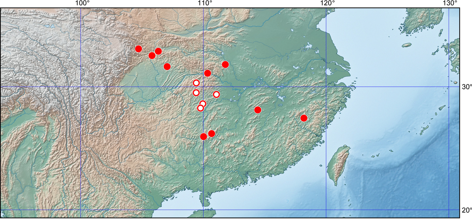
Distribution. The species is rare in collections; so far it is known only from central and southern China, from the Qingling to the Nanling and Wuyi Mountains ( Fig. 29 View FIGURE 29 ). It is apparently restricted to the zone of the subtropical evergreen broadleaf forests. The available information on the distribution of the species is summarized as follows: CHINA. Gansu: Wen County!; Hubei: Nanzhang!, Badong County!, Enshi ( Zhang et al. 1990, as P. hubeiensis), Laifeng ( Zhang & Zhao 1988, as Prionaca sp.; Lei & Zhou 1998, as P. hubeiensis); Shaanxi: Ningqiang!; Hunan: Guzhang ( Zhang et al. 1992, as P. hunanensis ), Xiangxi ( Zhang et al. 1992, as P. jiangxiensis), Cili ( Lin et al. 1993, as P. hunanensis ); Jiangxi: Wanzai!; Sichuan: Pingchang!, Guangyuan County!; Guizhou ( Rider et al. 2002); Guangxi: Longsheng!, Ziyuan!; Fujian: Jiangyang!
Type material examined. Tmetopis chinensis Kiritshenko, 1947 . Holotype: ♂, [gold-coloured circle without text], “84794.” [hw], “China \ Pjassetzky 1876.” [hw], “Tmetopis n. g. [hw] \ chinensis n. sp. [hw] \ Kiritshenko det. [pr]”, “HOLOTYPUS” [red, hw]; pinned, left antenna, right flagellum, tarsus of right fore leg, tibiae and tarsi of right fore and hind legs, and all the remaining legs lacking; deposited in ZMAS.
Prionaca jiangxiensis Lin & Zhang, 1989. Holotype: ♂, “<Tibet> [ch, pr] [in error] <Jiangxi Wanzai> [ch, hw, written over the previous word] \ 19 [pr] 79. 6. 25. [hw] \ <collected:> [ch, pr] <provincial forestry census team> [ch, hw]”, “HOLOTYPE” [red], “Prionaca [hw] \ jiangxiensis [hw] \ Zhang et Lin [hw] \ <identified: Lin Yu Jian> [ch, pr]” [with pr black frame]. Pinned, left and right mid tarsi, right hind tarsus, and left hind leg lacking, ventral surface of body mouldy; deposited in IZAS ( Figs. 6–8 View FIGURES 3 – 8 ).
Prionaca hunanensis Lin & Zhang, 1989 . Holotype: ♂, “<collecting number:> [ch, pr] <Qian [= Guizhou, in error] Tongdao> [ch, hw] \ <collecting locality:> [ch, pr] <Suoli> [ch, hw] \ <host plant:> [ch, pr] 1981-IV- [hw] <middle> [ch, hw] \ <time, person> [ch, pr] <at light> [ch, hw]”, “HOLOTYPE” [red], “Prionaca [hw] \ hunanensis [hw] \ Zhang et Lin [hw] \ <identified: Lin Yu Jian> [ch, pr]” [with pr black frame]. Pinned, left and right flagellum, segments II–III of left fore and right mid tarsi, and right hind tarsus lacking; deposited in IZAS ( Figs. 3–5 View FIGURES 3 – 8 ).
Prionaca hubeiensis Zhang, Lin & Zhao, 1990. Holotype: ♀, “<Hunan Province forestry disease and pest specimen> [ch, pr] ♀ [hw] \ <Daping Nanzhang> [ch, hw, written across three lines of pre-printed ch text] \ <collecting date:> 81 [hw] <year> [ch, pr] 6 [hw] <month> [ch, pr] 13 [hw] <day> [ch, pr] \ <collector:> [ch, pr] <Xiao Qiming> [ch, hw]” [with pr black frame], “<Hunan Forestry Institute> \ 2105” [hw], “HOLOTYPE” [red], “Prionaca [hw] \ hubeiensis [hw] \ sp. nov. [hw] \ <identified: Lin Yu Jian> [ch, pr]” [with pr black frame]. Pinned, right antenna, right fore leg, segments II–III of right mid and hind and left hind tarsi lacking, ventral surface of body mouldy; deposited in IZAS ( Figs. 9–11 View FIGURES 9 – 14 ).
FIGURES 24–28. Tmetopis chinensis Kiritshenko, 1947 . Fig. 24, terminalia of female, posteroventral view; Fig. 25, ectodermal female genitalia, dorsal view after removal of abdominal tergum; Fig. 26, distal part of spermatheca; Fig. 27, head, dorsal view; Fig. 28, same, lateral view. Lettering: fz = flexible zone of intermediate part of spermatheca; gy = gynatrium; lt9 = laterotergite IX; pof = postorbital field; vf8, vf9 = valvifers VIII and IX; v6, v7, v10 = ventrites VI, VII and X. Scales in mm.
Additional specimens examined. CHINA. Gansu: Wen County: Chengguan Township, 13.vii.1990 (1 ♂ NKUM), Wen County : Xinba, 30.vii.1988, from “huánglì” [in Chinese; = Cyclobalanopsis delavayi (Franch.) Schott. ] (1 ♂ NKUM) ; Hubei: Badong County, vi.1960, IOZ(E) 1314804 (1 ♀ IZAS) ; Shaanxi: Ningqiang , middle of v.1981, from “hétáo” [in Chinese; = Juglans regia L.], leg. Y.Z. Sun (2 ♂♂ 1 ♀ NKUM) ; Sichuan: Pingchang, 26.v.1980, IOZ(E)1313420 (1 ♂ IZAS), Guangyuan County, 5.vii. [19]80, IOZ(E)1313418 (1 ♂ IZAS), Guangyuan County , Weizi, 30.vii.1984, leg. Z.H. Tang (1 ♂ 1 ♀ NKUM), without locality, 1956 (1 ♀ NKUM) ; Guangxi: Longsheng, Cujiang , 23.viii.1964, leg. L.C. Wang (1 ♀ NKUM), Ziyuan (1 ♀ NKUM) ; Fujian: Jianyang, Huangkeng, Dazhulan , 900–1170 m, 5.vii.1960, leg. S.Q. Jiang, IOZ(E)1314775 (1 ♂ IZAS) .


No known copyright restrictions apply. See Agosti, D., Egloff, W., 2009. Taxonomic information exchange and copyright: the Plazi approach. BMC Research Notes 2009, 2:53 for further explanation.
|
Kingdom |
|
|
Phylum |
|
|
Class |
|
|
Order |
|
|
Family |
|
|
Genus |
Tmetopis chinensis Kiritshenko, 1947
| Rédei, Dávid 2017 |
hunanensis: Zhang et al. (1992 : 245 )
| Rider 2006: 336 |
| Rider 2002: 144 |
| Hua 2000: 179 |
| Lin 1993: 180 |
| Zhang 1992: 245 |
Prionaca hunanensis
| Lin 1989: 333 |
chinensis:
| Rider 2006: 312 |
| Rider 2002: 145 |
| Hua 2000: 180 |
| Stichel 1962: 224 |
| Stichel 1961: 743 |
Tmetopis chinensis
| Kiritshenko 1947: 73 |