
Synergus rutulus McCracken & Egbert, 1922
|
publication ID |
https://doi.org/ 10.11646/zootaxa.4906.1.1 |
|
publication LSID |
lsid:zoobank.org:pub:09383AAD-8E30-4E50-A533-C6DA4D00E33C |
|
DOI |
https://doi.org/10.5281/zenodo.4433986 |
|
persistent identifier |
https://treatment.plazi.org/id/03BF702A-955E-FF9F-FDE5-FB6C72A2FF86 |
|
treatment provided by |
Plazi |
|
scientific name |
Synergus rutulus McCracken & Egbert, 1922 |
| status |
|
Synergus rutulus McCracken & Egbert, 1922
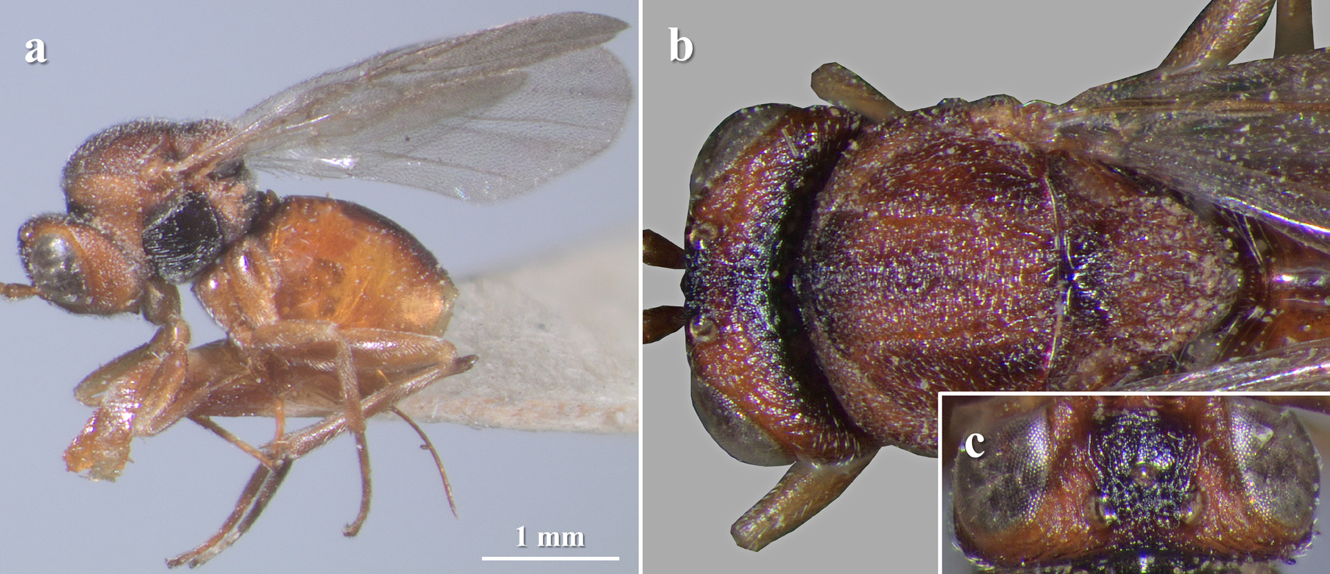
( Figure 37 View FIGURE 37 )
Synergus rutulus McCracken & Egbert, 1922 . Stanford Univ., Pubs., Univ. Ser. Biol. Sci. 3 (1): 60. Type material: CAS.
Type material (examined). HOLOTYPE ♀ with the following labels: ‘ L.S.Jr. U., Lot. 579, Sub. 25’ (white label) / ‘ Congress springs, Vickery col., Oct. 1919 ’ (white label, handwritten) / ‘Stanford Univ., Coll. L.A.C.M, Access’d 1964’ (white label) / ‘ Type rutulus’ (red label, ‘ rutulus’ is handwritten) / ‘ Synergus rutulus McC & Egb.’ (white label, handwritten) / ‘ California Academy of Sciences , Type No. 5816’ (white label) / ‘ Holotype ♀ Synergus rutulus McCracken & Egbert, 1922 ’ (red label).
Diagnosis. Synergus rutulus belongs to a group of species characterized by having the mesopleuron entirely sculptured, covered with parallel and fine transverse striae; hyaline wings and radial cell of fore wing at most 2.5 times as long as wide; malar space 0.6 or more times as long as height of eye; transfacial line as long as height of eye; POL longer than OOL; OOL 1.2–1.3 times as long as diameter of lateral ocelli; gena not broadened behind eye; F1 as long as F2, antenna 14-segmented in females; lateral pronotum wrinkled or carinated, at least ventrally; mesoscutum with transversal carinae, interspaces sculptured; notauli complete, reaching the posterior margin of pronotum; tarsal claws bidentate, with a distinct and conspicuous basal lobe; first metasomal segment completely sulcate dorsally and laterally; syntergum posteriorly with a complete wide band of micropunctures extended laterally more than 1/3 of its length, weakly to not dorsodistally incised in females; and body of females with some dark surfaces, never completely yellow. Synergus rutulus is morphologically close to S. oneratus , whose main differences have already been treated (see the diagnosis of S. oneratus ).
Redescription
FEMALE. Length. Body length 3.2 mm (n = 1).
Color ( Fig. 37 View FIGURE 37 ). Reddish yellow with some dark areas. Head mainly reddish yellow; frons posteriorly, ocellar area and occiput, black. Antenna yellow, the tips darker according to the original description (tips are broken). Mesosoma mainly reddish yellow to dark yellow; pronotum dorsomedially, mesoscutum anteriorly, scutellar foveae, propodeum and mesopleuron, black or dark brown; tegulae, yellow. Metasoma dark yellow to amber, dorsally with a black stripe. Legs yellow, meso- and metacoxae sometimes with a dark spot basally. Wings hyaline, veins dark yellow.
Head. In frontal view subtrapezoid, about 1.3 times as wide as high, gena not broadened behind eye. Face faintly pubescent, lower face with striae radiating from clypeus. Clypeus indistinct, ventral margin slightly projected over mandibles. Malar space almost 0.6 times as long as height of eye. Anterior tentorial pits visible; pleurostomal and epistomal sulcus absent. Transfacial about as long as height of eye. Toruli situated mid-height of eye; distance between torulus and eye shorter than diameter of torulus; distance between toruli clearly shorter than diameter of toruli. Frons coriaceous with some weak wrinkles; frontal carinae branched in their whole length, reaching lateral ocelli. Head in dorsal view ( Fig. 37c View FIGURE 37 ) is about 2.1 times as wide as long. Vertex ( Fig. 37c View FIGURE 37 ) wrinkled, with some punctures. POL: OOL: LOL = 8: 6: 4.5 and diameter of lateral ocelli, 4.5. Occiput weakly wrinkled, without punctures.
Antenna. 14-segmented according to the original description (tip of antenna broken); filiform, not broadened apically; pubescence dense and short. Scape plus pedicel about as long as F1; pedicel 1.3 times as long as wide; F1 about as long as F2, F2 and F3 subequal; the following segments progressively shorter.
Mesosoma. About 1.1 times as long as high in lateral view, including nucha, with short and not dense pubescence ( Fig. 37a View FIGURE 37 ). Ratio of length of pronotum medially/laterally: 0.4. Pronotal plate indistinct. Lateral pronotum carinated to imbricate; lateral carina absent, lateral margins of pronotum rounded seen from above. Mesoscutum ( Fig. 37b View FIGURE 37 ) about 1.1 times as wide as long, strongly carinated, carinae more or less continuous, interspaces alutaceous; anterior parallel lines inconspicuous; notauli complete, well impressed and wide, sometimes interrupted by carinae; median mesoscutal line visible at most as a small triangular incision; parapsidal lines inconspicuous. Mesoscutellum ( Fig. 37b View FIGURE 37 ) about 1.2 times as long as wide, strongly wrinkled; circumscutellar carina well defined but somewhat obscured by wrinkles; scutellar foveae ovate, shallow, weakly sculptured bottom, the posterior margin more or less well defined and separated by a narrow carina. Mesopleuron finely and regularly striated, interspaces weakly alutaceous. Metapleural sulcus reaching 3/4 of mesopleural height. Propodeum pubescent and alutaceous; propodeal carinae narrow, slightly curved and convergent posteriorly. Nucha sulcate dorsally and laterally.
Legs. Tarsal claws with a basal lobe.
Wings. Fore wing pubescent with short marginal setae, slightly longer than the body length. Radial cell closed, about 2.5 times as long as wide; areolet visible, basal vein not well pigmented. Rs+M weakly pigmented, not reaching the basal vein. Basal cell with sparsely spaced setae.
Metasoma. Slightly shorter than head plus mesosoma and about 1.2 times as long as high in lateral view ( Fig. 37a View FIGURE 37 ). First metasomal segment sulcate dorsally and laterally. Syntergum smooth, anterolateral pubescence composed of a few setae and posteriorly with a wide band of micropunctures occupying its distal half; not incised, not pointed. Hypopygial spine about as long as wide and with a few lateral setae; without apical setae.
MALE. Unknown.
Distribution. USA: California ( McCracken & Egbert 1922).
Biology. Reared from galls of Disholcaspis plumbella Kinsey, 1920 on Q. dumosa ( McCracken & Egbert 1922) .
Remarks. Synergus rutulus was described from a single female ( McCracken & Egbert 1922: 60), which has been located and examined.
No known copyright restrictions apply. See Agosti, D., Egloff, W., 2009. Taxonomic information exchange and copyright: the Plazi approach. BMC Research Notes 2009, 2:53 for further explanation.
|
Kingdom |
|
|
Phylum |
|
|
Class |
|
|
Order |
|
|
Family |
|
|
Genus |
Synergus rutulus McCracken & Egbert, 1922
| Lobato-Vila, Irene & Pujade-Villar, Juli 2021 |
Synergus rutulus
| McCracken & Egbert 1922 |