
Synergus, Hartig, 1840
|
publication ID |
https://doi.org/10.11646/zootaxa.4906.1.1 |
|
publication LSID |
lsid:zoobank.org:pub:09383AAD-8E30-4E50-A533-C6DA4D00E33C |
|
DOI |
https://doi.org/10.5281/zenodo.4450030 |
|
persistent identifier |
https://treatment.plazi.org/id/03BF702A-9563-FFB2-FDE5-FDC37206FF33 |
|
treatment provided by |
Plazi |
|
scientific name |
Synergus |
| status |
|
Key to Synergus View in CoL species from the New World
1 Radial cell of fore wing completely open (R1 and Rs veins do not reach the wing margin) or partially open (R1 vein partially running along the wing margin).......................................................................... 2
- Radial cell of fore wing closed along the wing margin (sometimes ambiguously, weakly pigmented)................... 3
2 Tarsal claws simple, without a basal lobe. Radial cell of fore wing 2.1 times as long as wide. F1 somewhat more than 1.5 times as long as F 2 in females, almost 2.0 times in males. F1 of males slightly curved and incised medially, not expanded apically nor basally. Syntergum of females not dorsodistally incised and with a pointed apex (see Pujade-Villar et al. 2015b: Figs 1–3 View FIGURE 1 View FIGURE 2 View FIGURE 3 ; Lobato-Vila et al. 2019: Figs 49–50)............................................................ S. mexicanus
- Tarsal claws bidentate, with a strong basal lobe. Radial cell of fore wing about 3.5 times as long as wide. F1 and F2 subequal in females, F1 about 1.5 times as long as F 2 in males. F1 of males curved and incised medially, more expanded apically than basally. Syntergum of females dorsodistally incised and with a non-pointed apex (see Lobato-Vila et al. 2017: Figs 1–3 View FIGURE 1 View FIGURE 2 View FIGURE 3 ; Lobato-Vila et al. 2020a: Figs 17–21 View FIGURE 17 View FIGURE 18 View FIGURE 19 View FIGURE 20 View FIGURE 21 )................................................................ S. pedroi
3 Mesopleuron partially smooth, with parallel or subparallel transverse striae or wrinkles not extended into the speculum, which is always entirely not sculptured, glabrous, and shining; sometimes, mesopleuron only has a few transversal striae medially, the rest of its surface being smooth, glabrous, and shining like the speculum (see Lobato-Vila & Pujade-Villar 2017: Figs 1e View FIGURE 1 ; 3b View FIGURE 3 ; 4d View FIGURE 4 ; 5i View FIGURE 5 ; 6g View FIGURE 6 ; 7j View FIGURE 7 )........................................................................................ 4
- Mesopleuron entirely sculptured, with parallel or subparallel transverse striae or wrinkles covering all of the surface, including the speculum (as in Figs 6g View FIGURE 6 ; 14 View FIGURE 14 h–i; 15b; 22g), which rarely appears very finely aciculate (as in Fig. 18h View FIGURE 18 ); sometimes, a small smooth patch can be found in the middle of or under the speculum (as in Figs 3a View FIGURE 3 ; 9g View FIGURE 9 ), but the rest of its surface is always covered with striae...................................................................................... 24
4 Lateral frontal carinae present (see Lobato-Vila & Pujade-Villar 2017: Figs 2a View FIGURE 2 , c–d; 6b–c). Sometimes very short, running just a little from the posterior margin of toruli and eclipsed by antennae, rarely appearing as multiple striae running from lower face and toruli to ocelli (as in S. striatifrons , see Lobato-Vila & Pujade-Villar, 2017: Fig. 7a View FIGURE 7 , c–d), but always visible. Syntergum with or without punctures; if present, they can form either a posterodorsal patch or a band............................ 5
- Lateral frontal carinae completely absent (see Lobato-Vila et al. 2020c: Fig. 3 View FIGURE 3 a–b). Syntergum with or without punctures, but if present, they form at most a posterodorsal patch, never a band............................................... 17
5 Syntergum posteriorly without punctures or just with some micropunctures sometimes forming a posterodorsal patch (except in males of S. stelluli , which have a narrow band, but in this case the radial cell of fore wing is 2.1 times as long as wide and vertex has some wrinkles in the ocellar area)................................................................ 6
- Syntergum posteriorly with a more or less extended band of punctures, occupying from 1/5 to 1/2 of its length.......... 12
6 Mesoscutum with strong, widely spaced discontinuous transversal carinae. Lateral pronotum strongly transversely carinate. Notauli complete and well impressed in their whole length, sometimes interrupted by transversal carinae. Pedicel as long as wide. Reared from tuberous galls (see Lobato-Vila et al. 2019: Figs 36–37 View FIGURE 36 View FIGURE 37 )............................... S. mendax
- Mesoscutal sculpture different; finely coriaceous or coriaceous, coriaceous to imbricated or just with weak, discontinuous and usually dense transversal elements not forming true carinae (see Lobato-Vila et al. 2020c: Figs 1b View FIGURE 1 ; 4d View FIGURE 4 ). Lateral pronotum finely coriaceous, coriaceous or imbricated, sometimes with some carinae in the basal half. Notauli incomplete and usually shallow, faint anteriorly, not reaching the posterior margin of pronotum (except in S. castanopsidis , which are complete but less impressed anteriorly, but in this case the mesoscutum never has strong transversal carinae). Pedicel longer than wide. Reared from different morphotypes of galls, except from tuberous..................................................... 7
7 Radial cell of fore wing very short, 2.1 times as long as wide. Vertex slightly wrinkled in the ocellar area. POL as long as OOL. Malar space 0.5 times as long as height of eye. Scutellar foveae inconspicuous (see Burnett 1976: Figs A–I)...... S. stelluli
- Radial cell of fore wing longer, at least 2.3 times as long as wide. Vertex coriaceous, with or without small piliferous punctures, but always without wrinkles (see Lobato-Vila et al. 2020c: Figs 1b View FIGURE 1 ; 4d View FIGURE 4 ). POL longer than OOL (except in females of S. striatifrons , which is subequal, but in this case frons is covered with striae projecting from toruli and lower face). Malar space longer, at least 0.6 times as long as height of eye. Scutellar foveae sometimes small, shallow and/or not well defined posteriorly, but always visible........................................................................................ 8
8 Frons covered with multiple fine striae projecting from lower face and toruli to ocelli. Malar space 0.8 times as long as height of eye. Head in frontal view more or less quadrate, the transfacial line long, 1.3 times as long as height of eye. In females, POL about as long as OOL. Circumscutellar carina projected and upturned (see Lobato-Vila & Pujade-Villar 2017: Fig. 7 View FIGURE 7 )….............................................................................................. S. striatifrons
- Frons coriaceous, without striae. Malar space shorter. Head in frontal view round or trapezoid, the transfacial line as long as height of eye or just slightly longer. In females, POL longer than OOL. Circumscutellar carina weak, neither projected nor upturned............................................................................................ 9
9 Notauli complete, reaching the posterior margin of pronotum. Median mesoscutal line appearing as a short basal sulcus. Scutellar foveae large, subquadrate, weakly sculptured bottom. Female syntergum dorsodistally incised (see Pujade-Villar & Melika 2005: Fig. 1 View FIGURE 1 )............................................................................ S. castanopsidis
- Notauli incomplete, faint in the anterior third of the mesoscutum or before reaching the pronotal margin. Median mesoscutal line absent. Scutellar foveae small, ovate or subtriangular, smooth bottom. Female syntergum not dorsodistally incised.... 10
10 Transfacial line slightly longer than height of eye. Mesoscutum finely coriaceous to reticulated, without transversal elements. Mesopleuron basally and medially with spaced striae. Body mainly brownish yellow to yellow (see Lobato-Vila et al. 2020c: Fig. 1 View FIGURE 1 a–b)................................................................................. S. agrifoliae
- Transfacial line as long as height of eye. Mesoscutum coriaceous or imbricated, with weak, discontinuous transversal elements (see Lobato-Vila et al. 2020c: Fig. 4d View FIGURE 4 ). Mesopleuron basally and medially with dense striae. Body mainly black, dark brown or rufous, with some yellowish areas (see Lobato-Vila et al. 2020c: Fig. 4 View FIGURE 4 c–d)...................................... 11
11 Head yellow, except for a black spot in the ocellar area. Vertex with some small punctures. OOL 1.7 times as long as diameter of lateral ocelli. Lateral pronotum dorsally and medially imbricated, weakly wrinkled basally. Scutellar foveae small, ovate. Metasoma about 1.2 times as long as high in lateral view, the syntergum with a posterodorsal patch of micropunctures somewhat laterally extended. Hypopygial spine almost 3.0 times as long as wide (see Lobato-Vila et al. 2020c: Fig. 4 View FIGURE 4 c–d)................................................................................................. S. confertus
- Head dark brown to black. Vertex without punctures. OOL 1.4 times as long as diameter of lateral ocelli. Lateral pronotum coriaceous. Scutellar foveae subtriangular, smooth bottom. Metasoma about as long as high in lateral view, the syntergum posteriorly without punctures or just with a few, not forming a true patch. Hypopygial spine about as long as wide (see Lobato-Vila et al. 2020b: Figs 53–54)........................................................................ S. walshii
12 Head and body mainly black, except for a yellow or brownish to chestnut halo surrounding the oral fovea (except in males of S. punctatus , which have the head yellow with a black spot in the ocellar area) (see Lobato-Vila & Pujade-Villar 2017: Fig. 8 View FIGURE 8 a–b). Frons and vertex coriaceous or reticulated, without punctures. OOL about as long as diameter of lateral ocelli. Circumscutellar carina inconspicuous or absent.......................................................................... 13
- Head and body never almost completely black, at most yellowish brown with some dark areas, brownish black or rufous (see Lobato-Vila et al. 2020c: Figs 4 View FIGURE 4 a–b, e–f; 5a–b, e–f). Frons and vertex finely coriaceous to coriaceous, with some small punctures. OOL longer than diameter of lateral ocelli. Circumscutellar carina weak, but always visible….................. 14
13 In females, head black except for a well delimited yellow halo around the oral fovea; in males, head yellow except for a black spot in the ocellar area. F1 1.4 times as long as F 2 in males. Notauli incomplete, faint in the anterior third of the mesoscutum. Syntergum with a complete band of micropunctures occupying the distal half of its length (see Lobato-Vila & Pujade-Villar 2017: Figs 6 View FIGURE 6 ; 8a View FIGURE 8 )............................................................................. S. punctatus
- Head black in both sexes, except for a not well defined brownish to chestnut halo around the oral fovea. F1 slightly longer than F 2 in males. Notauli complete and visible in their whole length, less impressed anteriorly. Syntergum with an incomplete band of micropunctures occupying about 1/3 of its length (see Lobato-Vila & Pujade-Villar 2017: Figs 2 View FIGURE 2 ; 3 View FIGURE 3 ; 8b View FIGURE 8 )........ S. gilletti
14 Notauli complete, reaching the posterior margin of pronotum, but narrower and less impressed anteriorly (see Lobato-Vila et al. 2020c: Figs 4b View FIGURE 4 ; 5f View FIGURE 5 )................................................................................ 15
- Notauli incomplete, faint in the anterior half or third of the mesoscutum, not reaching the posterior margin of pronotum (see Lobato-Vila et al. 2020c: Figs 4f View FIGURE 4 ; 5b View FIGURE 5 ).................................................................... 16
15 POL about as long as OOL. F1 1.5 times as long as F2. Lateral pronotum strongly imbricated to weakly carinated, especially basally (see Lobato-Vila et al. 2020c: Fig. 5 View FIGURE 5 e–f).................................................. S. succinipedis
- POL longer, about 1.8 times as long as OOL. F1 and F2 subequal. Lateral pronotum completely coriaceous (see Lobato-Vila et al. 2020c: Fig. 4 View FIGURE 4 a–b)........................................................................ S. campanula
16 Scutellar foveae large, subquadrangular. In females, syntergum weakly dorsodistally incised and last flagellar segment more than 4.0 times as long as wide. Males almost completely yellow, with the malar space about 0.7 times as long as height of eye and POL 1.7 times as long as OOL (see Lobato-Vila et al. 2020c: Fig. 4 View FIGURE 4 e–f)................................ S. flavens
- Scutellar foveae ovate to subtriangular. In females, syntergum not dorsodistally incised and last flagellar segment about 3.0 times as long as wide. Males mainly black, with the malar space about 0.5 times as long as height of eye and POL 2.2 times as long as OOL (see Lobato-Vila et al. 2020c: Fig. 5 View FIGURE 5 a–b)…............................................. S. pacificus
17 Head strongly transverse in dorsal view, more than 2.5 times as wide as long (see Nieves-Aldrey & Medianero 2011: Fig. 3C View FIGURE 3 ). Malar space about as long as height of eye (see Nieves-Aldrey & Medianero 2011: Fig. 1B View FIGURE 1 ).............. S. laticephalus
- Head in dorsal view about 2.0 times as wide as long. Malar space at most 0.8 times as long as height of eye............. 18
18 In females, body mainly black, head black except for a brownish orange surface under eyes and around the oral fovea; in males, lower face, gena and antennae (except the last segments), yellowish orange. OOL almost 3.0 times as long as diameter of lateral ocelli. Radial cell of fore wing about 2.7 times as long as wide in females, with R1 vein weakly pigmented and Rs vein strongly projected beyond the end of the radial cell along the wing margin in both males and females (see Lobato-Vila et al. 2020c: Figs 2–3 View FIGURE 2 View FIGURE 3 )...................................................................................... S. aurofacies
- In both sexes, body mainly yellow, yellowish brown, brown or black and yellow, with head at least with lower face and gena yellow or yellowish brown (see Lobato-Vila et al. 2020c: Figs 4 View FIGURE 4 g–h; 5c–d). OOL about 2.0 or less times as long as diameter of lateral ocelli. Radial cell of fore wing about 2.4 times as long as wide in females, with R1 vein either weakly pigmented or not, and Rs not so strongly projected in both sexes.............................................................. 19
19 Body yellow to yellowish brown or brown. Scutellar foveae large, subquadrate to ovate. Circumscutellar carinae weak, not well defined (see Lobato-Vila et al. 2020c: 5 c–d)....................................................... S. pomiformis
- Body brown or black with yellowish areas. Scutellar foveae absent, inconspicuous or small and shallow. Circumscutellar carina well defined, sometimes upturned and projected............................................................ 20
20 Mesoscutum alutaceous to finely coriaceous, with weak, discontinuous transversal elements not forming true carinae. Notauli complete, reaching the posterior margin of pronotum, narrow. Scutellar foveae small, circular, separated by a wide septum. Syntergum punctuation absent (see Lobato-Vila et al. 2020c: 4 g–h).................................... S. laeviventris
- Mesoscutum alutaceous to coriaceous or weakly imbricated, never with transversal elements. Notauli incomplete, faint in the anterior third or before reaching the pronotal margin (except in S. longimalaris , which are complete, but in this case the malar space is 0.8 times as long as height of eye and the transfacial line, 1.3 times as long as height of eye). Scutellar foveae shallowly impressed and almost inconspicuous or absent. Syntergum punctuation composed at most by some micropunctures either forming or not a posterodorsal patch......................................................................... 21
21 Malar space 0.6 times as long as height of eye............................................................. 22
- Malar space longer, 0.8 times as long as height of eye........................................................ 23
22 Scape long, longer than F 1 in females and about as long as F 1 in males. F1 as long as F 2 in females and just slightly longer in males. Transfacial line about as long as height of eye (see Lobato-Vila & Pujade-Villar 2017: Fig. 5 View FIGURE 5 )........ S. longiscapus
- Scape shorter than F1 and F1 1.3 times as long as F 2 in both sexes. Transfacial line about 1.3 times as long as height of eye (see Lobato-Vila et al. 2020b: Figs 9–12 View FIGURE 9 View FIGURE 10 View FIGURE 11 View FIGURE 12 )............................................................ S. citriformis
23 POL as long as OOL. OOL 2.2 times as long as diameter of lateral ocelli. F1 1.5 times as long as F2. Notauli complete. Mesoscutellum alutaceous to finely coriaceous or reticulated. Syntergum with a few weak posterodorsal micropunctures forming a small patch; following segments and hypopygium, which is completely covered by the syntergum, not punctate (see Lobato-Vila & Pujade-Villar 2017: Fig. 4 View FIGURE 4 )............................................................. S. longimalaris
- POL 1.5 times as long as OOL. OOL 1.5 times as long as diameter of lateral ocelli. F1 1.3 times as long as F2. Notauli almost complete, faint before reaching the pronotal margin. Mesoscutellum anteriorly coriaceous, posteriorly imbricated. Syntergum with a conspicuous posterodorsal patch of micropunctures; following segments and hypopygium, which is visible and projected (not covered by the syntergum), punctate (see Lobato-Vila & Pujade-Villar 2017: Fig. 1 View FIGURE 1 ).................... S. cibriani
24 Wings smoky ( Figs 3b View FIGURE 3 ; 12b View FIGURE 12 ), or hyaline but with a shaded infuscated area on the radial cell (see Nieves-Aldrey & Medianero 2011: Figs 17E View FIGURE 17 ; 19 View FIGURE 19 A–B)............................................................................... 25
- Wings entirely hyaline, without infuscated areas (as in Figs 2a View FIGURE 2 ; 4a View FIGURE 4 ; 8a View FIGURE 8 ).......................................... 27
25 Wings hyaline, but with a shaded infuscated area on the radial cell. Mesoscutum with strong and widely spaced discontinuous transversal carinae (see Nieves-Aldrey & Medianero 2011: Fig. 8F View FIGURE 8 ). Female mesosoma mostly yellowish orange, metasoma black (see Nieves-Aldrey & Medianero 2011: Fig. 19A View FIGURE 19 ).......................................... S. nicaraguensis
- Wings smoky, without a shaded infuscated area on the radial cell. Mesoscutum with weak to moderately strong, dense discontinuous transversal carinae ( Figs 3c View FIGURE 3 ; 12c View FIGURE 12 ). Female mesosoma black, metasoma amber ( Figs 3b View FIGURE 3 ; 12b View FIGURE 12 )................... 26
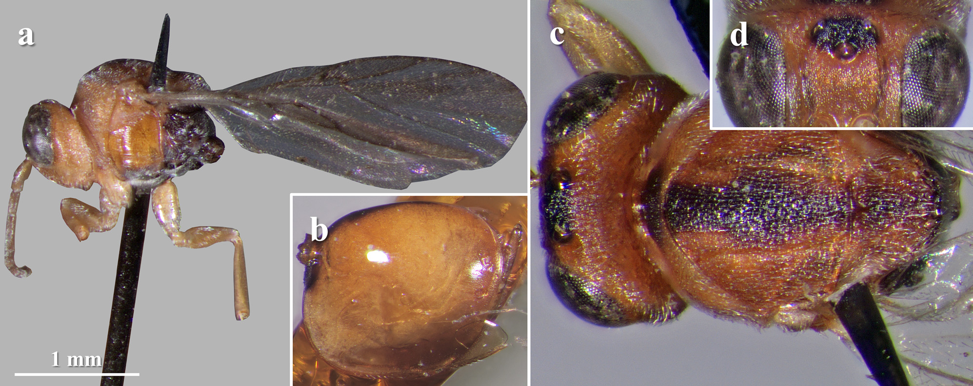
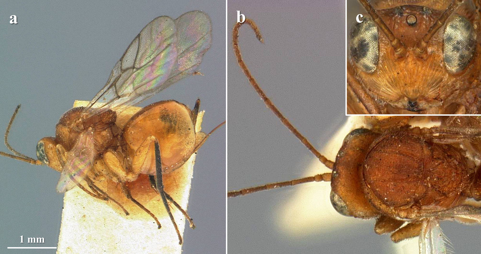
26 Female antenna with 15 segments ( Fig. 3c View FIGURE 3 ). Vertex coriaceous, with small piliferous punctures ( Fig. 3c View FIGURE 3 ). Mesopleuron with regular, dense and well-marked transversal striae covering all of the surface, except for a small aciculate, almost smooth, spot under the speculum ( Fig. 3a View FIGURE 3 ). In females, syntergum strongly dorsodistally incised ( Fig. 3b View FIGURE 3 ). Head amber, except frons, vertex and occiput, black ( Fig. 3 View FIGURE 3 b–d). Legs entirely amber ( Fig. 3b View FIGURE 3 ).......................................... S. atripennis
- Female antenna with 14 segments ( Fig. 12b View FIGURE 12 ). Vertex wrinkled, with some punctures ( Fig. 12c View FIGURE 12 ). Mesopleuron with regular, widely spaced and well-marked transversal striae covering all of the surface ( Fig. 12b View FIGURE 12 ). In females, syntergum weakly to not dorsodistally incised ( Fig. 12a View FIGURE 12 ). Head entirely black ( Fig. 12 View FIGURE 12 b–c). Legs amber, coxae and trochanters mostly black ( Fig. 12b View FIGURE 12 ) S. distinctus
27 Notauli incomplete, faint in the anterior half or third of the mesoscutum or just before reaching the posterior margin of pronotum (as in Figs 4b View FIGURE 4 ; 18j View FIGURE 18 ; 36l View FIGURE 36 ; 39b View FIGURE 39 ), rarely absent ( Fig. 1g View FIGURE 1 )..................................................... 28
- Notauli complete; sometimes narrower or less impressed and interrupted by the sculpture of the mesoscutum anteriorly, but always reaching the posterior margin of pronotum (as in Figs 6i View FIGURE 6 ; 22i View FIGURE 22 ; 24i View FIGURE 24 ; 27h View FIGURE 27 )................................... 33
28 Notauli absent; mesoscutum coriaceous-punctate, punctures conspicuous and covering all of the surface; mesoscutellum medially coriaceous, laterally and posteriorly wrinkled ( Fig. 1g View FIGURE 1 )................................... S. ashmeadi , sp. nov.
- Notauli present, incomplete, faint in the anterior half or third of the mesoscutum or just before reaching the posterior margin of pronotum; mesoscutum coriaceous, imbricated or transversely carinated, never coriaceous-punctate; mesoscutellum entirely coriaceous, carinated or wrinkled (as in Figs 4b View FIGURE 4 ; 18j View FIGURE 18 ; 36l View FIGURE 36 ; 39b View FIGURE 39 )................................................ 29
29 Mesoscutum strongly transversely carinated, either with closely or widely spaced carinae (as in Fig. 4b View FIGURE 4 ). Scutellar foveae inconspicuous to absent. Mesopleuron with well-impressed transversal, but non-parallel, striae or wrinkles covering all of the surface, sometimes reticulated. Radial cell of fore wing short, 2.2 times as long as wide (as in Fig. 4a View FIGURE 4 ). First metasomal segment smooth or with striae reaching dorsally only the half of its length (as in Fig. 4b View FIGURE 4 ). Hypopygial spine at least 1.5 times as long as wide............................................................................................... 30
- Mesoscutum coriaceous, imbricated or weakly and densely transversely carinated. Scutellar foveae visible and traceable, either well defined and well-marked or shallow and less defined posteriorly ( Figs 18j View FIGURE 18 ; 36l View FIGURE 36 ; 39b View FIGURE 39 ). Mesopleuron finely, regularly and densely transversely striated, speculum medially very finely aciculate to almost smooth ( Figs 20h View FIGURE 20 ; 36i View FIGURE 36 ; 39a View FIGURE 39 ). Radial cell of fore wing at least 2.5 times as long as wide ( Figs 18g View FIGURE 18 ; 36h View FIGURE 36 ). First metasomal segment sulcate dorsally and laterally, with complete striae ( Figs 18i, k View FIGURE 18 ; 36j, m View FIGURE 36 ; 39a View FIGURE 39 ). Hypopygial spine as long as wide.............................................. 31
30 Frons sharply and finely striated beneath toruli, striae running from toruli to lateral ocelli, without punctures ( Fig. 4c View FIGURE 4 ). Vertex finely striated, with small piliferous punctures. OOL 2.0 times as long as diameter of lateral ocelli ( Fig. 4c View FIGURE 4 ). Mesoscutum with strong, widely spaced transversal carinae ( Fig. 4b View FIGURE 4 ). Tarsal claws with a strong basal lobe. In males, F1 2.0 times as long as F2; F1 long, slightly curved and slightly expanded apically and basally. In both sexes, frons, vertex and occiput, black, the rest of the head lighter ( Fig. 4 View FIGURE 4 a–c), and hind legs almost completely black ( Fig. 4a View FIGURE 4 )............................... S. atripes
- Frons and vertex wrinkled and punctate, punctures on vertex stronger. OOL 1.5 times as long as diameter of lateral ocelli. Mesoscutum with strong and dense transversal carinae. Tarsal claws with a very small basal lobe. In males, F1 1.5 times as long as F2; F1 strongly curved, kidney shaped. In females, frons, vertex and occiput, black, the rest of the head lighter; in males, head yellow except for a dark spot in the ocellar area. In both sexes, hind legs yellow, basal half of metacoxae dark (see Lobato-Vila et al. 2019: Figs 1–2 View FIGURE 1 View FIGURE 2 )......................................................................... S. batatoides
31 In both sexes, head, mesosoma and metasoma completely ochre or amber, antennae yellow, legs pale yellow ( Fig. 39 View FIGURE 39 a–c). Gena slightly broadened behind eye, visible in frontal view. Frons and vertex alutaceous to finely coriaceous, without punctures nor wrinkles ( Fig. 39b View FIGURE 39 ). Frontal carinae very short, running just a little from toruli, obscured by antennae. F1 and F2 subequal in females (1.1), at most 1.2 in males. Syntergum posteriorly with a wide band of micropunctures occupying almost 1/3 of its length........................................................................................ S. virentis
- In both sexes, head, mesosoma, metasoma, antennae and legs black or at least with some dark surfaces, body never completely yellow, ochre or amber ( Figs 17 View FIGURE 17 a–b; 35a–b). Gena not broadened behind eye, not visible in frontal view ( Figs 18a View FIGURE 18 ; 36a View FIGURE 36 ). Frons and vertex finely coriaceous with some small punctures, sometimes also with very weak wrinkles. Frontal carinae absent ( Figs 18 View FIGURE 18 a–b; 36a–b). F1 at least 1.3 times as long as F 2 in females, almost 2.0 times in males. Syntergum with a posterodorsal small patch of micropunctures, sometimes inconspicuous ( Figs 18 View FIGURE 18 k–l; 36j–k, m)....................................... 32
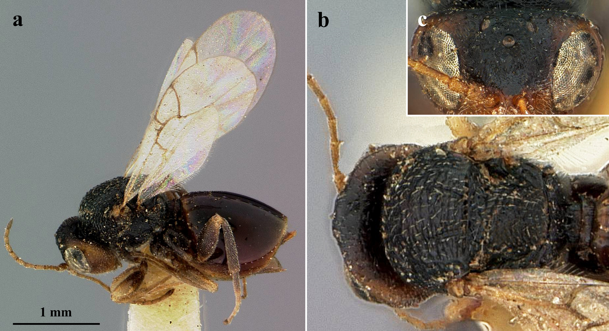
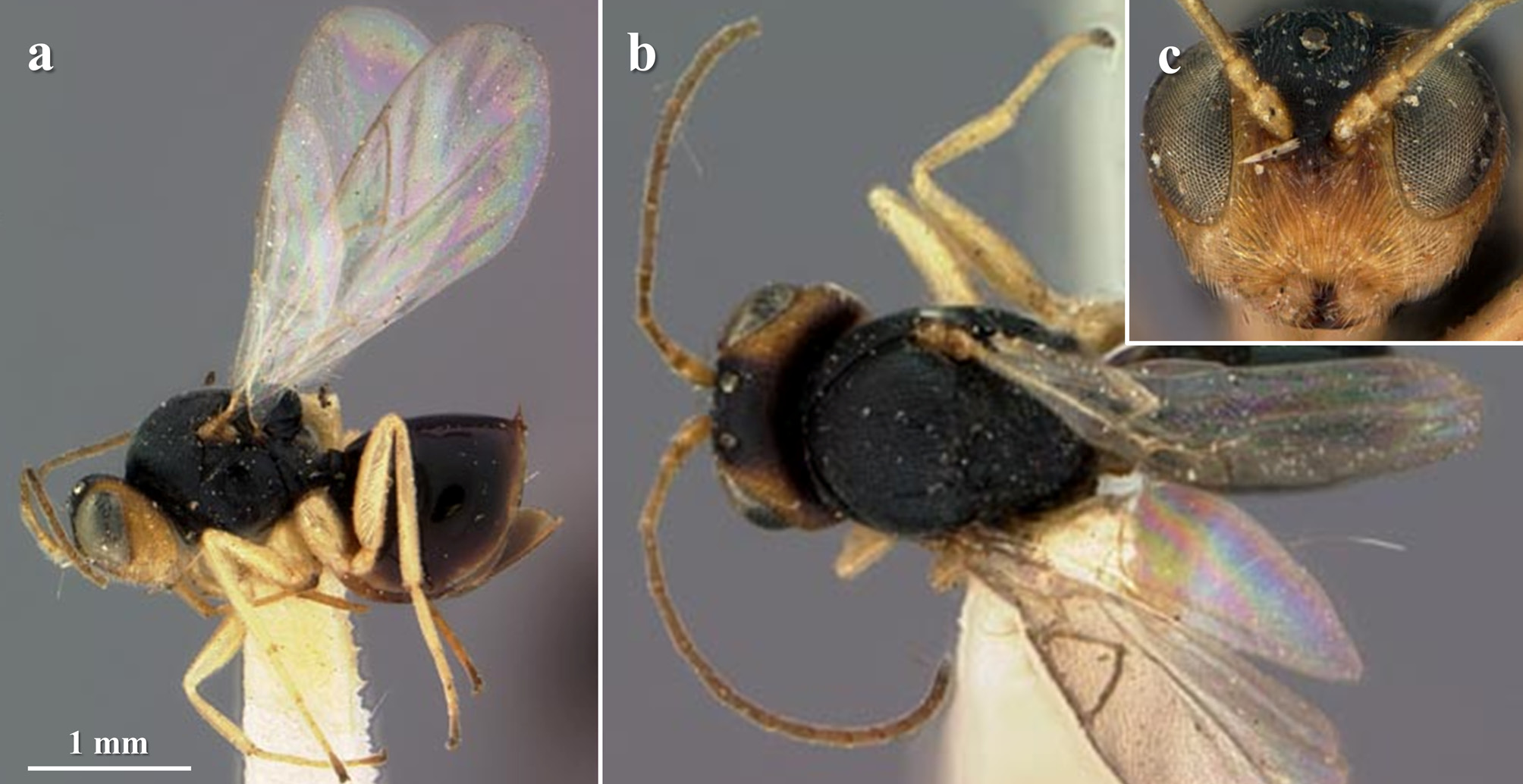
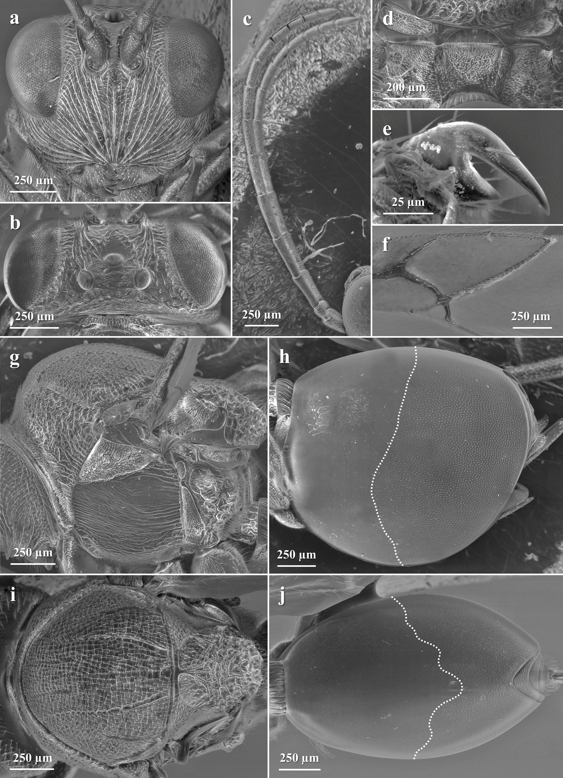
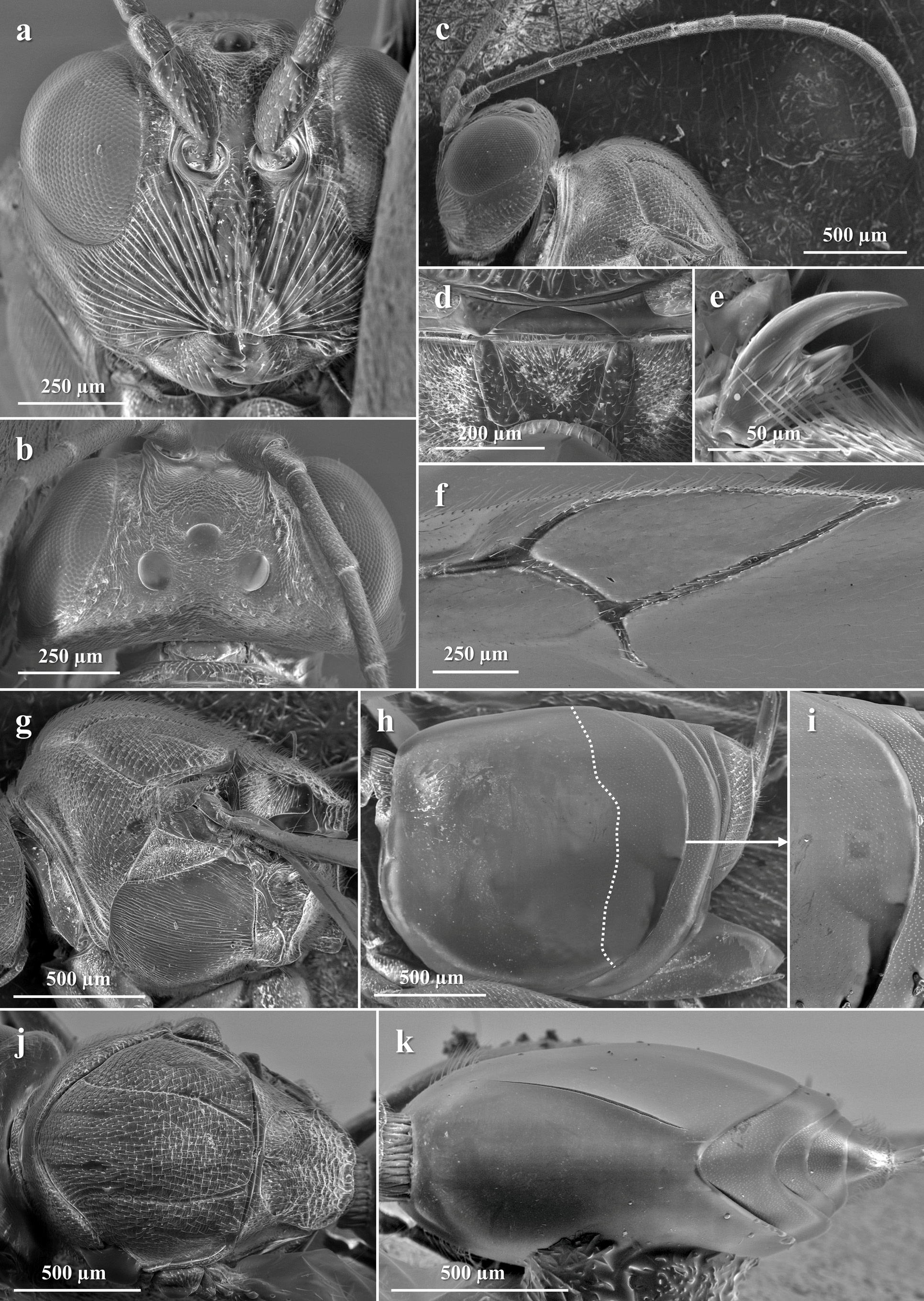
32 In both sexes, head, mesosoma and metasoma dark brown or black ( Fig. 17 View FIGURE 17 a–b). Transfacial line as long as height of eye ( Fig. 18a View FIGURE 18 ). POL about 2.0 times as long as OOL ( Fig. 18b View FIGURE 18 ). F1 1.3 times as long as F 2 in females ( Fig. 18c View FIGURE 18 ), almost 2.0 times in males ( Fig. 18d View FIGURE 18 ). Mesoscutum and mesoscutellum coriaceous; scutellar foveae not well defined, with a weakly sculptured bottom and separated by a narrow carina; circumscutellar carina well defined, slightly projected and upturned ( Fig. 18j View FIGURE 18 ). Radial cell of fore wing 2.5 times as long as wide ( Fig. 18g View FIGURE 18 ). Female syntergum not dorsodistally incised ( Fig. 18i View FIGURE 18 , k–l).... S. ebenus , sp. nov.
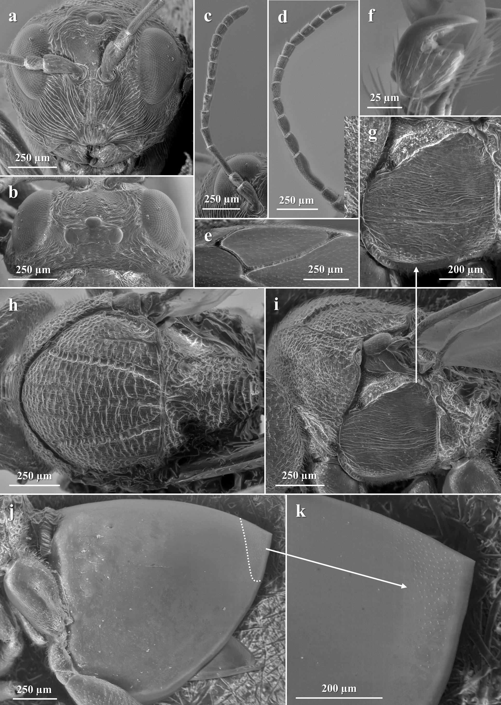
- In females, head mostly orange, the rest of the body black or dark ( Fig. 35a View FIGURE 35 ); males not as dark, mesosoma black with some yellowish orange surfaces ( Fig. 35b View FIGURE 35 ). Transfacial line 1.2 times as long as height of eye ( Fig. 36a View FIGURE 36 ). POL 1.5 times as long as OOL ( Fig. 36b View FIGURE 36 ). F1 almost 2.0 times as long as F 2 in both sexes ( Fig. 36 View FIGURE 36 d–e). Mesoscutum weakly but densely transversely discontinuously carinated; mesoscutellum weakly transversely carinated; scutellar foveae well defined, bottom with longitudinal carinae and separated by a wide septum; circumscutellar carina weak, obscured by the sculpture ( Fig. 36l View FIGURE 36 ). Radial cell of fore wing 3.0 times as long as wide ( Fig. 36h View FIGURE 36 ). Female syntergum dorsodistally incised ( Fig. 36m View FIGURE 36 )... ... S. ruficephalus , sp. nov.
33 POL shorter than OOL................................................................................ 34
- POL as long as OOL or longer.......................................................................... 35
34 Female antenna with 15 segments. Frons coriaceous. Mesoscutellum weakly wrinkled and with a median longitudinal depression. Circumscutellar carina well defined, upturned and projected. Mesopleuron irregularly transversely striated, medially with some punctures in both sexes, and with the speculum very finely aciculate in females. Areolet of fore wing absent or very elongated. In males, malar space 0.5 times as long as height of eye. Females ranging from 5.0 to almost 7.0 mm and males, from 3.5 to 4.5 mm (see Lobato-Vila et al. 2019: Figs 28–31 View FIGURE 28 View FIGURE 29 View FIGURE 30 View FIGURE 31 )................................................ S. kinseyi
- Female antenna with 14 segments. Frons coriaceous, but with some fine striae radiating from toruli to ocelli. Mesoscutellum strongly wrinkled, median longitudinal depression absent. Circumscutellar carina weak, obscured by wrinkles. Mesopleuron with fine, regular and dense transverse striae covering all of the surface, without punctures. Areolet of fore wing always present and easily traceable, never elongated. In males, malar space 0.7 times as long as height of eye. Females ranging from 3.0 to 4.0 mm and males, from almost 3.0 to 3.7 mm (see Lobato-Vila et al. 2019: Figs 38 View FIGURE 38 –48)…............... S. mesoamericanus
35 Radial cell of fore wing from 2.0 to about 2.5 times as long as wide............................................ 36
- Radial cell of fore wing at least 2.7, but usually longer....................................................... 58
36 Mesoscutum with widely spaced transversal carinae, the interspaces between carinae very finely alutaceous to smooth, glossy (as in Fig. 10c View FIGURE 10 )....................................................................................... 37
- Mesoscutum with dense, rarely widely spaced, transversal carinae, the interspaces between carinae alutaceous or coriaceous, never smooth nor glossy; mesoscutum rarely strongly coriaceous with some weak and dense transversal elements or imbricated (as in Figs 6i View FIGURE 6 ; 22i View FIGURE 22 ; 29c View FIGURE 29 )................................................................................ 38
37 Female antenna with 13 segments. F1 1.7 times as long as F 2 in females, 2.2 in males. Gena slightly broadened behind eye, visible in frontal view. Malar space 0.6 times as long as height of eye. POL 1.6 times as long as OOL, OOL 2.0 times as long as diameter of lateral ocelli. Circumscutellar carina weak, not well defined, obscured by wrinkles. Syntergum always with a conspicuous posterodorsal patch of micropunctures (see Lobato-Vila et al. 2019: Figs 3–4 View FIGURE 3 View FIGURE 4 )..................... S. bellus
- Female antenna with 14 segments. F1 as long as F 2 in females ( Fig. 10b View FIGURE 10 ), F1 1.3 times as long as F 2 in males. Gena not broadened behind eye, not visible in frontal view ( Fig. 10a View FIGURE 10 ). Malar space 0.7 times as long as height of eye. POL about 2.0 times as long as OOL, OOL 1.4 times as long as diameter of lateral ocelli ( Fig. 10a View FIGURE 10 ). Circumscutellar carina well defined, somewhat upturned ( Fig. 10c View FIGURE 10 ). Syntergum posteriorly without micropunctures, rarely with an inconspicuous incomplete narrow band............................................................................................ S. coniferae
38 F1 and F2 equal or subequal in both sexes. F1 of females usually with 14 segments, rarely 15 (never 13)............... 39
- F1 longer than F2, F1 always 1.2 or more times as long as F 2 in both sexes. F1 of females usually with 13 segments, rarely 14 (never 15).......................................................................................... 49
39 Female ranging from 4.5 to 5.0 mm. Female antenna with 15 segments ( Fig. 15e View FIGURE 15 ). Tarsal claws with an inconspicuous to absent basal lobe ( Fig. 15g View FIGURE 15 ). First metasomal segment with striae reaching dorsally only the half of its length ( Fig. 15a, c View FIGURE 15 ). Syntergum posteriorly without micropunctures................................................................ S. dorsalis
- Female at most 4.0 mm in length, but usually smaller. Female antenna with 14 segments, rarely 15 (in this case, syntergum posteriorly with a narrow band of micropunctures). Tarsal claws with a basal lobe, which can be small or acute and strong, but always distinct (as in Figs 6e View FIGURE 6 ; 22e View FIGURE 22 ). First metasomal segment sulcate dorsally and laterally, with complete striae (as in Figs 6h, j View FIGURE 6 ; 22h, j View FIGURE 22 ). Syntergum posteriorly with a band of micropunctures, which can be either complete (reaching the ventral margin of the tergite), incomplete, narrow or wide, but always present and visible......................................... 40
40 Lateral pronotum coriaceous, without transversal carinae nor wrinkles ( Fig. 38a View FIGURE 38 ). Mesoscutum weakly sculptured, imbricated, without carinae nor transversal elements, with some punctures ( Fig. 38b View FIGURE 38 ). Speculum medially with a small smooth spot. Female antenna with 15 segments ( Fig. 38b View FIGURE 38 )................................................................ S. villosus
- Lateral pronotum wrinkled or carinated, sometimes coriaceous but at least with some carinae or wrinkles ventrally (as in Figs 6g View FIGURE 6 ; 22g View FIGURE 22 ). Mesoscutum strongly or weakly, but always densely, transversely carinated, rarely coriaceous with weak transversal elements not forming true carinae (as in Figs 6i View FIGURE 6 ; 22i View FIGURE 22 ). Speculum completely transversely striated or wrinkled, without a smooth spot medially (as in Figs 6g View FIGURE 6 ; 22g View FIGURE 22 ). Female antenna with 14 segments (except in S. ochreus , which has 15; in this case, body of females is completely ochre, POL is 1.3 times as long as OOL and, in females, the last flagellar segment is subequal to the penultimate)......................................................................................... 41
41 Female with head and mesosoma entirely yellowish brown (ochre), metasoma entirely yellow ( Fig. 29 View FIGURE 29 a–c). Female antenna with 15 segments. Gena slightly broadened behind eye, visible in frontal view. Syntergum posteriorly with a very narrow band of micropunctures extended laterally at most 1/6 of its length........................................... S. ochreus
- Female with head, mesosoma and/or metasoma darker; head at least with a dark spot in the ocellar triangle, usually with frons, vertex and occiput black (rarely only with some dark surfaces surrounding each ocellus, Fig. 20c View FIGURE 20 ); mesosoma darker, usually with more dark surfaces or entirely black; metasoma yellowish and with a more or less extended dorsal black stripe, or entirely black ( Figs 5 View FIGURE 5 a–b, e; 16a–c; 19a–c; 21; 18a–c; 25a–d; 30a–j; 37a–c). Female antenna with 14 segments. Gena not broadened behind eye, not visible in frontal view (except in S. erinacei ). Syntergum posteriorly with a more widespread band of micropunctures, extended laterally from 1/4 to more than 1/2 of its length (except in S. duricorius )......................... 42
42 Head mostly rufous, only with some dark surfaces surrounding each ocellus at least in females ( Fig. 20c View FIGURE 20 ). Mesosoma rufous or mostly rufous ( Fig. 20 View FIGURE 20 a–b). Vertex deeply punctate. POL about as long as OOL ( Fig. 20 View FIGURE 20 a–c)................. S. ficigerae
- Head mainly yellowish, with a black spot in the ocellar area or with frons, vertex and occiput, black, at least in females. Mesosoma yellow, dark yellow and/or yellowish orange with some black surfaces or completely black ( Figs 5 View FIGURE 5 a–b, e; 16a–c; 19a–c; 21; 25a–d; 30a–j; 37a–c). Vertex coriaceous with some small piliferous punctures and/or weak wrinkles or weakly wrinkled, never deeply punctate (as in Figs 6b View FIGURE 6 ; 22b View FIGURE 22 ). POL at least 1.3 times as long as OOL, but usually longer.................. 43
43 Mesopleuron covered with non-parallel (irregular), dense, transverse striae, somewhat reticulated anteriorly (as in Figs 6g View FIGURE 6 ; 22g View FIGURE 22 )............................................................................................... 44
- Mesopleuron covered with parallel (regular), fine and dense, transverse striae, not reticulated anteriorly (as in Figs 25a View FIGURE 25 ; 30g View FIGURE 30 )................................................................................................... 46
44 Syntergum posteriorly with a narrow band of micropunctures extended laterally at most 1/5 of its length (less widespread in males). Clypeus slightly projected over mandibles. Tarsal claws with a small basal lobe. In females, syntergum strongly dorsodistally incised ( Fig. 16a View FIGURE 16 ). Head of females yellow, except for a black spot in the ocellar area and around the occipital foramen ( Fig. 16c View FIGURE 16 ); mesosoma yellow, only black between coxae and propodeum ( Fig. 16 View FIGURE 16 a–b)…................... S. duricorius
- Syntergum posteriorly with a complete wide band of micropunctures extended laterally somewhat more than 1/2 of its length ( Figs 6h View FIGURE 6 ; 22h View FIGURE 22 ). Clypeus straight, not projected over mandibles ( Figs 6a View FIGURE 6 ; 22a View FIGURE 22 ). Tarsal claws with a strong basal lobe ( Figs 6e View FIGURE 6 ; 22e View FIGURE 22 ). In females, syntergum weakly dorsodistally incised ( Figs 6j View FIGURE 6 ; 22j View FIGURE 22 ). Head of females with frons, vertex and occiput, black, the rest yellow (sometimes with a black stripe running between and below toruli) ( Fig. 5e View FIGURE 5 ); mesosoma with more dark surfaces ( Figs 5 View FIGURE 5 a–b; 21)...................................................................................... 45
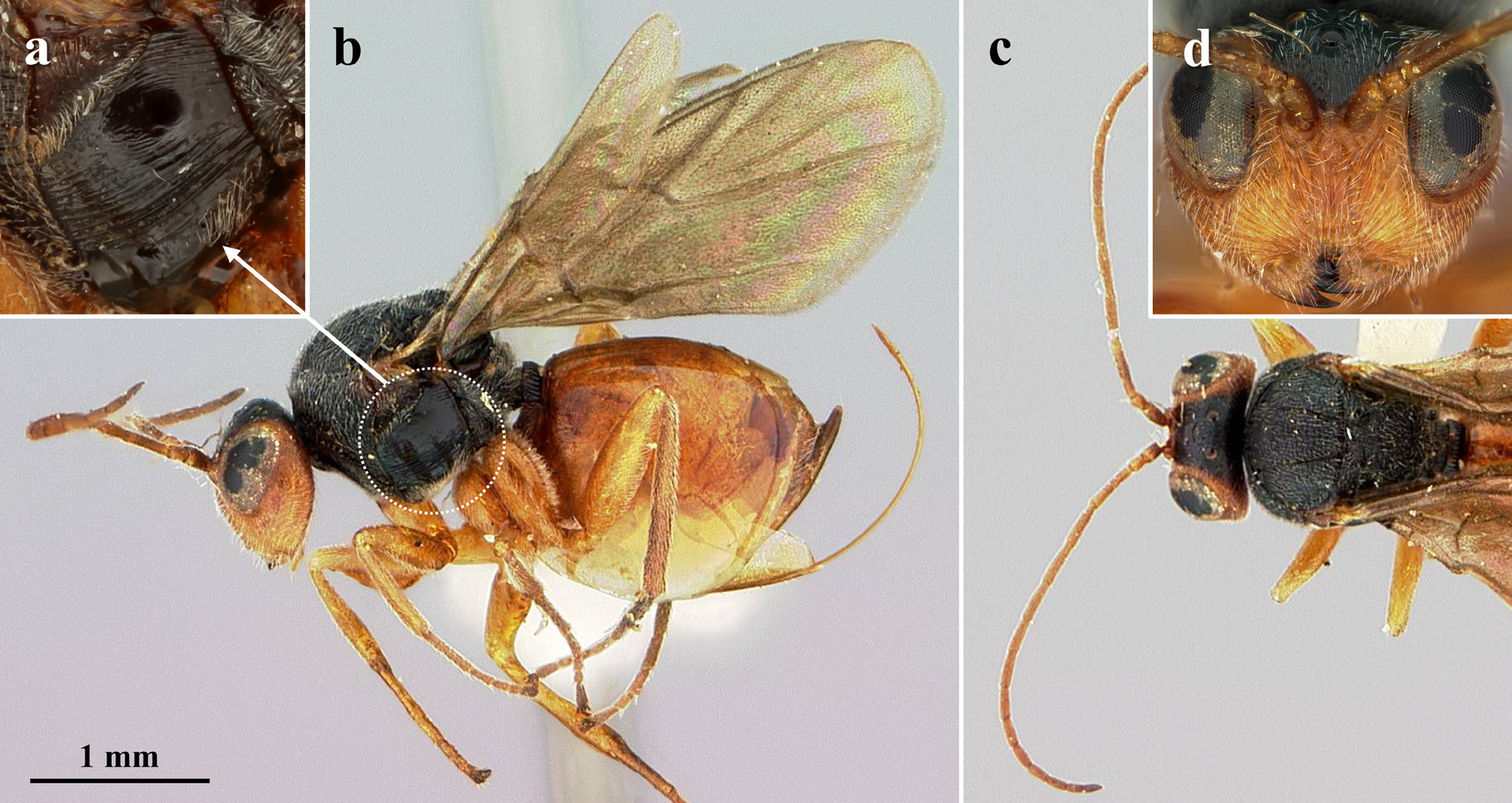
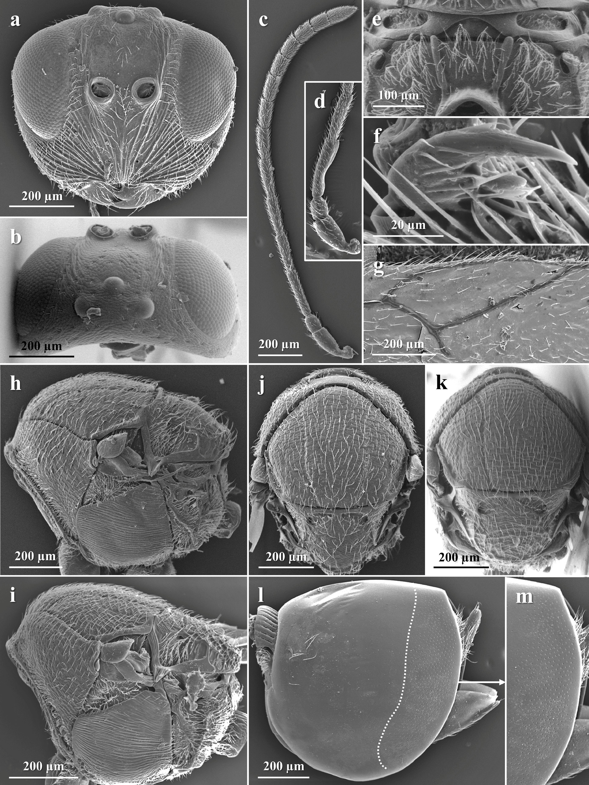
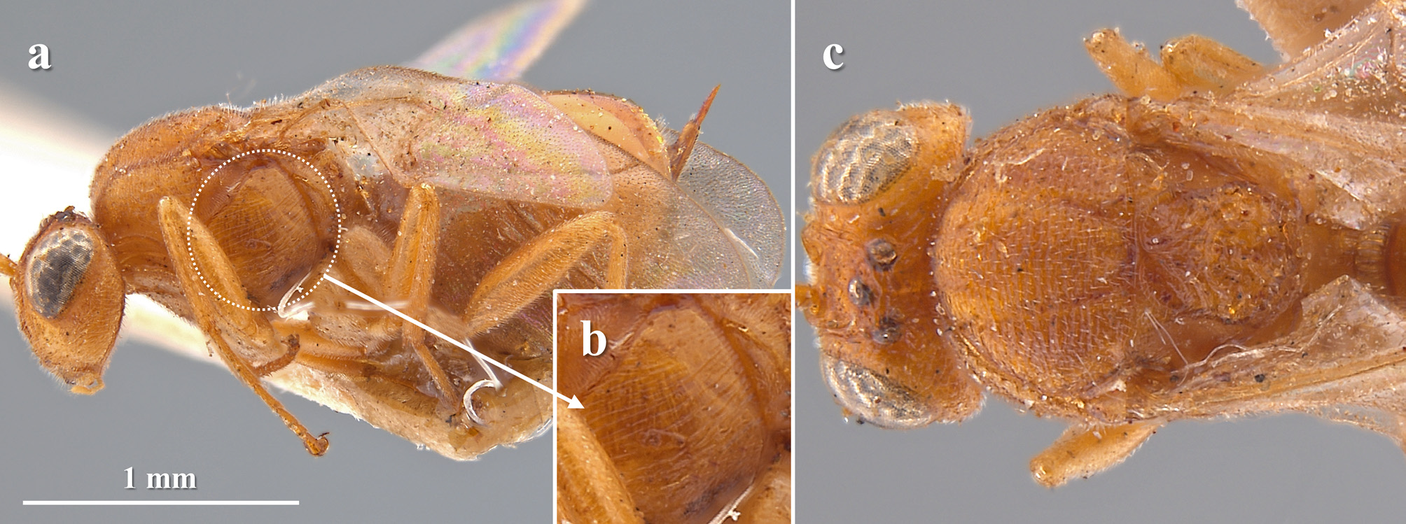
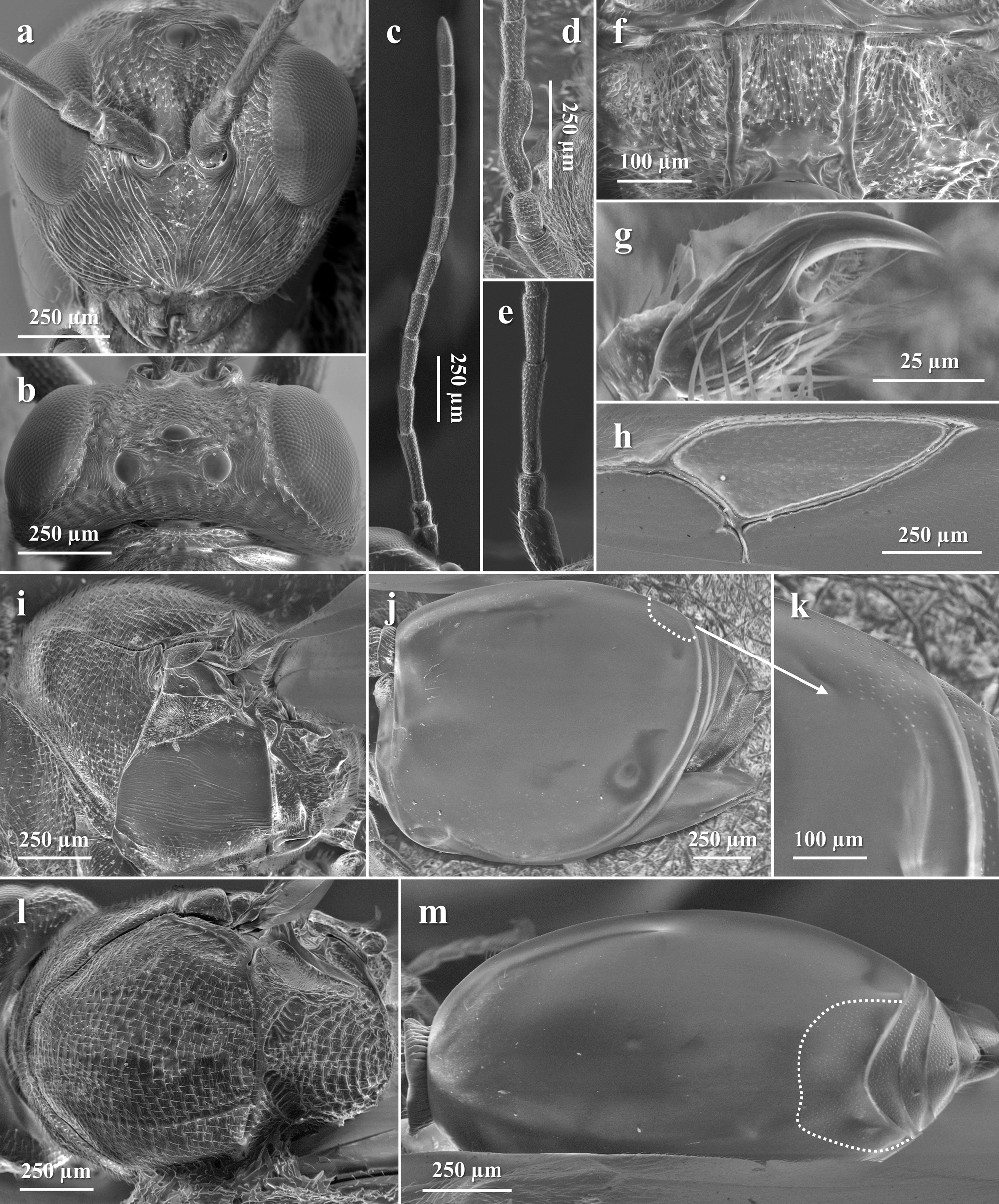
45 Lower face with widely spaced striae radiating from clypeus; malar space at most 0.5 times as long as height of eye; transfacial line as long as height of eye or slightly shorter; lateral frontal carinae thick and branched only distally, just before reaching lateral ocelli ( Fig. 6a View FIGURE 6 ). Head in dorsal view 2.1 times as wide as long ( Fig. 6b View FIGURE 6 ). Flagellomeres, especially the first ones, slender and long, F1 about 9 times as long as wide; scape plus pedicel 0.7 times as long as F1 ( Fig. 6c View FIGURE 6 ). Median mesoscutal line absent ( Fig. 6i View FIGURE 6 )......................................................................... S. beutenmulleri , sp. nov.
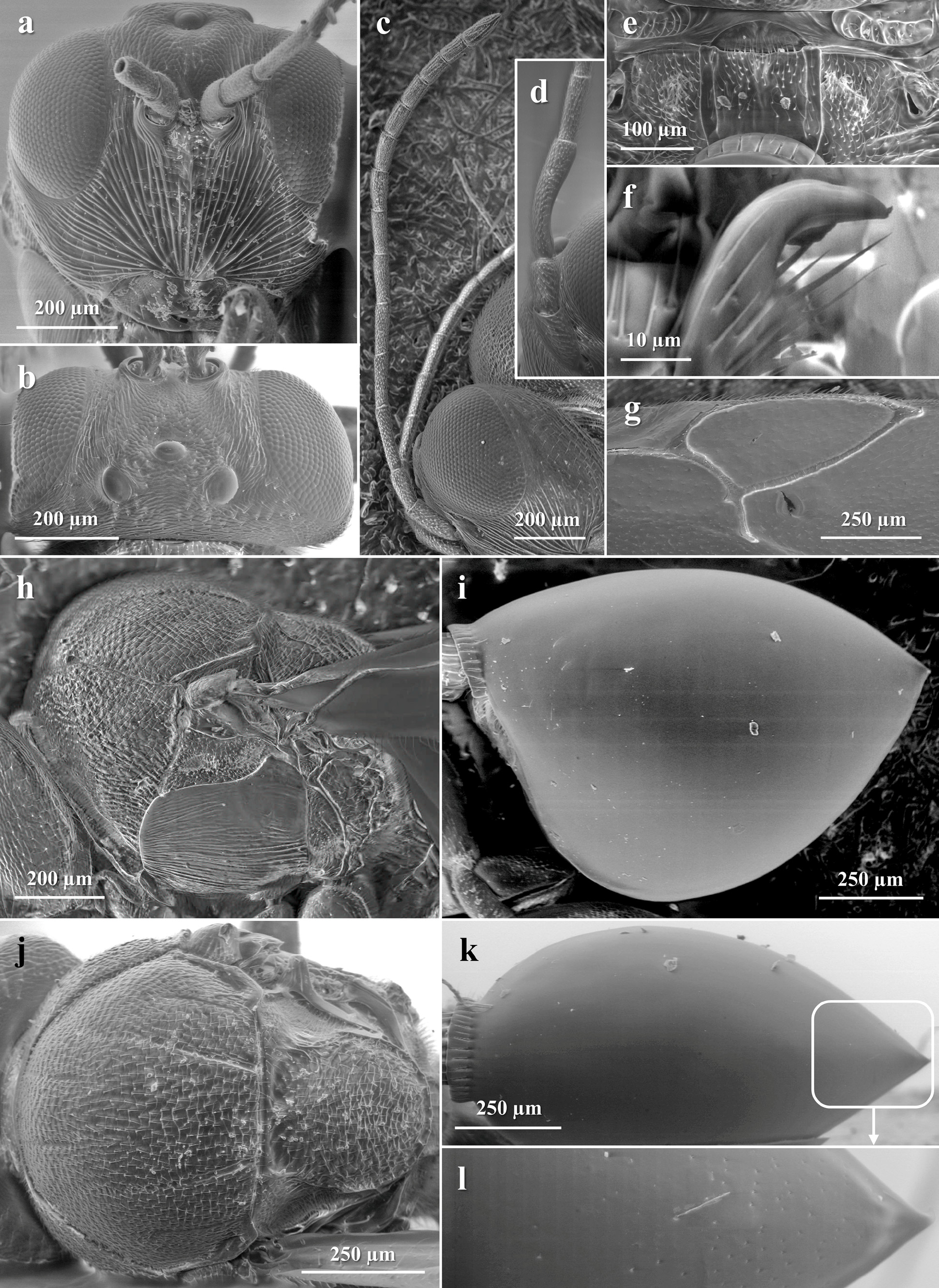
- Lower face with dense striae radiating from clypeus; malar space 0.6 times as long as height of eye; transfacial line slightly longer than height of eye; lateral frontal carinae narrow and branched in their entire length, reaching lateral ocelli ( Fig. 22a View FIGURE 22 ). Head in dorsal view 2.3 times as wide as long ( Fig. 22b View FIGURE 22 ). Flagellomeres not as slender and long, F1 about 5 times as long as wide; scape plus pedicel as long as F1 ( Fig. 22c View FIGURE 22 ). Median mesoscutal line shallow and interrupted by the transversal sculpture, sometimes appearing as a small incision, but always visible ( Fig. 22i View FIGURE 22 )............................... S. linnei , sp. nov.
46 In females, both mesosoma and metasoma entirely black ( Fig. 19 View FIGURE 19 a–b). Gena slightly broadened behind eye, visible in frontal view ( Fig. 19c View FIGURE 19 ). OOL about 1.7 times as long as diameter of lateral ocelli in females, equal in males. Pedicel 1.8 times as long as wide ( Fig. 19c View FIGURE 19 ). Syntergum of females not dorsodistally incised, with a pointed apex ( Fig. 19a View FIGURE 19 )............. S. erinacei
- In females, mesosoma and metasoma mostly yellow, dark yellow or reddish yellow, always with some black surfaces ( Figs 25a, c View FIGURE 25 ; 30 View FIGURE 30 a–g, j; 37a–b). Gena not broadened behind eye, not visible in frontal view. OOL about 1.2 or 1.3 times as long as diameter of lateral ocelli in both sexes. Pedicel at most 1.5 times as long as wide. Syntergum of females weakly to strongly dorsodistally incised; if not incised, then the metasoma does not end in a pointed apex ( Figs 25b View FIGURE 25 ; 30a, e, g View FIGURE 30 ; 37a View FIGURE 37 ).................... 47
47 Malar space 0.4 times as long as height of eye. Transfacial line slightly shorter than height of eye ( Fig. 25d View FIGURE 25 ). Head in dorsal view 1.8 times as wide as long. Tarsal claws with a very small, almost inconspicuous, basal lobe. Syntergum posteriorly with an incomplete band of micropunctures extended laterally about 1/4 of its length (almost 1/ 3 in males). In females, syntergum strongly dorsodistally incised ( Fig. 25b View FIGURE 25 )....................................................... S. nigroornatus
- Malar space about 0.6 times as long as height of eye. Transfacial line as long as height of eye. Head in dorsal view 2.1 times as wide as long. Tarsal claws with a conspicuous basal lobe. Syntergum posteriorly with a complete wide band of micropunctures extended laterally more than 1/3 of its length in both sexes. In females, syntergum weakly to not dorsodistally incised ( Figs 30a, e, g View FIGURE 30 ; 37a View FIGURE 37 )........................................................................................... 48
48 Clypeus straight, not projected over mandibles. POL 1.6 times as long as OOL ( Fig. 30c View FIGURE 30 ). Mesoscutum with weak, dense and discontinuous transversal carinae; median mesoscutal line absent ( Fig. 30f, j View FIGURE 30 ). Tarsal claws with a small basal lobe. Radial cell of fore wing 2.3 times as long as wide. Syntergum of females weakly dorsodistally incised ( Fig. 30a, e, g View FIGURE 30 )...... S. oneratus
- Clypeus slightly projected over mandibles. POL 1.3 times as long as OOL ( Fig. 37c View FIGURE 37 ). Mesoscutum with strong, dense, more or less continuous transversal carinae; median mesoscutal line appearing as a small incision ( Fig. 37b View FIGURE 37 ). Tarsal claws with a regular, acute basal lobe. Radial cell of fore wing 2.5 times as long as wide. Syntergum of females not dorsodistally incised ( Fig. 37a View FIGURE 37 )........................................................................................... S. rutulus
49 Syntergum posteriorly with a complete band of micropunctures extended laterally from 1/4 to 1/2 of its length.......... 50
- Syntergum posteriorly without micropunctures or just with a posterodorsal patch, rarely somewhat extended laterally..... 51
50 Body black and yellow. Frontal carinae narrow, branched before reaching lateral ocelli. Head in dorsal view 2.3 times as wide as long. F1 1.2–1.3 times as long as F2. Circumscutellar carina weak, obscured by wrinkles. Scutellar foveae separated by a narrow carina. Tarsal claws with a small basal lobe. Syntergum posteriorly with a complete wide band extended laterally 1/2 of its length; in females, syntergum strongly dorsodistally incised (see Lobato-Vila et al. 2019: Figs 26–27 View FIGURE 26 View FIGURE 27 )......... S. incisus
- Body, including head, antennae and legs, almost entirely black. Frontal carinae not well defined; especially in females, frons covered with multiple fine striae running from lower face and toruli towards ocelli. Head in dorsal view 2.0 times as wide as long. F1 1.5–1.6 times as long as F2. Circumscutellar carina well defined, moderately strong and upturned. Scutellar foveae separated by a wide septum. Tarsal claws with a strong basal lobe. Syntergum posteriorly with a complete band extended laterally 1/4 of its length (1/3 dorsally); in females, syntergum not dorsodistally incised, metasoma with a slightly pointed apex (see Lobato-Vila et al. 2019: Figs 63–72)............................................................ S. tenebrosus
51 Head in frontal view quadrate. OOL 2.0 times as long as diameter of lateral ocelli. Female antenna with 14 segments. Lateral pronotum coriaceous to imbricated, without carinae nor wrinkles. Scutellar foveae quadrate, shallow, not well defined posteriorly. Mesopleuron finely, regularly and densely striated, with a small smooth spot in the basal half of the speculum. Syntergum posterodorsally with a conspicuous patch of micropunctures somewhat laterally extended; in females, syntergum weakly dorsodistally incised (see Lobato-Vila et al. 2019: Figs 51–62)......................................... S. shorthousei
- Head in frontal view trapezoid to rounded. OOL usually less than 1.5 times as long as diameter of lateral ocelli, rarely 1.7. Female antenna with 13 segments. Lateral pronotum either with carinae or wrinkles. Scutellar foveae subtriangular or ellipsoidal, either well defined or not, rarely inconspicuous. Mesopleuron either finely and regularly striated or not, but always without a smooth spot under the speculum. Syntergum posterodorsally without micropunctures or just with a small, sometimes inconspicuous, patch, never laterally extended; in females, syntergum not dorsodistally incised........................... 52
52 Frons sharply and finely striated beneath toruli, striae running from toruli towards lateral ocelli ( Fig. 2d View FIGURE 2 ). POL more than 2.0 times as long as OOL. Notauli complete but less impressed and interrupted by carinae anteriorly ( Fig. 2c View FIGURE 2 ). Metasoma, at least in females, as high as long in lateral view ( Fig. 2b View FIGURE 2 ). Male antenna with 14 segments........................... .. S. atra
- Frons coriaceous, weakly wrinkled and/or with punctures, but never with sharp striae running from toruli. POL less than 2.0 times as long as OOL. Notauli complete and well defined in their entire length (as in Fig. 7c View FIGURE 7 ). Metasoma, at least in females, longer than high in lateral view. Male antenna always with 15 segments.......................................... 53
53 Gena broadened behind eye, visible in frontal view. Margins of pronotum swollen aside seen from above, sometimes somewhat sharp (as in Fig. 7c View FIGURE 7 )..………………………………………………… 54
- Gena not broadened behind eye, not visible in frontal view. Margins of pronotum not swollen aside seen from above, margins rounded.......................................................................................... ... 55
54 Transfacial line slightly longer than height of eye ( Fig. 7d View FIGURE 7 ). Frons finely wrinkled and with sparse, small piliferous punctures; frontal carinae inconspicuous to absent ( Fig. 7d View FIGURE 7 ). Vertex coriaceous, with some small punctures. POL 1.6 times as long as OOL. Margins of pronotum strongly swollen aside and somewhat sharp seen from above ( Fig. 7c View FIGURE 7 ). Mesopleuron covered with irregular, non-parallel, transversal striae ( Fig. 7b View FIGURE 7 )..................................................... S. bicolor
- Transfacial line as long as height of eye. Frons and vertex with broad punctures, interspaces coriaceous; frontal carinae very narrow, slightly branched, not reaching lateral ocelli. POL almost 2.0 times as long as OOL. Margins of pronotum slightly swollen aside but rounded seen from above. Mesopleuron anteriorly coriaceous, the rest of its surface more or less regularly and parallel, transversely striated (see Lobato-Vila et al. 2019: Figs 34–35 View FIGURE 34 View FIGURE 35 )............................... S. medullae
55 In females, coxae yellow. POL 1.2 times as long as OOL; OOL 1.7 times as long as diameter of lateral ocelli ( Fig. 11c View FIGURE 11 ). Scape plus pedicel as long as F1; pedicel as long as wide. In females, metasoma as long as head plus mesosoma. In males, malar space 0.7 times as long as height of eye............................................................... S. dimorphus
- In females, coxae basally black. POL more than 1.5 times as long as OOL; OOL 1.3 times as long as diameter of lateral ocelli. Scape plus pedicel longer than F1; pedicel at least 1.5 times as long as wide. In females, metasoma shorter than head plus mesosoma. In males, malar space at most 0.6 times as long as height of eye......................................... 56
56 Head in frontal view subtrapezoid, as wide as high. Toruli situated mid-height of eyes. Pedicel 1.5 times as long as wide. Scutellar foveae inconspicuous, separated by a narrow carina. First metasomal segment entirely sulcate dorsally and laterally. In males, F1 almost 1.5 times as long as F2; F1 slightly expanded apically and basally (see Lobato-Vila et al. 2019: Figs 9–11 View FIGURE 9 View FIGURE 10 View FIGURE 11 )........................................................................................... S. digressus
- Head in frontal view rounded, about 1.2 times as wide as high. Toruli situated under mid-height of eyes. Pedicel 2.0 times as long as wide. Scutellar foveae shallow but visible, not well defined posteriorly, separated by a wide septum. First metasomal segment dorsally with striae reaching the half of its length, anterior half smooth. In males, F1 almost 2.0 times as long as F2; F1 strongly expanded apically, weakly basally............................................................. 57
57 Frons and vertex coriaceous, with small and sparse piliferous punctures; frontal carinae weak but visible, not reaching lateral ocelli. Fore wing with moderately long marginal setae, the basal cell with sparse spaced setae, and the radial cell ambiguously closed along the wing margin, the R1 vein appearing weakly pigmented. Mesosoma completely black in both sexes (see Lobato-Vila et al. 2019: Figs 5–6 View FIGURE 5 View FIGURE 6 )............................................................... S. brevicornis
- Frons and vertex weakly wrinkled, with small and sparse piliferous punctures; frontal carinae very short, running just a little from the posterior margin of toruli, almost inconspicuous. Fore wing with short marginal setae, the basal cell with dense, closely spaced setae, and the radial cell clearly closed along the wing margin, the R1 vein appearing well-pigmented. Mesosoma with pronotum mainly yellowish orange, mesopleuron dorsally and medially orange and basally black in both sexes (see
Lobato-Vila et al. 2019: Figs 32–33 View FIGURE 32 View FIGURE 33 )............................................................. S. lignicola
58 Syntergum posteriorly without punctures or just with a few posterodorsal micropunctures forming at most a small patch... 59
- Syntergum posteriorly with a band of micropunctures, which can be either incomplete (not reaching the ventral edge of the metasoma) or complete (reaching the ventral edge of the metasoma), narrow or wide............................... 66
59 Tarsal claws simple, at most with an inconspicuous basal prominence not forming a lobe ( Fig. 33b, g View FIGURE 33 ). First metasomal segment dorsally with striae reaching the half of its length, anterior half smooth; sometimes completely smooth ( Fig. 33d, i View FIGURE 33 )........................................................................................... S. reniformis (part) *
- Tarsal claws always bidentate, either with a small or a strong basal lobe, but always conspicuous (as in Figs 24e View FIGURE 24 ; 27f View FIGURE 27 ). First metasomal segment completely sulcate dorsally and laterally (as in Figs 24h, j View FIGURE 24 ; 27j View FIGURE 27 )................................ 60
60 Female antenna with 15 segments. F1 as long as F2. Mesoscutum imbricated, at most with very weak transversal elements not forming true carinae (see Lobato-Vila et al. 2020b: Figs 51–52)..................................... S. quercuslana
- Female antenna with 13 or 14 segments. F1 longer than F2, at least 1.2 times as long. Mesoscutum with transversal carinae, either weak or strong, but conspicuous (as in Figs 24i View FIGURE 24 ; 27h View FIGURE 27 )................................................... 61
61 Body, including head and legs, almost completely black in females; face of males somewhat lighter. Median mesoscutal line weak but visible, reaching 1/3 or 1/2 of the mesoscutal length (as in Fig. 27h View FIGURE 27 ).................................... 62
- Body lighter, lower face and gena always yellowish in both sexes. Median mesoscutal line absent.................... 63
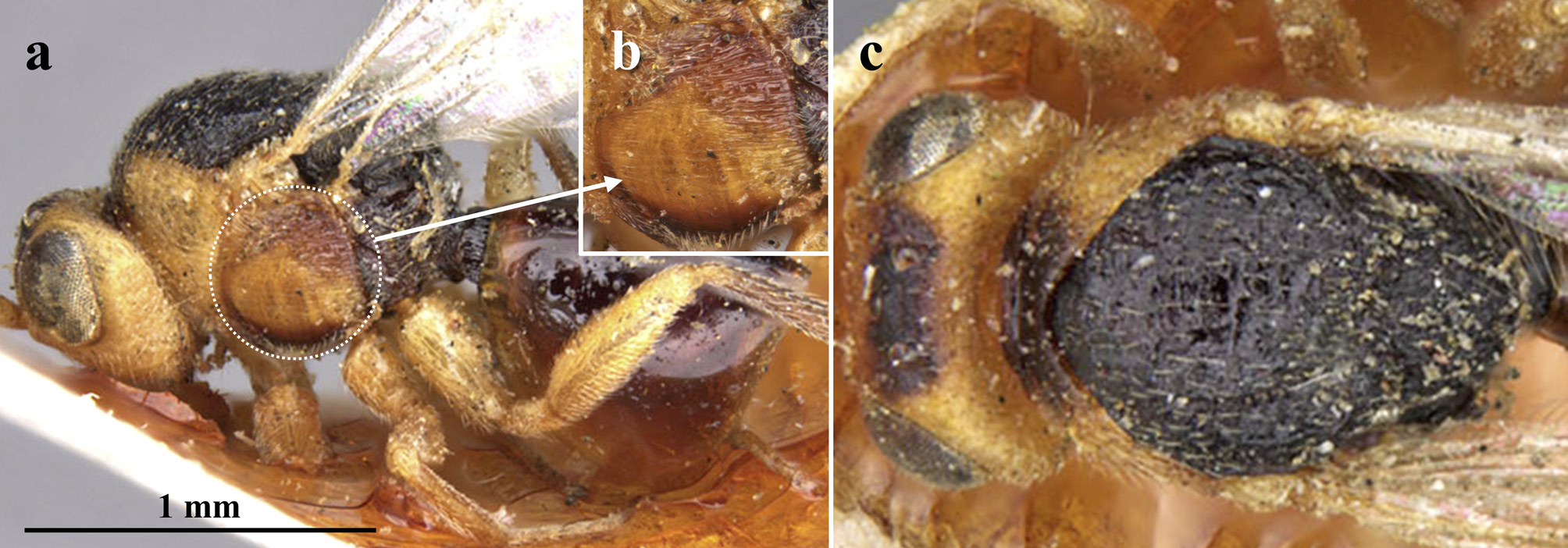
62 Transfacial line 1.3 times as long as height of eye ( Fig. 27a View FIGURE 27 ). Frons and vertex coriaceous, at most with a few small piliferous punctures and some weak wrinkles between ocelli ( Fig. 27 View FIGURE 27 a–b). OOL about 2.0 times as long as diameter of lateral ocelli ( Fig. 27b View FIGURE 27 ). F1 2.0 times as long as F 2 in females ( Fig. 27c View FIGURE 27 ), 2.3 in males ( Fig. 27d View FIGURE 27 ). Areolet of the fore wing very small or absent ( Figs 26 View FIGURE 26 a–b; 27e). In females, metasoma as high as long in lateral view ( Fig. 27j View FIGURE 27 ) and antenna with 13 segments. In males, face and gena dark brown ( Fig. 26b View FIGURE 26 )....................................................... S. oaxaquensis , sp. nov.
- Transfacial line as long as height of eye (see Nieves-Aldrey & Medianero 2011: Fig. 1C View FIGURE 1 ). Frons and vertex deeply punctate (see Nieves-Aldrey & Medianero 2011: Figs 1C View FIGURE 1 ; 2F View FIGURE 2 ). OOL 1.3 times as long as diameter of lateral ocelli (see Nieves-Aldrey & Medianero 2011: Fig. 2F View FIGURE 2 ). F1 about 1.5 times as long as F 2 in both sexes (see Nieves-Aldrey & Medianero 2011: Figs 5C View FIGURE 5 ; 6G View FIGURE 6 ). Areolet of the fore wing visible, well defined and pigmented (see Nieves-Aldrey & Medianero 2011: Fig. 17C View FIGURE 17 ). In females, metasoma longer than high in lateral view (see Nieves-Aldrey & Medianero 2011: Fig. 13F View FIGURE 13 ) and antenna with 14 segments. In males, face and gena yellowish (see Nieves-Aldrey & Medianero 2011: Fig. 18E View FIGURE 18 )........................... S. ramoni
63 Female antenna with 13 segments. Malar space 0.6 or more times as long as height of eye (except in males of S. estradae , which is 0.5). Transfacial line 1.2 or more times as long as height of eye. Toruli situated clearly under mid-height of eyes, almost in line with the base of the eyes (as in Fig. 24a View FIGURE 24 ). Radial cell of fore wing undoubtfully closed along the wing margin, R1 vein well pigmented; 2.7 or 2.8 times as long as wide................................................................ 64
- Female antenna with 14 segments. Malar space at most 0.5 times as long as height of eye in both sexes. Transfacial line about as long as height of eye. Toruli situated just slightly under mid-height of eyes. Radial cell of fore wing ambiguously closed along the wing margin, R1 vein not well pigmented; 3.0 or more times as long as wide................................... 65
64 Head in frontal view trapezoid. Lateral frontal carinae narrow, weak, branched, not reaching lateral ocelli, but visible. POL 2.0 times as long as OOL in females, 3.0 in males; OOL 1.5 times as long as diameter of lateral ocelli in females, equal in males. Pedicel slightly longer than wide. Margins of pronotum not swollen aside, pronotum margins rounded seen from above. In females, syntergum not dorsodistally incised. In males, F1 2.0 times as long as F2 (see Pujade-Villar et al. 2016: Fig. 2 View FIGURE 2 a–j)…........................................................................................... S. estradae
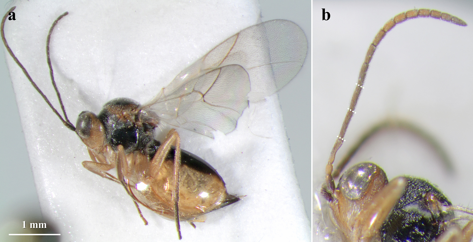
- Head in frontal view rounded ( Fig. 24a View FIGURE 24 ). Lateral frontal carinae absent, frons covered with very fine striae ( Fig. 24 View FIGURE 24 a–b). POL 1.2 or 1.3 times as long as OOL; OOL 2.0 times as long as diameter of lateral ocelli in both sexes ( Fig. 24b View FIGURE 24 ). Pedicel 2.3 times as long as wide ( Fig. 24a, c View FIGURE 24 ). Margins of pronotum swollen aside, pronotum margins somewhat sharp seen from above ( Fig. 24i View FIGURE 24 ). In females, syntergum dorsodistally incised ( Fig. 24j View FIGURE 24 ). In males, F1 about 1.5 times as long as F2 ( Fig. 23d View FIGURE 23 ).......................................................................................... S. macrackenae , sp. nov.
65 Lateral frontal carinae strong, not branched, almost reaching lateral ocelli (see Nieves-Aldrey & Medianero 2011: Fig. 2B View FIGURE 2 ). Pedicel 1.2 times as long as wide; F1 1.2 times as long as F 2 in females, 1.7 in males (see Nieves-Aldrey & Medianero 2011: Figs 5I View FIGURE 5 ; 6D View FIGURE 6 ). Scutellar foveae separated by a wide septum (see Nieves-Aldrey & Medianero 2011: Fig. 9D View FIGURE 9 ). Metasoma in lateral view 1.3 times as long as high (see Nieves-Aldrey & Medianero 2011: Fig. 14B View FIGURE 14 )......................... S. chiricanus
- Lateral frontal carinae weak, narrow, not branched and not reaching lateral ocelli. Pedicel 1.8 times as long as wide; F1 1.5 or 1.6 times as long as F 1 in both sexes. Scutellar foveae separated by a narrow carina. Metasoma in lateral view as high as long (see Lobato-Vila et al. 2019: Figs 12–25 View FIGURE 12 View FIGURE 13 View FIGURE 14 View FIGURE 15 View FIGURE 16 View FIGURE 17 View FIGURE 18 View FIGURE 19 View FIGURE 20 View FIGURE 21 View FIGURE 22 View FIGURE 23 View FIGURE 24 View FIGURE 25 ).......................................................... S. grahami
66 F1 as long as F2, rarely almost 1.1 times as long (subequal)................................................... 67
- F1 at least 1.2 or 1.3 times as long as F2, sometimes longer................................................... 76
67 Tarsal claws simple, at most with an inconspicuous basal prominence not forming a lobe ( Fig. 33b, g View FIGURE 33 ). First metasomal segment dorsally with striae reaching the half of its length, anterior half smooth; sometimes completely smooth ( Fig. 33d, i View FIGURE 33 )............................................................................................ S. reniformis (part) *
- Tarsal claws bidentate, with a more or less strong basal lobe (as in Figs 9e View FIGURE 9 ; 14f View FIGURE 14 ; 32e View FIGURE 32 ; 41e View FIGURE 41 ). First metasomal segment completely sulcate dorsally and laterally (as in Figs 9i View FIGURE 9 ; 14l View FIGURE 14 ; 32j View FIGURE 32 ; 41k View FIGURE 41 )..................................................... 68
68 Large specimens, females ranging from 5.0 to 7.0 mm in length and males, from 3.0 to 5.0 mm. POL as long as OOL in both sexes. Circumscutellar carina very strong and upturned. In females, metasoma very long in lateral view, much longer than head plus mesosoma and about 1.7 times as long as high (see Lobato-Vila et al. 2019: Figs 7–8 View FIGURE 7 View FIGURE 8 )…................ S. cultratus
- Specimens not as large, females measuring at most 4.0 mm in length and males, at most 3.0 mm. POL at least 1.2 times as long as OOL (except in females of S. forcadellae , but in this case the mesoscutum is coriaceous-punctate and the syntergum in females is from weakly to not dorsodistally incised). Circumscutellar carina weak, inconspicuous or absent (as in Fig. 32i View FIGURE 32 ). In females, metasoma not as long in lateral view, as long or just slightly longer than head plus mesosoma and at most 1.3 times as long as high......................................................................................... 69
69 Syntergum posteriorly with a complete wide band of micropunctures extending laterally 1/2 of its length. Radial cell of fore wing 3.2 times as long as wide. Head of females completely yellow, except for some dark spots close to ocelli ( Fig. 28 View FIGURE 28 a–c) S. obtusilobae
- Syntergum posteriorly either with a complete or an incomplete band, extended at most 1/3 of its length. Radial cell of fore wing 2.7 to 3.0 times as long as wide. Head of females at least with frons, vertex and occiput black, face and gena more or less yellowish............................................................................................. 70
70 Antenna at most surpassing the mesoscutellum and reaching the metasoma; basal flagellomeres about 4.0 or 5.0 times as long as wide. Gena slightly broadened behind eye, visible in frontal view............................................ 71
- Antenna long, as long as the body length or almost reaching the tip of the metasoma, and thin, with flagellomeres, especially the basal ones, more than 6.0 times as long as wide. Gena not broadened behind eye, not visible in frontal view.......... 72
71 Malar space 0.4 times as long as height of eye. Transfacial line 0.8 times as long as height of eye. Frons and vertex weakly wrinkled and punctate. Areolet of the fore wing very small to absent. Syntergum posteriorly with a complete band of micropunctures extended laterally about 1/3 of its length. Basal half of metacoxae black. Antenna and wing veins, dark brown. F1 of males from moderately to strongly expanded apically (see Nieves-Aldrey 2005: Figs 1 View FIGURE 1 ; 2 View FIGURE 2 ; 3 View FIGURE 3 a–b)…........ S. colombianus
- Malar space about 0.6 times as long as height of eye. Transfacial line as long as height of eye. Frons and vertex mostly coriaceous and punctate, rarely with a few weak wrinkles. Areolet of the fore wing conspicuous, however it is not delimited by distinct veins. Syntergum posteriorly with an incomplete band of micropunctures extended laterally at most 1/4 of its length. Metacoxae yellow, rarely with a small, dark spot. Antenna and wing veins, yellow to testaceous. F1 of males slightly expanded apically (see Lobato-Vila et al. 2020a: Figs 4–16 View FIGURE 4 View FIGURE 5 View FIGURE 6 View FIGURE 7 View FIGURE 8 View FIGURE 9 View FIGURE 10 View FIGURE 11 View FIGURE 12 View FIGURE 13 View FIGURE 14 View FIGURE 15 View FIGURE 16 )..................................................... S. dawnus
72 Basal half of lower face and basal half of gena, yellow to yellowish orange, the rest of the head, black (as in Fig. 31 View FIGURE 31 ); black surfaces more expanded in males. Lateral pronotum carinated to wrinkled or somewhat reticulated (as in Fig. 32g View FIGURE 32 ). Mesopleuron with reticulated wrinkles covering all of the surface (see Nieves-Aldrey & Medianero 2011: Fig. 11D View FIGURE 11 ) or, at least, somewhat reticulated anteriorly ( Fig. 32g View FIGURE 32 ).......................................................................... 73
- Lower face and gena entirely yellowish, the lower face sometimes with a median dark stripe under toruli (as in Fig. 8 View FIGURE 8 a–b). Lateral pronotum alutaceous to coriaceous, without carinae nor wrinkles (like Fig. 9g View FIGURE 9 ). Mesopleuron finely, regularly and densely transversely striated, usually with a small smooth spot under toruli (as in Fig. 9g View FIGURE 9 ).................................. 74
73 Mesopleuron finely, regularly, and densely striated, somewhat reticulated anteriorly, interspaces smooth, glabrous, and shining ( Fig. 32g View FIGURE 32 ). Transfacial line 1.1 to 1.2 times as long as height of eye ( Fig. 32a View FIGURE 32 ). Frons and vertex coriaceous, vertex with some small piliferous punctures and weak wrinkles ( Fig. 32 View FIGURE 32 a–b). Areolet of fore wing visible ( Fig. 32f View FIGURE 32 ).…. S. personatus , sp. nov.
- Mesopleuron with transversal striae obscured by reticulated wrinkles, especially anteriorly, interspaces coriaceous (see Nieves-Aldrey & Medianero 2011: Fig. 11D View FIGURE 11 ). Transfacial line slightly shorter than height of eye (see Nieves-Aldrey & Medianero 2011: Fig. 2D View FIGURE 2 ). Frons and vertex with reticulated wrinkles (see Nieves-Aldrey & Medianero 2011: Figs 2D View FIGURE 2 ; 4 View FIGURE 4 G–H). Areolet of fore wing small to inconspicuous (see Nieves-Aldrey & Medianero 2011: Fig. 17F View FIGURE 17 )...................... S. rufinotaulis
74 Mesoscutum coriaceous-punctate, sometimes with very weak and discontinuous transversal elements not forming true carinae. Mesopleuron entirely finely, regularly and densely transversely striated, without a small smooth spot under the speculum. Radial cell of fore wing ambiguously closed, R1 vein not well pigmented. In females, mesoscutellum medially finely coriaceous, posteriorly with some weak wrinkles, metasoma shorter than head plus mesosoma and syntergum not dorsodistally incised (rarely weakly incised) (see Lobato-Vila et al. 2020b: Figs 19–42 View FIGURE 19 View FIGURE 20 View FIGURE 21 View FIGURE 22 View FIGURE 23 View FIGURE 24 View FIGURE 25 View FIGURE 26 View FIGURE 27 View FIGURE 28 View FIGURE 29 View FIGURE 30 View FIGURE 31 View FIGURE 32 View FIGURE 33 View FIGURE 34 View FIGURE 35 View FIGURE 36 View FIGURE 37 View FIGURE 38 View FIGURE 39 View FIGURE 40 View FIGURE 41 View FIGURE 42 )................................... S. forcadellae
- Mesoscutum coriaceous to weakly transversely carinated, without punctures. Mesopleuron finely, regularly and densely transversely striated, usually with a small smooth spot under the speculum (as in Fig. 9g View FIGURE 9 ). Radial cell of fore wing conspicuously closed, R1 vein well pigmented. Mesoscutellum entirely wrinkled in both sexes (as in Fig. 9h View FIGURE 9 ). In females, metasoma longer than head plus mesosoma and syntergum moderately to strongly dorsodistally incised (as in Fig. 9i View FIGURE 9 )................... 75
75 Base of metacoxae with a small black spot, the rest yellow ( Fig. 8a View FIGURE 8 ). In females, scape plus pedicel shorter than F1 ( Fig. 9c View FIGURE 9 ). Mesoscutum finely coriaceous in females ( Fig. 9h View FIGURE 9 ), strongly coriaceous in males. Metasoma of females strongly laterally compressed ( Fig. 9i View FIGURE 9 ); in lateral view, about 1.3 times as long as head plus mesosoma and almost 1.4 times as long as high ( Fig. 9j View FIGURE 9 ). Syntergum of females posteriorly with an interrupted and incomplete band of micropunctures extended laterally about 1/4 of its length ( Fig. 9 View FIGURE 9 j–k). F1 of males slightly expanded apically and basally ( Fig. 8b View FIGURE 8 )................ S. compressus , sp. nov.
- Metacoxae entirely yellow. In females, scape plus pedicel longer than F1. Mesoscutum from strongly coriaceous to weakly carinated in females, more strongly carinated in males. Metasoma of females ovate seen from above, not strongly compressed; about as long as head plus mesosoma or just slightly longer and about 1.2 times as long as high in lateral view. Syntergum of females posteriorly with a complete wide band of micropunctures extended laterally about 1/3 of its length. F1 of males strongly expanded apically, not expanded basally (see Ritchie & Shorthouse 1987: Figs 2 View FIGURE 2 , 6 View FIGURE 6 , 10 View FIGURE 10 , 14 View FIGURE 14 , 20 View FIGURE 20 ; Lobato-Vila et al. 2020b: Figs 13–18 View FIGURE 13 View FIGURE 14 View FIGURE 15 View FIGURE 16 View FIGURE 17 View FIGURE 18 ).................................................................................... .. S. filicornis
76 Female antenna with 13 segments. Malar space 0.8 times as long as height of eye in females, 0.6 in males. Transfacial line 1.2 times as long as height of eye in females, 1.1 in males. Mesoscutum with strong, widely spaced and continuous transversal carinae. Yellow legs with black coxae. Females ranging from 4.1 to 4.5 mm in length and males, from 3.2 to 3.7 mm (see Pujade-Villar et al. 2016: Fig. 1 View FIGURE 1 a–h)…............................................................ .. S. equihuai
- Female antenna with 14 segments. Malar space 0.5 to 0.6 times as long as height of eye in both sexes. Transfacial line as long as height of eye or shorter in both sexes. Mesoscutum coriaceous (as in Figs 14 View FIGURE 14 j–k; 41j) or weakly to strongly transversely carinated, but carinae always discontinuous and densely spaced (see Nieves-Aldrey & Medianero 2011: Fig. 9 View FIGURE 9 A–C; Lobato-Vila et al. 2018: Fig. 5a View FIGURE 5 ). Legs entirely yellow, including coxae. Females less than 4.0 mm in length and males, less than 3.0 mm.... .................................................................................................. 77
77 Lateral frontal carinae strong, high, not branched, almost reaching lateral ocelli (see Nieves-Aldrey & Medianero 2011: Fig. 2C View FIGURE 2 ). OOL as long as diameter of lateral ocelli (see Nieves-Aldrey & Medianero 2011: Fig. 4F View FIGURE 4 ).............. S. baruensis
- Lateral frontal carinae weak, narrow or moderately strong, but always entirely or distally branched, almost reaching lateral ocelli ( Figs 14a View FIGURE 14 , 41b View FIGURE 41 ; see Nieves-Aldrey & Medianero 2011: Fig. 2A, F View FIGURE 2 ; Lobato-Vila et al. 2018: Fig. 4a View FIGURE 4 ). OOL at least 1.3 times as long as diameter of lateral ocelli.................................................................. 78
78 Gena not broadened behind eye, not visible in frontal view (see Nieves-Aldrey & Medianero 2011: Fig. 2A View FIGURE 2 ). Syntergum posteriorly with a minute, obsolete narrow band of micropunctures (see Nieves-Aldrey & Medianero 2011: Fig. 14A View FIGURE 14 ). Radial cell of fore wing 3.4 times as long as wide (see Nieves-Aldrey & Medianero 2011: Fig. 17G View FIGURE 17 ). Females small, ranging from 1.5 to 1.7 mm in length................................................................................ .. S. gabrieli
- Gena slightly broadened behind eye, visible in frontal view. Syntergum posteriorly with a conspicuous band of micropunctures extended laterally from 1/4 to 1/3 of its length. Radial cell of fore wing 2.8 to 3.2 times as long as wide. Females ranging from 2.0 to 4.0 mm........................................................................................ 79
79 OOL 1.8 times as long as diameter of lateral ocelli ( Fig. 14b View FIGURE 14 ). In females, antenna subclavate, slightly broadened apically ( Fig. 14c View FIGURE 14 ), and metasoma shorter than head plus mesosoma and about as long as high in lateral view (slightly longer if we include the hypopygium, with is projected beyond the apex of the metasoma) ( Figs 13a, c View FIGURE 13 ; 14l View FIGURE 14 ). Also in females, syntergum weakly dorsodistally incised ( Fig. 14l View FIGURE 14 )........................................................ S. diversicolor , sp. nov.
- OOL at most 1.4 times as long as diameter of lateral ocelli. In females, antenna filiform, not broadened apically, and metasoma as long as head plus mesosoma or longer and longer than high in lateral view. Also in females, syntergum moderately to strongly dorsodistally incised........................................................................... 80
80 Lateral pronotum alutaceous to finely coriaceous, without carinae nor wrinkles ( Fig. 41g View FIGURE 41 ). Mesoscutum alutaceous to finely coriaceous, with small piliferous punctures, posteriorly sometimes with a few very weak transversal elements not forming true carinae ( Fig. 41j View FIGURE 41 ). Circumscutellar carina strong, slightly upturned and projected ( Fig. 41g, j View FIGURE 41 ).......... … S. weldi , sp. nov.
- Lateral pronotum imbricated to weakly carinated or wrinkled. Mesoscutum strongly coriaceous with weak transversal elements to strongly and densely transversely carinated. Circumscutellar carinae visible but weak, obscured by wrinkles, or inconspicuous to absent, never projected nor upturned (see Nieves-Aldrey & Medianero 2011: Figs 9B View FIGURE 9 , 11E View FIGURE 11 ; Lobato-Vila et al. 2018: Fig. 5a, b View FIGURE 5 )…............................................................................................ 81
81 Transfacial line as long as height of eye (see Nieves-Aldrey & Medianero 2011: Fig. 2F View FIGURE 2 ). Pronotum rounded and not swollen aside seen from above; mesoscutellum strongly wrinkled; circumscutellar carina inconspicuous to absent (see Nieves-Aldrey & Medianero 2011: Fig. 9B View FIGURE 9 ). Radial cell 3.2 times as long as wide and ambiguously closed, R1 vein not well pigmented (see Nieves-Aldrey & Medianero 2011: Fig. 17I View FIGURE 17 ). Syntergum posteriorly with a complete wide band of micropunctures extended laterally 1/3 of its length (see Nieves-Aldrey & Medianero 2011: Fig. 14D View FIGURE 14 ). In males, F1 2.0 times as long as F2................................................................................................... S. medianeroi
- Transfacial line 0.8 times as long as height of eye (see Lobato-Vila et al. 2018: Fig. 4a View FIGURE 4 ). Pronotum rounded but slightly swollen aside seen from above; mesoscutellum strongly coriaceous to weakly transversely carinated; circumscutellar carina weak but visible, somewhat obscured by wrinkles (see Lobato-Vila et al. 2018: Fig. 5a View FIGURE 5 ). Radial cell 2.8 times as long as wide and conspicuously closed, R1 vein well pigmented. Syntergum posteriorly with an incomplete band of micropunctures extended laterally about 1/4 of its length (see Lobato-Vila et al. 2018: Fig. 5 View FIGURE 5 c–d). In males, F1 1.2 times as long as F2.................................................................................................. S. pseudofilicornis
* Synergus reniformis (= S. magnificus ) is keyed out twice as it can have a posterior band of micropunctures on the syntergum or just a small posterodorsal patch (which is sometimes inconspicuous).
Unplaced species


No known copyright restrictions apply. See Agosti, D., Egloff, W., 2009. Taxonomic information exchange and copyright: the Plazi approach. BMC Research Notes 2009, 2:53 for further explanation.
|
Kingdom |
|
|
Phylum |
|
|
Class |
|
|
Order |
|
|
Family |