
Holoengythrips, Mound & Tree, 2014
|
publication ID |
https://doi.org/10.11646/zootaxa.3860.2.2 |
|
publication LSID |
lsid:zoobank.org:pub:EFF3B789-5578-4E7B-B6C7-3895511E0CE4 |
|
DOI |
https://doi.org/10.5281/zenodo.4929811 |
|
persistent identifier |
https://treatment.plazi.org/id/03BF7339-FF90-8D6F-FF05-74D5FA14FEE1 |
|
treatment provided by |
Felipe |
|
scientific name |
Holoengythrips |
| status |
gen. nov. |
Holoengythrips View in CoL gen.n.
Diagnosis. Usually apterous; antennae 8-segmented with suture between VII and VIII complete, III with 2 or 3 sensoria, IV with 2, 3 or 4 sensoria; maxillary stylets slender, about 3 microns in diameter, retracted to level of eyes, close together medially for full length of head; head longer than wide, slightly to greatly elevated in midline, usually with a few small cheek setae; mouth cone long, pointed; prosternal basantra absent, mesopresternum sexually dimorphic; sternopleural sutures present or absent; notopleural sutures complete; pelta with paired campaniform sensilla; fore tarsal tooth present in male, sometimes absent in female; male fore tibia sometimes with an apical or subapical tooth; male tergite IX setae S2 shorter than S1, iS setae relatively long; male sternite VIII with pore plate; tube shorter than head.
Type species Holoengythrips maynardae sp.n.
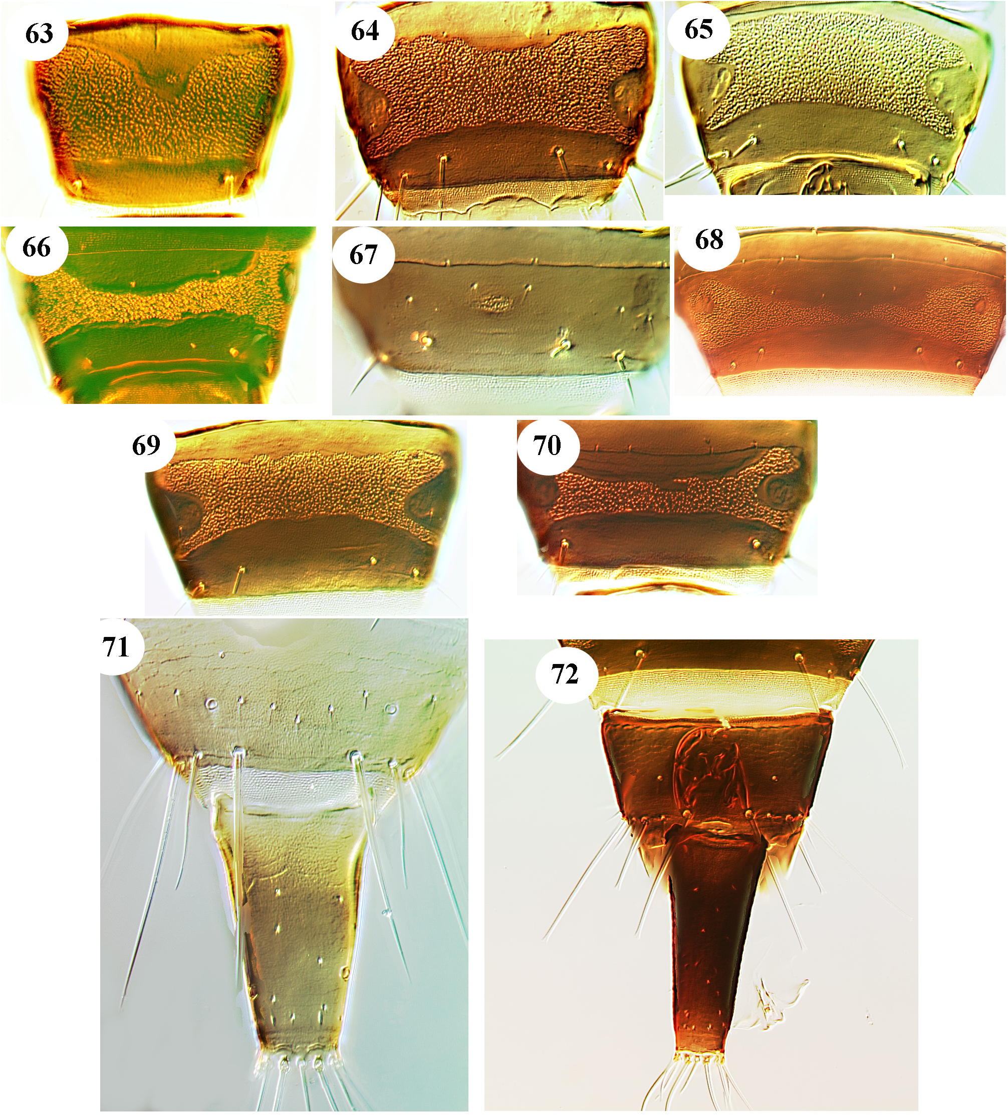
This genus is erected for a group of species from Australia that are similar in general appearance to many Holothrips species. That is, they have the maxillary stylets very long and close together medially for the full length of the head, the head is longer than wide and elevated dorsally to a greater or lesser extent, and antennal segments VII and VIII are broadly joined and sometimes form a unit with an almost continuous outline. However, in contrast to Holothrips species , the suture between antennal segments VII and VIII is fully complete dorsally and ventrally, the males have a pore plate on sternite VIII, males rarely have any reticulate areas on the intermediate sternites, the maxillary stylets are more slender than in Holothrips species , and on tergite IX setal pair iS between setae S1 and S2 are particularly long ( Fig. 71 View FIGURES 63–72 ). These character states suggest that this new genus is more likely to be related to the genus Hoplandrothrips in the Phlaeothrips -lineage than to Holothrips in the Docessissophothrips genus-group ( Dang et al. 2014).
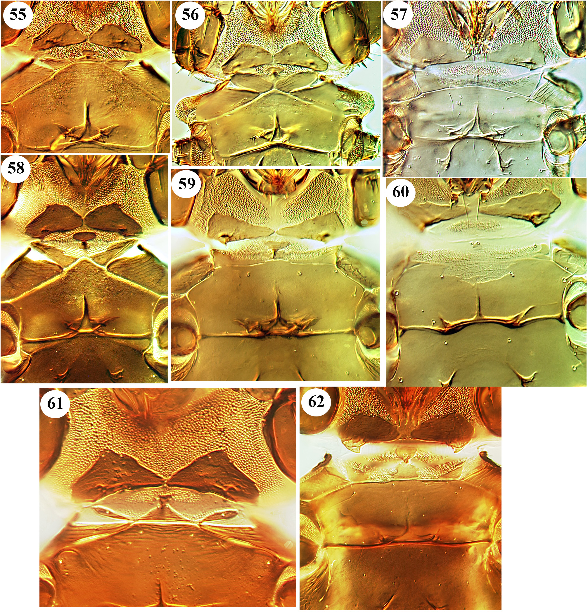
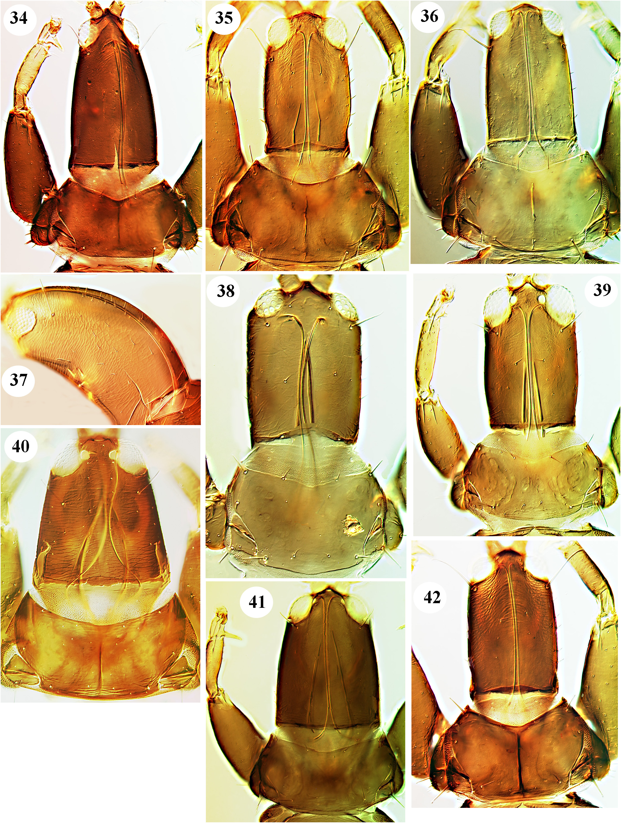
Sexual dimorphism is well developed among the species of Holoengythrips , as it is among the species of Hoplandrothrips ( Mound & Tree 2013) . Not only do males have a larger fore tarsal tooth than females, but the anterior margin of the mesoeusternum is much narrower in males than in females, with associated differences in the mesopresternum ( Figs 56–59 View FIGURES 55–62 ). In some of the species the postocular setae of males are not only longer than those of females, but have curiously flattened apices, not the normal capitate apices. Moreover, in some species the males have the major setae on tergites III–V broadly spatulate, in contrast to the normal setae of females. The pore plate on sternite VIII of the males is large in most of the species, but is small and circular in one species. Head shape varies amongst the species of this genus, with two species having the head exceptionally long and elevated medially, although this elevation is less among the other species ( Figs 34–42 View FIGURES 34–42 ).
Key to Holoengythrips species
1. Tergite IX major setae capitate ( Fig. 72 View FIGURES 63–72 ) [postocular setae capitate, almost as long as eye length; body and legs dark brown, tarsi yellow]............................................................................. padthawayi View in CoL sp.n.
-. Tergite IX major setae finely pointed, or rarely bluntly pointed.................................................. 2
2. Antennal segment III with 2 sensoria...................................................................... 3
-. Antennal segment III with 3 sensoria...................................................................... 5
3. Mesoeusternum anterior margin deeply eroded ( Fig. 60 View FIGURES 55–62 ); mesopresternum almost absent, represented by two very small lateral sclerites; antennal segment IV with 2 sensoria; male sternite VIII with small circular pore plate medially ( Fig. 67 View FIGURES 63–72 )................................................................................................... namadgi View in CoL sp.n.
-. Mesoeusternal anterior margin entire, often angulate ( Figs 56–57 View FIGURES 55–62 ); mesopresternum represented by two lateral triangles; antennal segment IV with 3 or 4 sensoria; where known, male sternite VIII with broadly transverse pore plate ( Figs 63–70 View FIGURES 63–72 )..... 4
4. Antennal segment IV with 4 sensoria; female with fore tarsal tooth shorter than tarsal width; female with mesoeusternal anterior margin weakly angulate and mesopresternum of two slender triangles ( Fig. 59 View FIGURES 55–62 ); male mesoeusternal anterior margin narrow and sharply angulate ( Fig. 58 View FIGURES 55–62 )........................................................... tallagandai View in CoL sp.n.
-. Antennal segment IV with 3 sensoria; female with fore tarsal tooth longer than tarsal width ( Fig. 41 View FIGURES 34–42 ); female with mesoeusternal anterior margin clearly angulate and mesopresternum of two well-defined triangles; male not known...... tarsalis View in CoL sp.n.
5. Mid and hind femora bicoloured, sharply yellow at apex.......................................... maynardae View in CoL sp.n.
-. Mid and hind femora not sharply bicoloured................................................................ 6
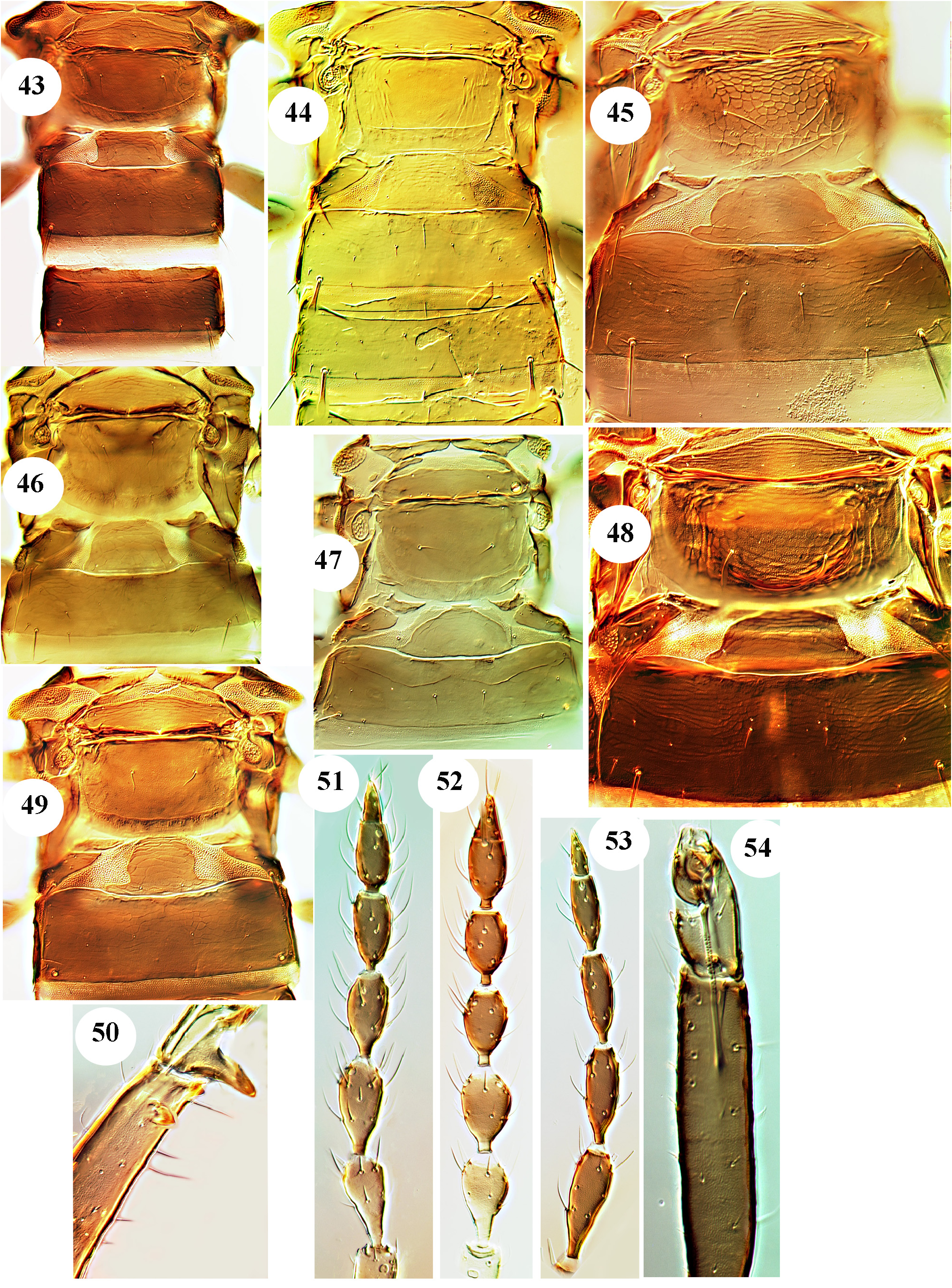
6. Antennal segments III–VI brown except III yellow at extreme base; male fore tibiae without subterminal tubercle or with small setal-bearing tubercle ( Fig. 54 View FIGURES 43–54 )........................................................................... 7
- Antennal segment III extensively yellow; male fore tibiae with prominent subterminal tubercle ( Figs 34 View FIGURES 34–42 , 50 View FIGURES 43–54 )............. 8
7. Tube medially with transverse yellow area; female microptera with minute fore tarsal tooth; male with broad pore plate on sternite VIII ( Fig. 64 View FIGURES 63–72 )........................................................................... kathyae View in CoL sp.n.
-. Tube brown, paler distally; female microptera with no fore tarsal tooth; male with slender transverse pore plate ( Fig. 70 View FIGURES 63–72 ).............................................................................................. turcoae View in CoL sp.n.
8. Male fore tibia with dorsal subapical tubercle ( Fig. 50 View FIGURES 43–54 ); sternite VIII with narrow transverse pore plate medially ( Fig. 66 View FIGURES 63–72 ); metanotum reticulate ( Figs 45, 48 View FIGURES 43–54 )............................................................. monteithi View in CoL sp.n.
-. Male fore tibia with subapical tubercle on inner lateral margin ( Fig. 34 View FIGURES 34–42 ); sternite VIII occupied by large pore plate except anteromedially ( Fig. 63 View FIGURES 63–72 ); metanotum without sculpture ( Fig. 43 View FIGURES 43–54 )...................................... barrinei View in CoL sp.n.
No known copyright restrictions apply. See Agosti, D., Egloff, W., 2009. Taxonomic information exchange and copyright: the Plazi approach. BMC Research Notes 2009, 2:53 for further explanation.
|
Kingdom |
|
|
Phylum |
|
|
Class |
|
|
Order |
|
|
Family |