
Proxima meloi, Laranjeira & Gonçalves & Domahovski & Takiya, 2022
|
publication ID |
https://doi.org/ 10.11646/zootaxa.5091.4.5 |
|
publication LSID |
lsid:zoobank.org:pub:5B5E7026-B0D7-4BCC-BD17-4EB7045CF26F |
|
DOI |
https://doi.org/10.5281/zenodo.5864228 |
|
persistent identifier |
https://treatment.plazi.org/id/AF3B2FDA-F96D-4522-8899-CCF27A55397B |
|
taxon LSID |
lsid:zoobank.org:act:AF3B2FDA-F96D-4522-8899-CCF27A55397B |
|
treatment provided by |
Plazi |
|
scientific name |
Proxima meloi |
| status |
sp. nov. |
Proxima meloi View in CoL sp. nov.
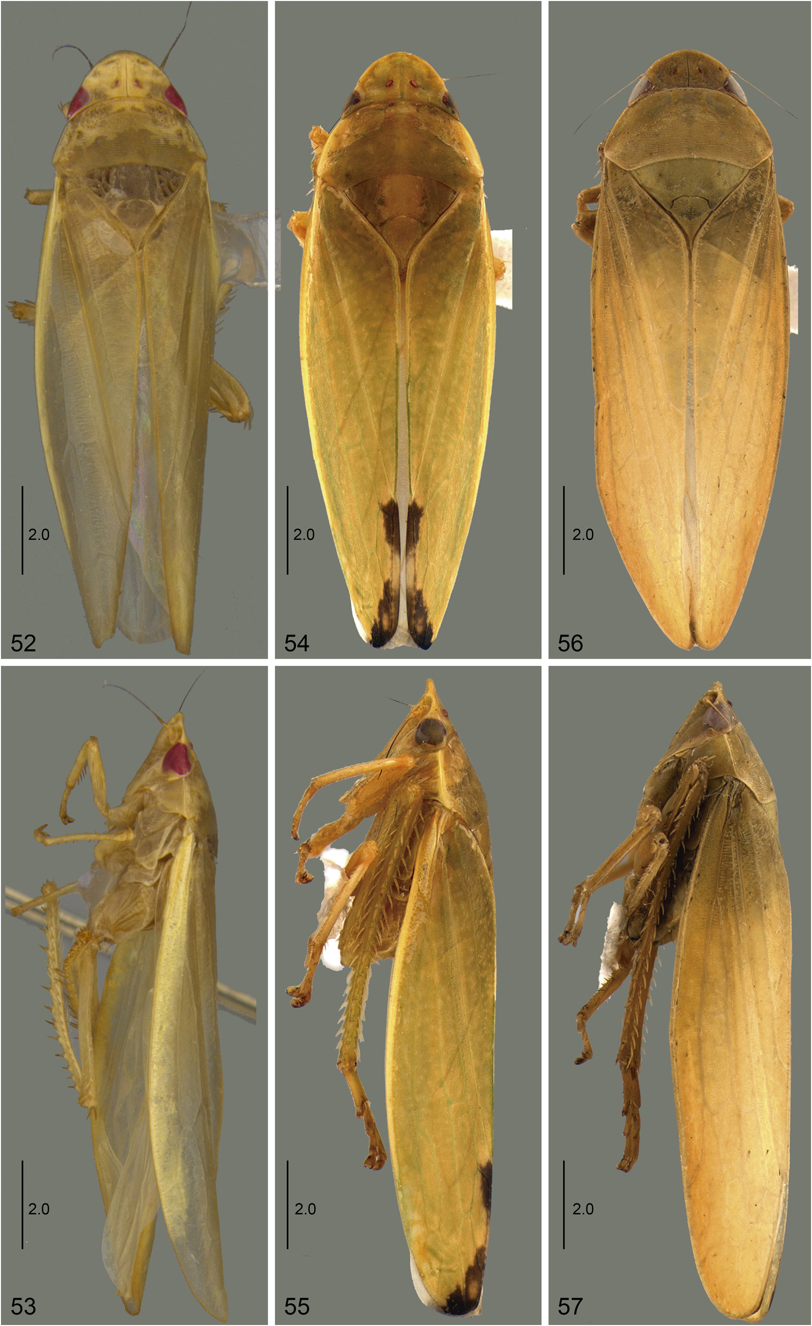
( Figs 33–51 View FIGURES 33–44 View FIGURES 45–51 , 56, 57 View FIGURES 52–57 )
Diagnosis. Forewing ( Fig. 36 View FIGURES 33–44 ) without dark maculae. Male pygofer ( Fig. 39 View FIGURES 33–44 ), in lateral view, with apical portion rounded, not curved or folded inwardly. Subgenital plate ( Fig. 40 View FIGURES 33–44 ), in ventral view, with inner margin strongly expanded. Style ( Fig. 42 View FIGURES 33–44 ), in lateral view, with blade approximately straight, higher at base and narrowing towards the apex; ventral margin weakly serrated. Aedeagus ( Figs 43, 44 View FIGURES 33–44 ) with atrial processes not bifid and as long as shaft; shaft without processes; apex bifid into acute processes directed anteriorly.
Measurements (mm). 11.8–12.9 mm (male); 12.8–13.5 mm (female).
Coloration. As in generic description.
External morphology. Crown-face transition ( Fig. 34 View FIGURES 33–44 ) with transverse striae only close to the eye, smooth on the median portion. Forewing ( Fig. 36 View FIGURES 33–44 ) with extra-numerary veins on discal, subapical and apical cells, sometimes apical half almost reticulate. Other characters as in generic description.
Male terminalia. Sternite VIII ( Fig. 37 View FIGURES 33–44 ), in ventral view, approximately 1.4 times wider than long, strongly convex; lateral margins parallel; posterior margin widely rounded, emarginated medially. Valve ( Fig. 38 View FIGURES 33–44 ), in ventral view, approximately 2.7 times wider than long; posterior margin slightly excavated medially. Pygofer ( Fig. 39 View FIGURES 33–44 ), in lateral view, approximately 1.8 times longer than high; internal surface of lateral lobe with a thickening of the integument at anterodorsal portion; ventral margin rounded and expanded ventrally; posterodorsal and posteroventral margins approximately straight and strongly convergent at apex; apex rounded. Subgenital plate ( Fig. 39 View FIGURES 33–44 ), in lateral view, strongly curved dorsally; in ventral view ( Fig. 40 View FIGURES 33–44 ), 2.5 times longer than wide; subtriangular, outer margin slightly excavated; inner margin expanded and widely rounded; apex rounded. Connective ( Fig. 41 View FIGURES 33–44 ), in dorsal view, large and wide; arms 1.2 times longer than stalk; anterior margin deeply excavated. Style ( Fig. 41 View FIGURES 33–44 ), in dorsal view, with lateral lobe rounded; in lateral view ( Fig. 42 View FIGURES 33–44 ), blade approximately straight, higher at base and narrowed towards apex; ventral margin weakly serrated; apex acute and curved dorsally. Aedeagus ( Figs 43, 44 View FIGURES 33–44 ) with preatrium reduced; dorsal apodeme moderately developed with dorsal margin deeply excavated, lateral margins slightly produced laterally; atrial processes long, wide at base and narrowing towards apex, as long as the shaft, apex rounded; shaft long and tubular, widely curved anterodorsally; apex bifid (from apical third of shaft) into acute processes directed anteriorly.
Female terminalia. Sternite VII ( Fig. 45 View FIGURES 45–51 ), in ventral view, approximately 1.4 times wider than long; posterolateral angle rounded; posterior margin slightly excavated near lateral angles, median portion strongly projected posteriorly with distinct median lobe deeply excavated medially. Pygofer ( Fig. 46 View FIGURES 45–51 ), in lateral view, approximately 1.3 times longer than high; macrosetae distributed at ventral half and apex; apex rounded. First valvifer ( Fig. 47 View FIGURES 45–51 ), in lateral view, subelliptic, slightly higher than wide; posterior margin truncated. First valvula ( Figs 47, 48 View FIGURES 45–51 ), in lateral view, approximately 8.6 times longer than high; apex subacute. Second valvula ( Figs 49, 50 View FIGURES 45–51 ), in lateral view, approximately 10.5 times longer than high; dorsal margin with four rounded teeth, basal tooth larger than others and apical tooth reduced; apex rounded.Second valvifer ( Fig. 51 View FIGURES 45–51 ), in lateral view, approximately 2.2 times higher than wide.Gonoplac ( Fig. 51 View FIGURES 45–51 ), in lateral view, approximately 3.6 times longer than high. Other characters as in generic description.
Etymology. The new species is named in tribute to Dr. Gabriel A. R. Melo, Hymenoptera specialist and one of the collectors of the type-series.
Material examined: Holotype male: “ Brasil, Bahia, 10 Km a\ NE de Encruzilhada,\ 15. 483ºS 40.824ºW,\ 830m 15.xii.2012,\ G. Melo & P. Grossi \Arm luminosa” ( DZUP) GoogleMaps . Paratypes: Bahia: 2 ♂, 3 ♀, same data as holotype ( DZUP); GoogleMaps 9 ♂, 1 ♀, “ Brasil, Bahia, 6 Km a\ SE de Encruzilhada,\ 15. 567ºS 40.870ºW,\ 910m 16.xii.2012,\ G. Melo & P. Grossi \ Arm luminosa” (4 ♂, 1 ♀, DZRJ DZRJ-AUCH 0232 , 0233 , 0234 , 0235 , 0236 ; 1 ♂, MZSP; 4 ♂, DZUP); GoogleMaps 4 ♂, 2 ♀, “ Brasil, Bahia,\ Encruzilhada \ XII.1980 \ M. Alvarenga leg.” ( DZUP); GoogleMaps 2 ♂, 1 ♀, “ Brasil, Bahia,\ Encruzilhada \ XI.1975 \ M. Alvarenga leg.” ( MNRJ); GoogleMaps 2 ♂, 2 ♀, “ Encruzilhada \ BA. Brasil XI/74\ M. Alvarenga ” ( DZUP); GoogleMaps 1 ♂, “ Encruzilhada \ BA. Brasil XII./80\ M. Alvarenga ” ( DZUP); GoogleMaps Minas Gerais: 1 ♂, “ Brasil, Minas Gerais, 12km a N de\ Águas Vermelhas, Faz.\ Faceiro, 15.640ºS \ 41.477ºW, 845m,\ 12.xii.2012 G. Melo & P.\ Grossi. Arm luminosa” ( DZUP); GoogleMaps 1 ♀, “ Brasil, Minas Gerais, 22km \ a NE de Pedra Azul , 15.819ºS 41.200ºW, 745m,\ 13.xii.2012, G. Melo &\ P. Grossi ” ( DZUP) GoogleMaps .
Notes. Proxima meloi sp. nov. can be distinguished from other species of Proxima by its subgenital plate ( Fig. 40 View FIGURES 33–44 ) with inner margin strongly expanded medially and by the aedeagus ( Fig. 43 View FIGURES 33–44 ) with atrial processes simple and as long as shaft.


No known copyright restrictions apply. See Agosti, D., Egloff, W., 2009. Taxonomic information exchange and copyright: the Plazi approach. BMC Research Notes 2009, 2:53 for further explanation.
|
Kingdom |
|
|
Phylum |
|
|
Class |
|
|
Order |
|
|
Family |
|
|
Genus |