
Dolinasorex glyphodon, Rofes & Cuenca-Bescós, 2009
|
publication ID |
https://doi.org/ 10.1111/j.1096-3642.2008.00470.x |
|
DOI |
https://doi.org/10.5281/zenodo.10546136 |
|
persistent identifier |
https://treatment.plazi.org/id/03BFB91B-337C-FF89-88EB-FF7436EECB6A |
|
treatment provided by |
Felipe |
|
scientific name |
Dolinasorex glyphodon |
| status |
sp. nov. |
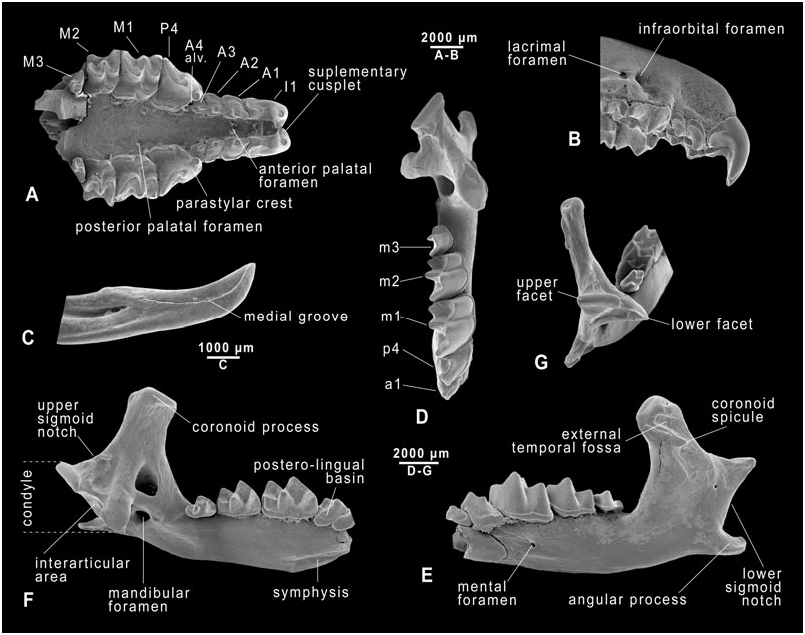
DOLINASOREX GLYPHODON SP. NOV.
FIGURE 3A–G View Figure 3
Type locality and age: Gran Dolina, Sierra de Atapuerca, Burgos ( Spain); stratigraphical levels TD4, TD5, and TD6, corresponding to the late Early Pleistocene.
Holotype: MPZ 2007 View Materials /699, left mandible with all lower teeth in situ but a broken incisor.
Paratypes: MPZ 2007 View Materials /700, incomplete skull with all upper teeth in situ, with the exception of both A4s ; MPZ 2007 View Materials /701, left lower incisor .
Location of types: Holotype and paratypes in the collection of the MPZ, University of Zaragoza ( Spain) .
Etymology: Derived from the Greek terms glyphos (groove) and odon (tooth), in reference to the grooved first lower incisor.
Measurements: See Table 1A and B and the morphometric analysis below.
Diagnosis: Although no unambiguous autapomorphies can be recognized in the material, the taxon can be diagnosed by the following unique combination of characters (as stated in the apomorphy lists of the phylogenetic analysis): there is no buccal cingulum in the first lower incisor and no entoconid crest in m1; the mandible has a deeply excavated external temporal fossa, an internal temporal fossa in a low position, and a horizontal bar dividing the latter in two sections is not present. Some other useful diagnostic traits are: large-sized shrew with stoutly built skull and mandible; teeth stained red to dark red; dental formula: 1-4-1-3/1-1-1-3; first upper incisor strongly bifid; four upper antemolars; M2 markedly trapezoidal in shape; first lower incisor acuspulate, strongly upturned and with a narrow but conspicuous groove in its medial face; shallow posterolingual basin of p4; talonid of m3 with hypoconid and entoconid; lower teeth, except for the first incisor, with a relatively broad and pronounced buccal cingulum; ascending ramus of the mandible leaning laterally; coronoid process long, broad, robust, and not leaning forward; coronoid spicule large and strongly pronounced; internal temporal fossa large and deeply pocketed; lower facet of the mandibular condyle very anteriorly placed and thus invisible in buccal view; mandibular foramen large and frequently connected to the internal temporal fossa; symphyseal fossa deep and elongated.
Description
In general, the dentition of the soricids consists of one incisor, three molars and small elements situated between the incisor and the molars, which are named, following Reumer (1984), the antemolars (A). In the upper dentition, the last antemolar is conventionally designated P4, the number of remaining antemolars varying from two to five. The antemolars of the Soricidae are often called ‘unicuspids’. This name is considered incorrect because quite often these teeth possess accessory cusps ( Reumer, 1984). In the lower dentition there is only one tooth called an antemolar (a1); as in the case of its upper counterpart, the last antemolar (‘a2’) is generally considered to be a premolar and named p4. The i1 of the soricids has a single cusp, known as the apex, and a series of serrations on the dorsal edge which may vary from nonexistent up to four. Reumer (1984) named these serrations ‘cuspules’ to distinguish them from the true cusps (cuspules having no connection with the pulp cavity). As in other soricines, all the dental elements of Dolinasorex are stained a dark red in the apical part of the crown.
I1 ( Fig. 3A, B View Figure 3 ): Strongly bifid, namely with a supplementary cusplet on the medial side, slightly divergent and separated by a wide, deep groove on the apical part of the tooth. The talon is broad mesiodistally, and trapezoidal to square-shaped in lateral view. The lateral root-crown junction line is nearly perpendicular to the dorsal edge of the tooth, or inclines slightly to the front. This line is either slightly or markedly undulated. The cingulum along the posterior buccal edge is only present behind the talon. The root is
| MPZ |
Museo Paleontologico de la Universidad de Zaragoza |
No known copyright restrictions apply. See Agosti, D., Egloff, W., 2009. Taxonomic information exchange and copyright: the Plazi approach. BMC Research Notes 2009, 2:53 for further explanation.
|
Kingdom |
|
|
Phylum |
|
|
Class |
|
|
Order |
|
|
Family |
|
|
Genus |