
Neochavesia lapollai, Williams, 2010
|
publication ID |
https://doi.org/10.1080/00222930310001657856 |
|
persistent identifier |
https://treatment.plazi.org/id/03C087B3-FFDC-FFAF-EDBC-79BB1CEF6ABA |
|
treatment provided by |
Felipe |
|
scientific name |
Neochavesia lapollai |
| status |
sp. nov. |
Neochavesia lapollai sp. nov.
( figures 3–6 View FIG View FIG View FIG View FIG )
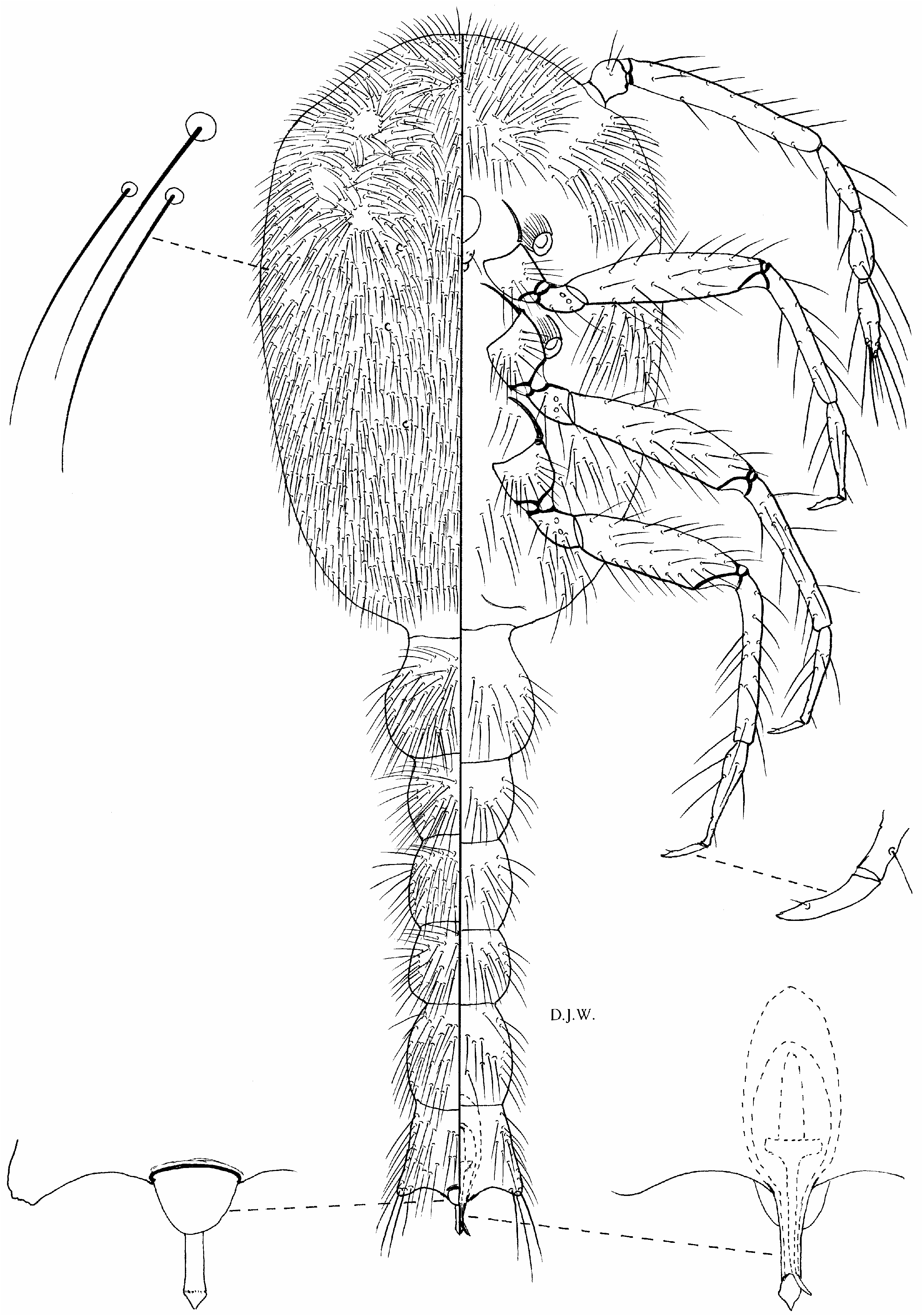
Description—adult female ( figure 3 View FIG )
Body of adult female elongate-pyriform, largest specimen 1.65 mm long, 0.73 mm wide; head and thorax broadly rounded, abdomen gently tapering to abdominal segment VIII. Anal lobe area almost rectangular, about 250–262 M m long, 110–140 M m wide, separated from abdominal segment VIII by intersegmental line, a little narrower at base than abdominal segment VIII then broadening slightly. Anal lobes rounded posteriorly, separated at apex by a short narrow notch about 50 M m long; inner apical edges of lobes not divergent. Each lobe sclerotized on dorsum except near medial area; weakly sclerotized ventrally mostly on margins. Dorsum of each anal lobe with numerous long slender setae, many about 250 M m long, except anteriorly where a few are 90 M m long. Ventral setae on posterior edges of anal lobes about 250 M m long, lobe setae anteriorly much shorter. Anal ring situated within intersegmental line dividing abdominal segment VIII and anal lobes, sometimes difficult to locate, represented dorsally by narrow slit-like opening, 50–70 M m wide, rim sclerotized anteriorly and barely perceptible posteriorly, represented by a thin line. Antennae 450–650 M m long, each with five segments; segment 2 longest with almost parallel sides, last segment more slender than others, broadening near base then tapering to apical point. Legs well developed. Hind trochanter z femur 220–310 M m long, tibia z tarsus 200–270 M m long; tarsus tapering abruptly to long slender claw, 62–80 M m long. Ratio of lengths of hind tibia z tarsus to trochanter z femur 0.87–0.90. Ratio of lengths of hind tibia to tarsus 1.52–1.85. Claw shorter than tarsus, with a pair of short setose digitules at base. Labium 160–200 M m long, about 65 M m wide, basal segment with two clusters each of four short setae, medial segment apparently without setae, apical segment with eight pairs of setae on anterior surface in addition to minute apical pair. Circuli numbering two, situated in middle of abdominal segments II and III. Each circulus
truncate-conical, about 25 M m in diameter at base, projecting about 40 M m from surface of derm, distal end about 12.5 M m in diameter with deep cylindrical inner cup, slightly surpassing circulus in length and tapering to blunt inner end. Spiracles each with narrow apodemes then widening to crescentic inner ends.
Dorsal surface with numerous flagellate setae of various lengths on abdominal segments III–VIII, longest about 100 M m long, in single rows across middle of segments, others 30–50 M m long. Head, thorax and abdominal segments I and II densely covered in slender setae, mostly 15 M m long, with minute setal collars, accompanied by fewer longer setae 20–25 long, with slightly wider collars; bands of setae well separated from intersegmental lines on abdominal segments I and II. A few long stout setae, 100–150 M m long, present near head margin.
Ventral surface with similar abdominal setae to those on dorsal segments III– VIII. Medial area of thorax with a few short setae, widely spaced. Similar setae present across abdominal segment II. Short setae, similar to those on dorsum of head and thorax, densely covering head margin and thorax lateral to spiracles. A few long setae present on head margin. Pores and ducts absent from dorsum and venter. Description—adult male ( figure 4 View FIG )
Body of unusual shape, strongly capitate, about 1.4 mm long. Head, thorax and abdominal segments I and II combined, oval, about 0.35 mm wide, widest at prothorax, tapering abruptly at abdominal segment III to extremely narrow elongate abdomen with segments clearly defined, terminating in a pair of laterally projecting angular anal lobes. Genital capsule between anal lobes, mostly internal, elongate-oval, about 190 M m long, 65 M m wide, tapering to elongate penial sheath, pointed at distal end and projecting well beyond anal lobes. Ventral slit apparently near tip of penial sheath only. Aedeagus long and slender, bluntly pointed. Dorsal apical margin of abdomen with anal ring 32 M m wide, represented by anterior sclerotized rim and lobe-like membranous rounded projection. Antennae about
510 M m long, each with five segments; second segment with almost parallel sides, about same length as last three segments combined. Terminal segment elongateoval, narrowing to a point. Legs well developed, slender. Hind trochanter z femur about 300 M m long, hind tibia z tarsus about 320 M m long. Ratio of lengths of hind tibia z tarsus to trochanter z femur 1.06. Ratio of lengths of hind tibia to tarsus 1.90. Tarsus slightly swollen near base then tapering to fairly stout elongate claw,
about 40 M m long, with a pair of short setose digitules situated towards distal end on a small prominence or denticle. Mouthparts represented by remains of clypeolabral shield and a small vestigial labium. Spiracular openings situated at tips of short sclerotized conical apodemes. Apical setae at tips of anal lobes about 105 M m long, accompanied by others on dorsum about 80 M m long; anterior dorsal setae on anal lobes each about 60 M m long. Most other dorsal setae each about 50 M m long, fairly numerous on abdominal segments, crowded on enlarged head, thorax and abdominal segments I and II, mostly with small setal collars, some with larger collars. Ventral setae on abdomen similar to those on dorsum but not so numerous. Setae on medial area of thorax sparse, numerous on head and lateral areas.
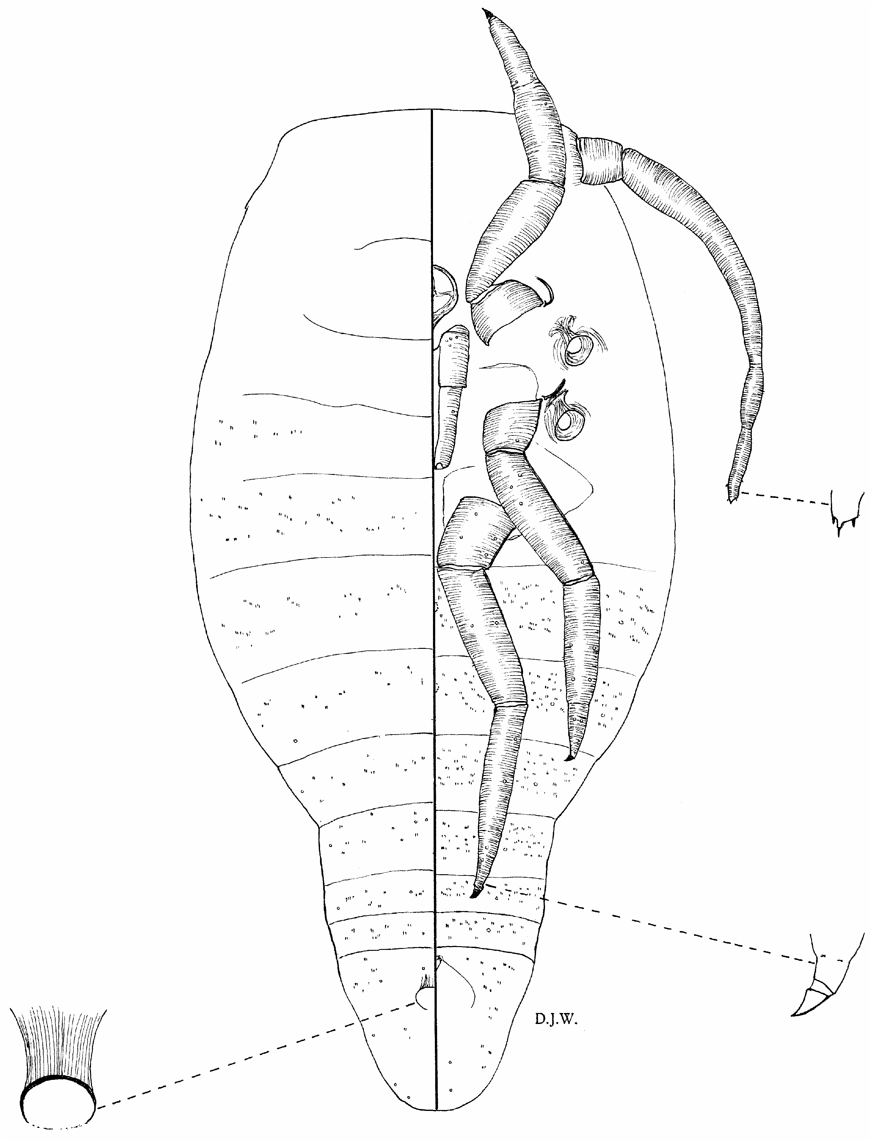
Description—female pupa ( figures 5A View FIG , 6 View FIG )
Body pyriform, 0.9–1.2 mm long, 0.50–0.75 mm wide, head margin almost straight, head and thorax expanded, widest at metathorax, then gently tapering to abdominal segment IV; remainder of abdomen much narrower, tapering to rounded posterior end, segmentation distinct. Antennae each about 320–500 M m long, showing slight constrictions in positions of developing segments 3–5, sclerotized,
terminal segment with three apical points. Legs sclerotized; hind trochanter z femur fused, 150–190 M m long, tibia z tarsus fused, 150–210 M m long; claw stout, almost triangular, sclerotized, about 20 M m long. Labium 120–160 M m long, subrectangular, much longer than developing clypeolabral shield. Spiracles well developed. Anal ring about 32.5 M m wide, subcircular, with anterior rim sclerotized, posterior rim sometimes indistinct, situated well forward on last abdominal segment. Developing circuli present in most specimens, represented by small indistinct circular areas in
middle of abdominal segments II and III. Dorsum of thorax and abdomen, and venter of abdomen with numerous minute microtrichia.
Material examined
HOLOTYPE: adult ”, Guyana, Mount Ayanganna , Falls Camp, 05 ‡ 22.332’N, 59 ‡ 57.563’W, 1134 m, 13 May 2002, associated with Acropyga sp. (coll. J. S. LaPolla) ( UGGG).
PARATYPES: Guyana, same data as holotype, eight adult ””, three third-instar ””, five ” pupae enclosing adult ””, one adult „, three „ pupae, three „ pupae enclosing adult „„, one „ prepupa, one prepupa „ enclosing pupa ( BMNH), four adult ””, two ” pupae enclosing adult ””, one ” pupa, one „ pupa enclosing adult „ ( USNM) .
Etymology
The species is named after the collector, John S. LaPolla, who has supplied some interesting specimens for study.
Comments
Adult females of N. lapollai are similar to those of N. trinidadensis in possessing antennae that are longer than the first legs. There is a distinct notch between the anal lobes in N. trinidadensis with the inner distal edges of the lobes diverging. In N. lapollai , the notch between the anal lobes is short and narrow and the inner edges of the anal lobes are parallel and do not diverge. Moreover, there are many long stout setae on the dorsum and venter of the abdomen in N. lapollai , and these are absent from N. trinidadensis .
Important features of the adult male are the extremely slender elongate abdomen with the anal lobes projecting laterally, and fairly stout claws with the digitules situated towards the apices.
The female pupa ( figures 5A View FIG , 6 View FIG ) differs from the male pupa ( figure 5B View FIG ) and prepupa in possessing a long, well-developed labium and a rounded apex to the abdomen. The male prepupa and pupa each have a rounded dorsal projection at the apex of the abdomen, and the developing penial sheath on the ventral apex is slightly longer in the pupa than in the prepupa. In the male prepupa and pupa, the anus is located in about the same position as in the female pupa but the mouthparts are degenerate and barely perceptible.
Collected with the mealybugs was also a single specimen of Mixorthezia near reynei (Laing), family Ortheziidae .
Neochavesia trinidadensis (Beardsley)
Chavesia trinidadensis Beardsley, 1970: 514 View in CoL . Holotype adult ”, Trinidad, San Rafael , 31 May 1935 (coll. N. A. Weber), in a log under a cacao tree (USNM).
Neochavesia trinidadensis (Beardsley) View in CoL , Williams and Granara de Willink, 1992: 236; Ben-Dov, 1994: 241.
Material examined
Trinidad, St Augustine , on roots of cacao, 12 May 1935 (coll. N. A. Weber) . Nicaragua, Chontales, 28 June 1940 (coll. R.- P. Roba) ( USNM) .
Comments
Each antenna of this species is five-segmented and longer than a first leg. Antennal segment 2 is by far the longest, about 168–185 M m long, and the terminal segment is almost bulbous. The anal lobes are well developed with the inner edges clearly defined on the dorsum and venter. Although the anal ring is placed at the base of the lobes, it is within the lobe area and not in the intersegmetal line. A single circulus was described originally but there are two present in the specimens at hand, located in the middle of abdominal segments II and III. All the dorsal setae on abdominal segments III –VII are noticeably slender, 15–50 M m long. Furthermore, there are about 50 setae on the dorsum of each anal lobe. The species is close to N. lapollai in its general shape but the anal lobes in N. trinidadensis are well separated at the distal end and the inner edges of the lobes are clearly defined. In N. lapollai , the anal lobes are not well separated and the notch at the apex is short and sometimes difficult to observe. Moreover, the inner distal edges of the anal lobes in N. trinidadensis are divergent, whereas in N. lapollai they are separated by a short notch with parallel sides. There are also long, thick setae on the abdomen, interspersed with shorter slender setae in N. lapollai but in N. trinidadensis all the setae in these positions are slender.
This mealybug was first discussed as a coccid by Weber (1944), associated with Acropyga (Rhizomyrma) berwicki Wheeler.
Chavesia weberi Beardsley, 1970: 517 View in CoL . Holotype adult ”, Guyana, Mazaruni River , associated with Acropyga (Rhizomyrma) paludii Weber (USNM).
Neochavesia weberi (Beardsley) View in CoL , Williams and Granara de Willink, 1992: 236; Ben- Dov, 1994: 241.
Material examined
Guyana, Mazaruni River , near forest settlement, 21 August 1935 (coll. N. A. Weber) ( USNM) .
Comments
This is a distinctive species, described with short four-segmented antennae, each shorter than a first leg. The anal lobes are well developed with the anal ring situated only a short distance from the base of the notch between the anal lobes. The single paratype available for study, and other paratypes kindly examined by Douglass R. Miller, show marked differences from the original illustration and description. The anal lobes are distinctly separated from abdominal segment VIII by an intersegmental line. Abdominal segment VIII is distinct, with long slender setae, mostly longer than the length of the segment. Similar setae are present on abdominal segments V and VI but minute setae, mostly about 15 M m long, are present on abdominal segments III and IV, not on abdominal segments II and III as shown in the original illustration. These setae are similar to those on the dorsum of the head, thorax and abdominal segments I and II, and on the venter of the head margin and thorax lateral to the spiracles. The medial ventral areas of the thorax and abdominal segment II possess long stout setae and the two circuli are situated in the middle of abdominal segments III and IV. The vulva is located between abdominal segments VII and VIII as in all mealybugs .
A single female pupa is available with identical collecting data. It possesses a rounded abdomen and the labium is long and well developed, characters similar to those of the female pupa of N. lapollai . N. weberi has not been collected again since its original discovery.


No known copyright restrictions apply. See Agosti, D., Egloff, W., 2009. Taxonomic information exchange and copyright: the Plazi approach. BMC Research Notes 2009, 2:53 for further explanation.
|
Kingdom |
|
|
Phylum |
|
|
Class |
|
|
Order |
|
|
Family |
|
|
Genus |
Neochavesia lapollai
| Williams, D. J. 2010 |
Neochavesia trinidadensis (Beardsley)
| WILLIAMS, D. J. & GRANARA DE WILLINK, M. C. 1992: 236 |
Neochavesia weberi (Beardsley)
| DOV, Y. 1994: 241 |
| WILLIAMS, D. J. & GRANARA DE WILLINK, M. C. 1992: 236 |
Chavesia trinidadensis
| BEARDSLEY, J. W. 1970: 514 |
Chavesia weberi
| BEARDSLEY, J. W. 1970: 517 |