
Parartemia auriciforma, Timms, Brian V & Hudson, Peter, 2009
|
publication ID |
https://doi.org/ 10.5281/zenodo.190741 |
|
DOI |
https://doi.org/10.5281/zenodo.6225429 |
|
persistent identifier |
https://treatment.plazi.org/id/03C38793-FFB8-814A-7BDA-3D82FC119833 |
|
treatment provided by |
Plazi |
|
scientific name |
Parartemia auriciforma |
| status |
sp. nov. |
Parartemia auriciforma View in CoL sp. nov.
(Figure 5)
Type material. Holotype. Male, SOUTH AUSTRALIA, Great Victoria Desert, Wyola Lake, (29o09’00”S, 130o14’30”E), 15 December 1994, P. Hudson, SAM C6794; Allotype. Female, same collecting data as holotype, SAM C6797; Paratypes. Two males and two females, same collecting data as holotype, SAM C6795, C6796.
Description. Male. Length 11.5 mm (head and thorax 5 mm and abdomen 6.5 mm).
Head (Fig. 5A) with first antenna filiform a little longer than eye plus peduncle. Basal antennomeres of second antenna fused proximally at an angle of about 50 degrees from body axis. Ventral margin with paired linear, ventral processes (VP, Fig 5A) clothed irregularly with small spines and tubercles mainly at lateral and medial corners. Overall dimensions about 3 times longer than deep with lateral edge about one third the length of medial edge. Lateral corner rounded and medial corner prominent and bluntly triangular. Area between ventral processes concave without any medial projection or doming. Frontal surface of basal antennomeres with paired ridges parallel to body axis and terminating in small triangular frontal processes (FP, Fig 5A). These frontal processes subequal in length to depth of ventral processes and length subequal to basal width. Second antennal distal antennomere curved, a little longer than basal segment and terminating in a small spine-like appendix. Labrum lacking a spine.
Each side of thorax with a near symmetrical lateral lobe on each segment and small lateral lobe on first genital segment (L, Fig.5B). Eleven pairs of thoracopods with first two and last three noticeably reduced. Fifth thoracopod structure as in P. acidiphila , though numbers of posterior setae slightly different (see above).
Gonopods (Fig. 5C) paired, basal parts fused together and about twice the diameter of tubular free apical parts. Basal part with a broad triangular process (DP, Fig. 5C) apically and apical tube with a finger-like process subapically. Neither process hooked.
Abdominal segments increasing in length and narrowing 1 to 6, particularly 5 and 6, so that 6th segment about twice the length of first. Telson about half the length of first abdominal segment and bearing fringed cercopods subequal in length to fifth abdominal segment.
Description. Female. Length 8.5 mm.
Head (Fig. 5D) with first antenna filiform and about half the length of the eye plus peduncle. Second antenna flattened and about twice the length of eye plus peduncle and terminating in a markedly narrowed, acute apex on the posterior side. Labrum with a prominent recurved spine.
Thorax with 10 pairs of thoracopods, similar in structure to those of male. Eleventh thoracic segment without appendages Tenth thoracopod (Fig. 5G) reduced: endites with few posterior setae, endopodite and exopod with about 12 and 20 such setae respectively, and epipodite smaller than both exopod and praepipodite. Posterior thoracic segments, particularly those of 7th to 10th segments, expanded laterally (Fig. 5E). Eight and ninth segments with tuberculate lateral lobes terminating in hollowed out auriculiform structures, about one-sixth segment width. Segments 5, 6 and especially 7th with simpler, less prominent lateral extensions. Segment 10 with lateral blunt triangular projection on each side and segment 11 with dorsolateral surface uneven and consisting of two triangular anteriorly pointed mounds (TM, Figs 5E,F) on each side. Surface of both segments 10 and 11 papillate.
Lateral brood pouches (Fig. 5F) almost spherical in shape but with small lobes ventrally and connected ventrally to a shared gonopore on a short tubular protrusion. Dorsal surface of brood chambers pigmented and each chamber with about 25 spherical smooth-surfaced eggs in mature ovigerous females.
Abdominal segments largely as in male, but with the telson proportionally larger and all segments papillate.
Etymology. The specific name reflects the auriculiform lateral lobes of the female.
Variability. Adult male lengths vary from 10.5 to 12mm and adult femal from 7.9 to 8.7 mm. The ventromedial surface of the male fused basal antennomeres is concave in most specimens, but is convex in some and rarely there is a small central knob. The antennal distal antennomere apex is occasionally not appendix like, but simply narrowing to a sharp point. In juvenile females the ear-like structures of the 8th and 9th lobes are undeveloped.The lobe on the 10th thoracic segment may be more rounded and symmetrical than asymmetrical and triangular.
Differential diagnosis. Parartemia auriciforma males are most similar to those of P. longicaudata , while the females have no close morphological similarities to other species. Like P. longicaudata , P. auriciforma has a wide space between the ventral processes without any medial process of any kind as in most other species of Parartemia . In P. longicaudata this space is convex, but in P. auriciforma it is usually concave. In both species the ventral processes are similarly shaped, but in P. longicaudata they are about twice as long as deep FIGURE 5. Parartemia auriciforma n.sp. Male A-C, Holotype; Female D-G, Allotype; both Wyola Lake, SA. A, anterior view of head with first and second antennae (VP = ventral processes, FP = frontal processes); B dorsal view of thorax segments 1-11, genital segments 1 and 2 and first abdominal segment (L = lobes); C, gonopods and genital segments (DP = digitiform process); D, lateral view of head; E, dorsal view of thoracic segments 5-11, genital segments, brood pouches and first abdominal segment (TM = triangular mounds); F, lateral view of brood pouch region and adjacent thorax; G, 10th thoracopod with anterior setae. Scale bars 1 mm.
compared to three times in P. auriciforma . The frontal processes also tend to be more prominent in P. longicaudata where in most specimens they are twice as wide as deep, compared to equal width and depth in P. auriciforma . Most P. a u r i c i f o r m a have a spine-like appendix at the apex of the second antennal antennomere, which is absent in P. longicaudata . Both the first antenna and cercopods are proportionally longer in P. longicaudata than in P. auriciforma and overall P. longicaudata tends to be a larger species usually 20–30 mm in length while P. auriciforma is a smaller species, 10–12 mm in length. Thoracic lateral lobes are unusual in males of Parartemia , and those of P. auriciforma are similar to those of P. triquetra n. sp. Besides differences between these two in lobe symmetry, there are many other differences in the distal antennal antennomere, ventral processes and frontal processes, as discussed later.
For female P. a u r i c i f o r m a the distinctive features are the lateral auriculiform lobes on many posterior thoracic segments, the twin spherical brood pouches, and to a lesser extent the triangular lateral lobe on segment 10. Its head and abdominal structures are not at all distinctive, and the lack of thoracopods on segment 11 is shared with a few other species (e.g. P. informis , P. serventyi ). Also shared with a few other species is the lack of swellings on posterior thoracic segments (i.e. 8 –11) (e.g. P. zietziana , P. minuta ) and lack of narrow sclerotised ridges on these later thoracic segments (e.g. P. cylindrifera ). A few species have lateral lobes on posterior segments (e.g. P. informis , P. longicaudata ) but not like in P. auriciforma , which is the only species with auriculiform lateral lobes. P. auriciforma shares with P. triquetra n. sp. (see later) in having two separate, almost spherical, brood pouches, though they are connected ventrally. In other species there is either one chamber (e.g. P. cylindrifera , P. minuta ) or two joined dorsally and usually extended posteriorly (e.g. P. zietziana , P. longicaudata , P. extracta , P. serventyi Linder, 1941 ). As in many species of Parartemia , the eggs are not distinctive microscopically.
Type locality. Wyola Lake is an unstudied episodic saline lake, difficult of access in the remote Great Victoria Desert. Specimens used in this study were reared from sediment collected from the lake.
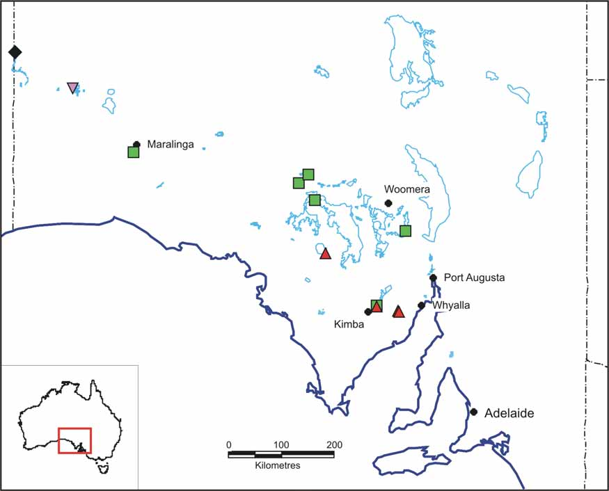
Distribution. P. a u r i c i f o r m a is known only from its type locality in the Great Victoria Desert ( Fig. 4 View FIGURE 4 ). It is not known how widespread it is, as lakes in the immediate vicinity have not been sampled, but some further away have different species―the Serpentine lakes 140 km to the northwest have P. triquetra n. sp. and the Yarle lakes, 172 km to the southeast support P. y a r l e e n s i s n. sp.
| SAM |
South African Museum |
No known copyright restrictions apply. See Agosti, D., Egloff, W., 2009. Taxonomic information exchange and copyright: the Plazi approach. BMC Research Notes 2009, 2:53 for further explanation.