
Puccinia caricis-hebeiensis Jing X. Ji, Zhuang Li, Y. Li & Kakish., 2022
|
publication ID |
https://doi.org/ 10.11646/phytotaxa.542.3.1 |
|
DOI |
https://doi.org/10.5281/zenodo.6421284 |
|
persistent identifier |
https://treatment.plazi.org/id/03C38793-FFC2-FFD6-FF29-F9B4BFFBCE96 |
|
treatment provided by |
Plazi |
|
scientific name |
Puccinia caricis-hebeiensis Jing X. Ji, Zhuang Li, Y. Li & Kakish. |
| status |
sp. nov. |
C7: Puccinia caricis-hebeiensis Jing X. Ji, Zhuang Li, Y. Li & Kakish. View in CoL sp. nov. ( Fig. 10 View FIGURE 10 )
MycoBank No: MB 838298.
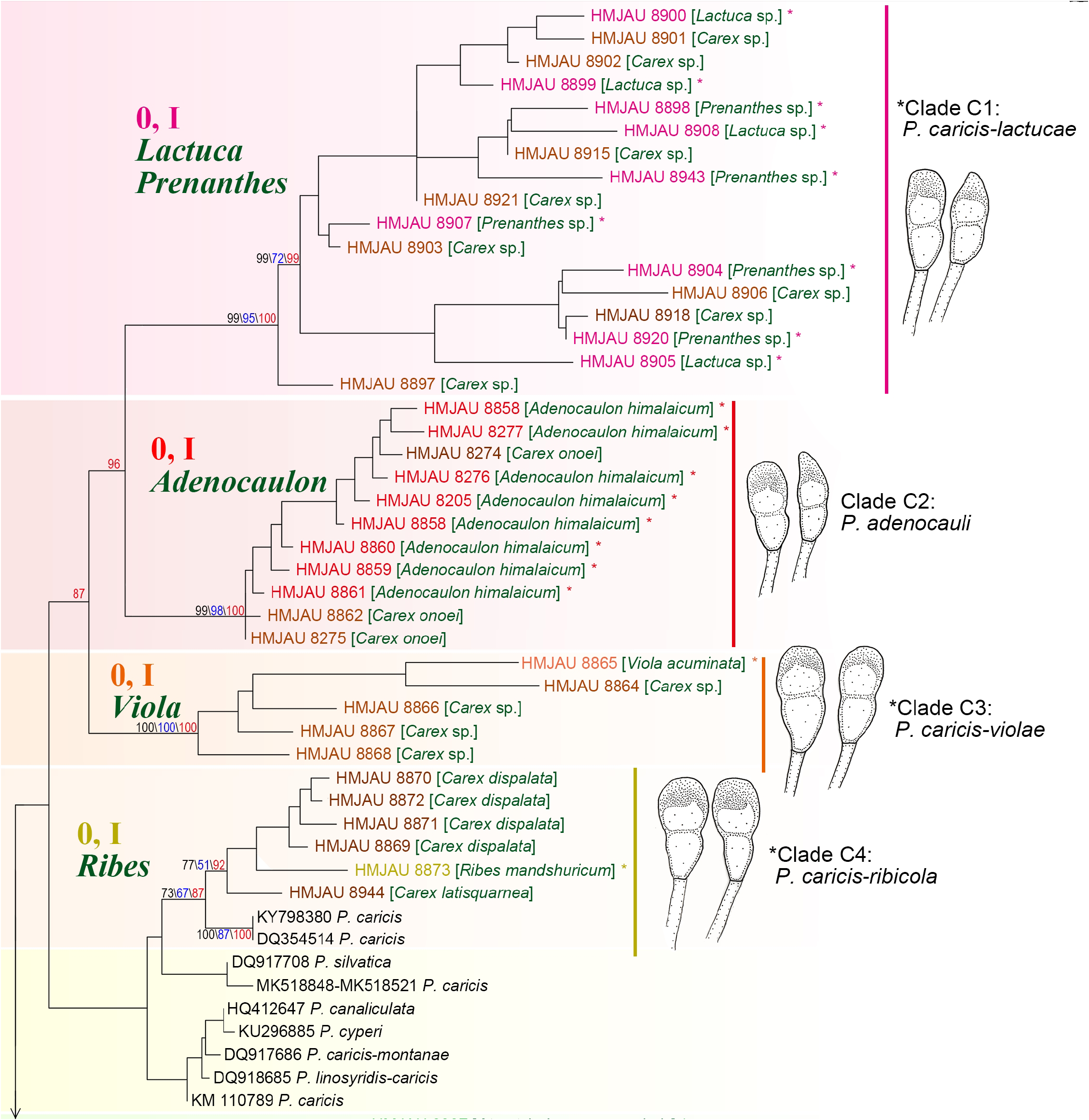
Diagnosis: —This species is morphologically similar to P. caricis-atractylodis and P. vaginatae , but phylogenetically separated from these species.
Typus: — CHINA, Hebei Province, Cangzhou, Cangxian , telia on Carex sp. , 17 November. 2018, J. X. Ji, HMJAU 8895 View Materials , Holotype.
Etymology: —Named after host plant genus of the telial stages, and the locality of collection, Hebei Province.
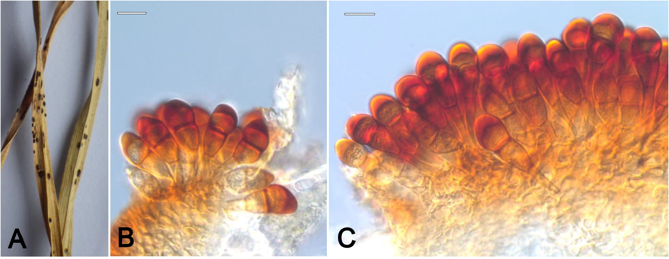
Description: — Spermogonia, aecia and uredinia not found. Telia hypophyllous, compact, black, rounded to broadly elliptic, erumpent. Teliospores broadly clavate, ellipsoid, mostly conical and rounded at apices, weakly constricted at the septa, attenuate at the bases, 34.0–51.0 × 17.0–21.0 µm (av. 43.0 × 19.5 µm); walls brown, 0.6–1.4 µm (av. 0.9 µm) thick at sides, 5.4–8.6 µm (av. 6.9 µm) at apices; pedicels persistent, hyaline, 19.5–48.0 µm (av. 32.0 µm) long.
Additional specimens examined from CHINA:— Hebei Province, Cangzhou , telia on Carex sp. , 12 May 2019, HMJAU 8894 View Materials .
Hosts and distribution in China: —Telia on Carex sp. (Hebei Province).
Note:— Two specimens of the telial stage are included in this clade and they are morphologically similar and phylogenetically close to P. caricis-atractylodis and P. vaginatae . However, they are clearly separated from clades of these species ( Fig. 3 View FIGURE 3 ), and are also slightly different in the shape and size of the teliospores, being slightly shorter and wider.
| J |
University of the Witwatersrand |
No known copyright restrictions apply. See Agosti, D., Egloff, W., 2009. Taxonomic information exchange and copyright: the Plazi approach. BMC Research Notes 2009, 2:53 for further explanation.