
AMPHISBAENIA
|
publication ID |
https://doi.org/10.5252/geodiversitas2020v42a20 |
|
publication LSID |
urn:lsid:zoobank.org:pub:8FF2A078-CE45-4BF1-A681-00136F57375E |
|
DOI |
https://doi.org/10.5281/zenodo.4486567 |
|
persistent identifier |
https://treatment.plazi.org/id/03C587C7-430C-FFEC-FEE8-FE844B4BF83E |
|
treatment provided by |
Felipe |
|
scientific name |
AMPHISBAENIA |
| status |
|
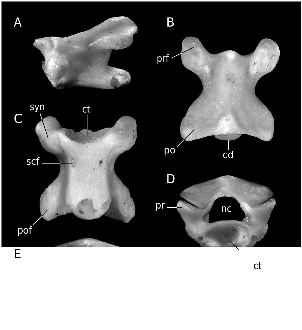
AMPHISBAENIA indet. ( Fig. 3 View FIG )
Blanus sp. – Ivanov et al. 2006: 229, table 2.
MATERIAL. — MWQ, early Miocene, Burdigalian, Orleanian, MN 4: 2/2003 Reptile Joint: One trunk vertebra (Pal. 1570).
DESCRIPTION
Trunk vertebra
A single trunk vertebra is preserved. It is small in size. A neural spine is absent, and the dorsal portion of the neural arch forms a median edge. In lateral view, the synapophysis is simple and large. The posterior portion of the neural arch is fused with the postzygapophyses, forming the dorsal roof (or lamina) between the left and right postzygapophyses. The neural canal is subtriangular with distinct lateral sinuses. The interzygapophyseal constriction is distinct and it occurs in the anterior half of the anteroposterior vertebral length. The dorsally tilted prezygapophyseal articular facets have an elliptical shape. A zygosphene is absent. The ventral side of the depressed centrum is flat, pierced by a pair of large subcentral foramina in the anterior 1/3 of the anteroposterior length. The lateral margins (subcentral ridges) are roughly parallel in ventral aspect. No constriction is developed at the base of the damaged condyle. The postzygapophyseal articular facets are oval and slightly enlarged posteriorly. The cotyle is distinctly laterally enlarged.
REMARKS
The vertebra described here can be attributed to Amphisbaenia based on the following combination of features (see Estes 1983): 1) the depressed centrum, having a flat ventral surface; 2) roughly parallel lateral margins in ventral aspect; 3) massive synapophyses; 4) the absence of a zygosphene (enabling distinction of amphisbaenians from scolecophidian snakes ( Estes 1983; Rage 1984); and 5) the sinusoidal neural arch lacking a neural spine.
Family level allocation of an isolated vertebra is limited by a lack of clear diagnostic features for identification ( Estes 1983; Augé 2005, 2012; Georgalis et al. 2016b).We can exclude rhineurids, which have a denticulate vertebral posterior margin. The same feature can be observed in trogonophiids as well ( Kearney 2003; Augé 2012; Čerňanský et al. 2016a). Based on the geographical position of the locality and the age of the sediments, this vertebra most likely represents a blanid taxon. According to cranial elements, amphisbaenians reported from the Central European late Oligocene and Miocene localities are almost exclusively identified as belonging to the clade Blanidae ( Roček 1984; Schleich 1988; Čerňanský & Venczel 2011; Čerňanský et al. 2016a). The morphology and dimensions of the vertebra described here are very similar to those of trunk vertebra of Blanus gracilis Roček, 1984 reported from the Czech early Miocene (MN 4b) Dolnice site ( Roček 1984: 5, table 16).
| MN |
Museu Nacional, Universidade Federal do Rio de Janeiro |
No known copyright restrictions apply. See Agosti, D., Egloff, W., 2009. Taxonomic information exchange and copyright: the Plazi approach. BMC Research Notes 2009, 2:53 for further explanation.
|
Kingdom |
|
|
Family |
AMPHISBAENIA
| Ivanov, Martin, Čerňanský, Andrej, Bonilla-Salomón, Isaac & Luján, Àngel Hernández 2020 |
Blanus
| Ivanov et al. 2006: 229 |