
Ferolocella tessellata (Berlese 1908)
|
publication ID |
https://doi.org/10.11646/zootaxa.3637.3.2 |
|
publication LSID |
lsid:zoobank.org:pub:AB22567C-9735-4809-BBE0-295A9E6D56B2 |
|
DOI |
https://doi.org/10.5281/zenodo.6153076 |
|
persistent identifier |
https://treatment.plazi.org/id/03C687AC-FF98-066F-A2BF-11F3323EF89F |
|
treatment provided by |
Plazi |
|
scientific name |
Ferolocella tessellata (Berlese 1908) |
| status |
|
Ferolocella tessellata (Berlese 1908) View in CoL
( Figs. 3 View FIGURE 3 , 4A,B View FIGURE 4 A, B , 5A–E View FIGURE 5 A – E )
Oribatella tessellata Berlese 1908 , Castagnoli & Pegazzano 1985, p. 414 Oribatella carolina Banks, 1947 , p. 112
Ferolocella carolina (Banks, 1947) , Grabowski 1971, p. 44, Norton & Kethley 1990, p. 477
Material examined. Topotypes, 2 males, 2 females, USA, Missouri, Boone Co., Hickson Ck., Columbia, S. U. Mo. Campus, 25.iv.1985, (J. Kethley, R.A. Norton) FMHD #85-128, from mixed leaf litter on ridge top (RNC); Ashland Wildlife Management Res., 26.iv.1985 (J. Kethley, R.A. Norton) FMHD #85-135, from litter with root mat (RNC); Roaring Rocks State Park, Trail to lookout tower, 36 0 35’N 93 0 48’W, 13.v.1999 (VBP) from sifted oak litter in oak hickory forest; from soil under rotting oak; Lake of the Ozarks State Park, Woodland trail, elv. 900 ft., 14.v.1999 (VBP) from oak litter by rotting log; Virginia, Mountain Lake, 11.ix.1968 (J.M. Campbell) from deciduous duff; Nelson Co., Washington National Forest, Blue Ridge Parkway, Wintergreen Trail, 1.x.1992 (VBP) from gilled mushroom on chestnut oak, from plants on forest floor in oak-hemlock forest, from litter in and under decaying tree trunk, from litter in oak-hemlock forest; West Virginia, Mercer Co., 1.5 mi N on Brushcock Falls Trail, 13.vi.1971 (W. Sheer) from deep litter near cliff; Randolph Co., Stuart Park Recreation Area, 8 km W Elkins, 30.vii.1986 (EEL) from mixed deciduous litter; Georgia, Clarke Co., Athens, Simonton Bridge Rd, Botanical Gardens, 1982 (D. Mallow); Wisconsin, Vilas Co., Northern Highland State Forest, Falliston Lake Nature Trail, 45 0 59’41”N 89 0 37’01”W, 23.x.1998 (VBP & M. Behan) from rotting log; Ohio, Ross Co., Scioto Trail St Park, 4.xi.1978 (L. Watrous) leaf litter along rotten log; Alabama, De Kalb Co., De Soto State Park, Rhododendron Trail , 27.ix.1992 (VBP) from moss at base of hemlock; Arkansas, Newton, Alum Cove Recreation Area, 11.xi.2002 (H.W. Robison); Texas: Bandera Co., Lost Maples State Natural Area, East Trail, 29° 48.984N 99° 34.599W; 28.ii.2007 (VBP) from oak, juniper and laurel litter, some lichens and bark; from Bigtooth maple litter along dry creek bed; from deep Bigtooth maple and oak litter.
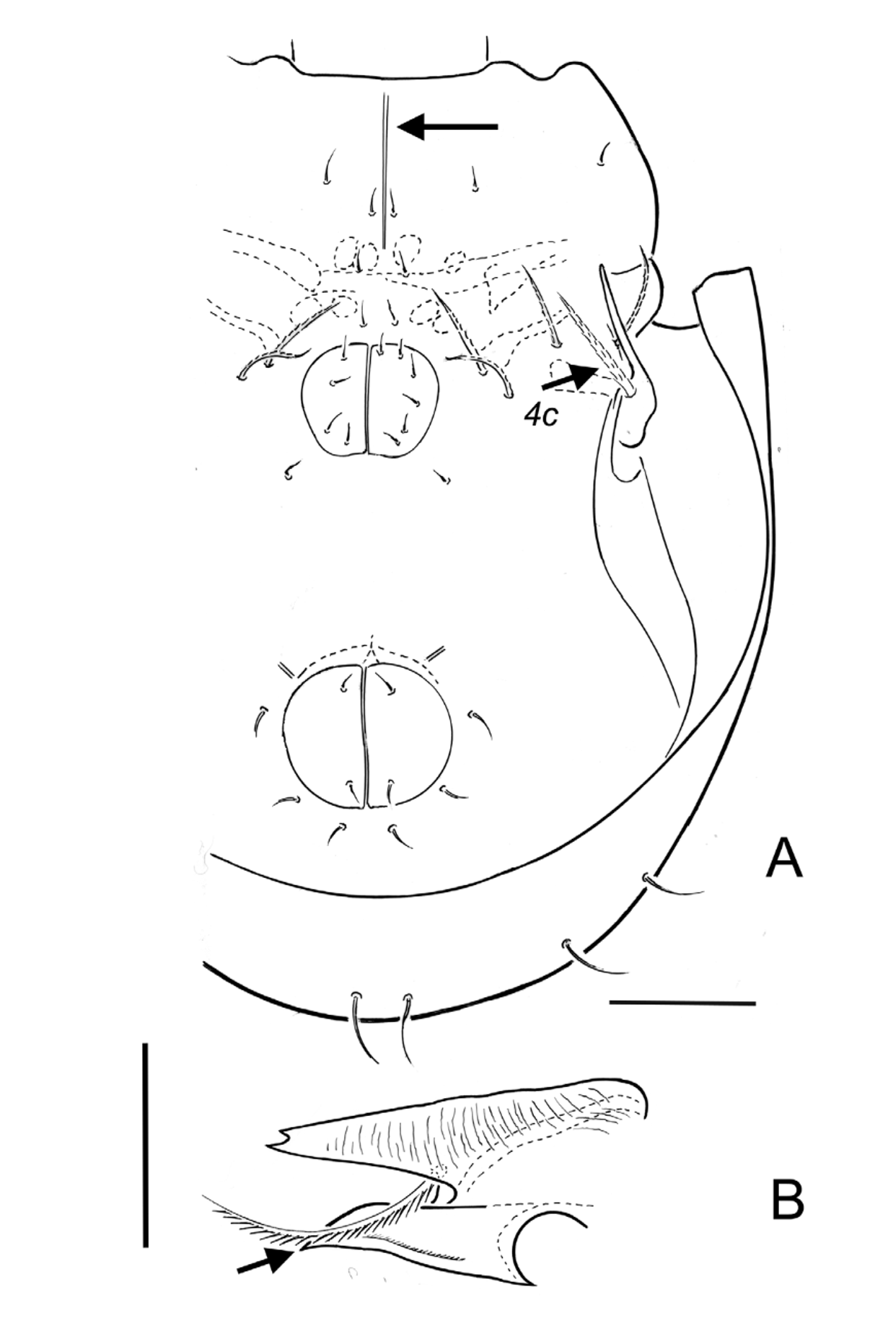
Diagnosis. Total length of adult 274–293 μm (topotype female ( n =1) 293 µm; topotype males ( n =2) 280 µm (274, 288); Length of Oribatella carolina given as 220 µm (Banks 1947) and 275.5 µm (Grabowski 1971)). Notogastral width about 192 µm. Prodorsum, notogaster, anal plates, genital plates, venter and mentum microtuberculate. Coxisternal region without striae laterally; pedotectum I with striae distally ( Fig. 3 View FIGURE 3 ), proximally with irregular transverse striae. Lamellae with distinct irregular longitudinal striae laterally and medially in interlamellar region; with curved, semi-transverse striae on lamellar cusp ( Fig. 3 View FIGURE 3 ). Distinct longitudinal line of thickened integument medially in coxisternal region, extending from gnathosoma to posterior of epimere II ( Fig. 4A View FIGURE 4 A, B , arrow). Rostral margin tridentate with narrow medial and lateral dentes ( Fig. 5C View FIGURE 5 A – E ); medial dens half to twothirds length lateral dens. Lamella 83–93 μm long, of which cusp 61–66 μm; about 36 μm wide at level of insertion of seta le. Lamellar cusps contiguous and parallel anteromedially, separated posteromedially, leaving prodorsum visible through oval opening (13–15 μm wide x about 19 μm long); translamella without tooth, about 13 μm at greatest width; pocket formed by translamella and medial edge of lamellar cusp very deep, extending posteriad of dorsosejugal scissure, almost square in shape about 34 μm wide at dorsosejugal scissure and about 36 μm deep ( Figs. 3 View FIGURE 3 , 5A View FIGURE 5 A – E ). Medial dens on lamellar cusp about 14 μm long, without teeth; lateral dens about 30–41 μm, without teeth; tip of lateral dens bifid in one specimen. Seta ro 76 µm long, strongly barbed along length, acuminate, curved anteromedially. Seta le 70–81 µm long, thick, heavily barbed, tapered, arising anteroventrally ( Figs. 3 View FIGURE 3 , 5A View FIGURE 5 A – E ). Insertion of lamellar seta expanded and flask-shaped. Seta in 100–121 µm long, thick (less so than le), barbed, tapered; mutual distance about 41 µm. Bothridial seta club-shaped, 89–93 µm long ( Fig 3 View FIGURE 3 ); head barbed, distally tapered, directed slightly anteromedially; stalk short, smooth. Exobothridial seta about 11 µm long, thin, smooth. Tutorium subrectangular, tapered distally, with dorso-ventral striae ( Figs. 4B View FIGURE 4 A, B , 5B View FIGURE 5 A – E ), about 116 μm long, of which cusp about 66 μm long; distal margin with 3–4 dentes. Custodium about 21 μm long ( Figs. 4 View FIGURE 4 A, B , 5D View FIGURE 5 A – E ). Porose area Al about 8 μm in diameter. Notogaster slightly wider than long. Anterior margin undulating, convex region lateral of bothridium with about 9 transverse ridges ( Fig. 3 View FIGURE 3 ). Pteromorph punctate, anteroventral margin serrate. Notogastral saccules indistinct, weakly developed, about 4 μm in diameter. Notogastral setae very weakly barbed, 15–36 μm long, with seta c longest, lm posteriad Aa, and lp anteriad A1; mutual distance p1–p1 about 14 μm, narrower than distance h1–h1 about 24 μm. Subtriangular lenticulus present. Epimeral setae mostly about 11–18 μm long, barbed, acuminate; 3c, 4a and 4b about 19–24 μm long, distinctly barbed; 4c longest epimeral seta, about 46 μm, thick and heavily barbed ( Figs. 4A View FIGURE 4 A, B , 5D View FIGURE 5 A – E ) (2 setae 4c on one side of one topotype specimen). Genital, aggenital, anal and adanal setae weakly barbed, 8–11 μm long; genital setae 2 + 4. Lyrifissure iad anterolateral of anal plate. Postanal porose area about 12 x 4 μm, not illustrated. Anterior margin of mentum without tectum or thickened transverse ridge. Length of chelicera about 67 μm. Axillary saccule about 6 x 2 Μm. Leg setation (I to IV): trochanters, 1-1-2- 1; femora, 5-5-2-2; genua, 3(1)-3(1)-1(1)-2; tibiae 4(2)-4(1)-3(1)-3(1); tarsi, 20(2)-15(2)-15-12. Seta l’ absent from femur III. Setae l” of genua I and II 21–23 and 17 –24 Μm, respectively; l” of tibia I and II about 33 and 21–24 Μm, respectively.
Immatures. Unknown.
Remarks. Norton & Kethley 1990 (p. 477) corrected the redescription of this species by Grabowski (1971), noting that body and setal porportions given by this author were imprecise, that there are 4 pairs of saccules, 10 pairs of notogastral setae, a postanal porose area, and that the auxillary dens of the lamellar cusp described by Grabowski (1971) is absent. I have examined topotypic material of Ferolocella tessalata housed in the collection of R.A. Norton for the Diagnosis and Description given above, and this material was used for Figures 3 View FIGURE 3 and 4 View FIGURE 4 A, B . The “auxillary dens of the lamellar cusp” as indicated in Grabowski (1971, his Fig, 3) clearly refers to the expanded integument around the insertion of seta le.
Distribution and Ecology. This species is known throughout eastern USA from Ohio to Texas and east to Virginia and Alabama, associated with rich forest litter. Gravid females carry two eggs. Gut contents included fungal hyphae.


No known copyright restrictions apply. See Agosti, D., Egloff, W., 2009. Taxonomic information exchange and copyright: the Plazi approach. BMC Research Notes 2009, 2:53 for further explanation.
|
Kingdom |
|
|
Phylum |
|
|
Class |
|
|
Order |
|
|
SubOrder |
Oribatida |
|
Family |
|
|
Genus |