
Leptoomidae Gibson, 2023
|
publication ID |
https://doi.org/ 10.11646/zootaxa.5318.2.2 |
|
publication LSID |
lsid:zoobank.org:pub:CF8E0B91-9AF7-4075-963D-9BC977B41852 |
|
DOI |
https://doi.org/10.5281/zenodo.8168989 |
|
persistent identifier |
https://treatment.plazi.org/id/967CDA5C-9735-4C80-884E-07F89177576F |
|
taxon LSID |
lsid:zoobank.org:act:967CDA5C-9735-4C80-884E-07F89177576F |
|
treatment provided by |
Plazi |
|
scientific name |
Leptoomidae Gibson |
| status |
fam. nov. |
Leptoomidae Gibson fam. nov.
http://zoobank.org/ urn:lsid:zoobank.org:act:967CDA5C-9735-4C80-884E-07F89177576F
Type genus. Leptoomus Gibson 2008 .
Included genera. Leptoomus and Neanaperiallus Gibson, 2009 .
Diagnosis. Antenna 12- or 13-segmented, with basal flagellomere not distinctly anelliform and clava 3- segmented. Mesoscutum with notaular furrows extending along length. Prepectus anteriorly rounded to angulate, extending to or slightly over posterolateral margin of pronotum; dorsal margin of prepectus intersecting base of the tegula distinctly anterior to and forming an almost right-angle with posterior margin of prepectus; posterior prepectal margin truncate along anterior margin of acropleuron.Acropleuron greatly enlarged, extending posteriorly to metapleuron and ventrally at least to anterodorsal margin of mesocoxa. In lateral view mesocoxa with anterior margin distinctly posterior to midline of acropleuron. Mesopectus with posterior margin abutting bases of mesocoxae. Mesotibia with 1 or 2 rows of apical pegs and mesotarsus with spine- to peg-like setae along both ventral margins. Fore wing with oblique bare band beyond basal fold.
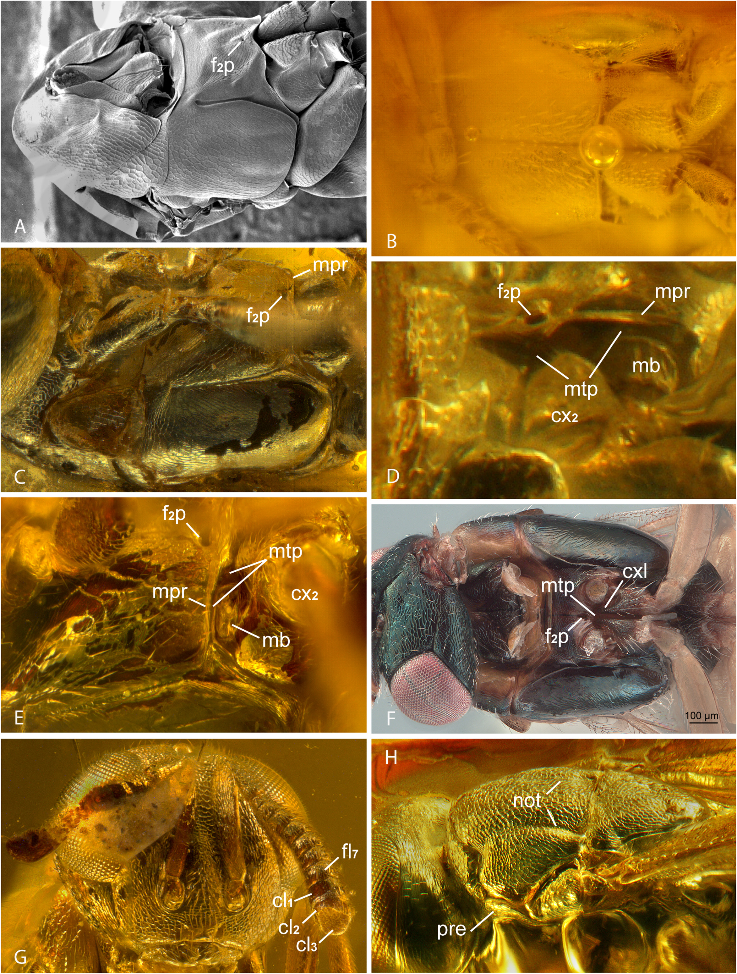
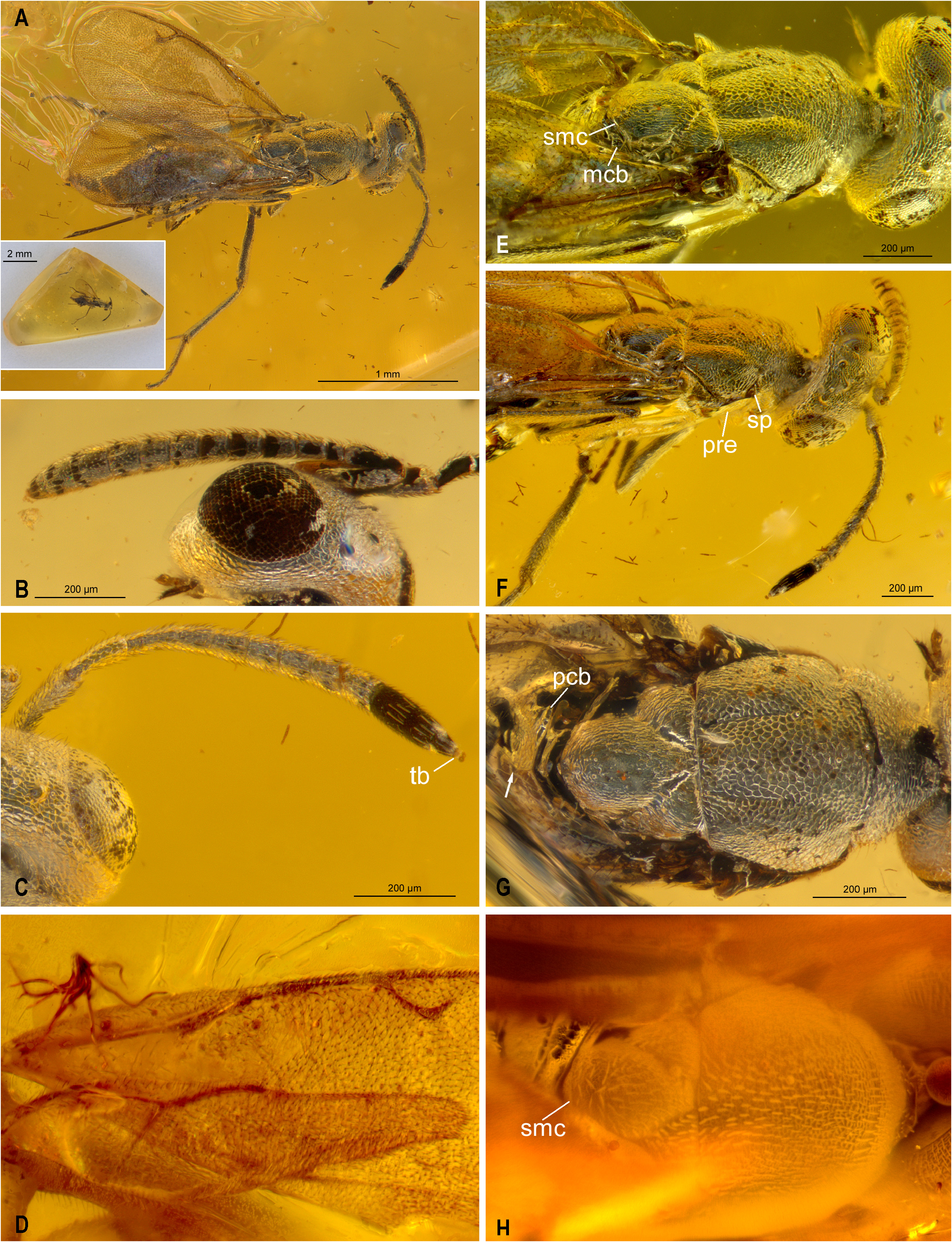
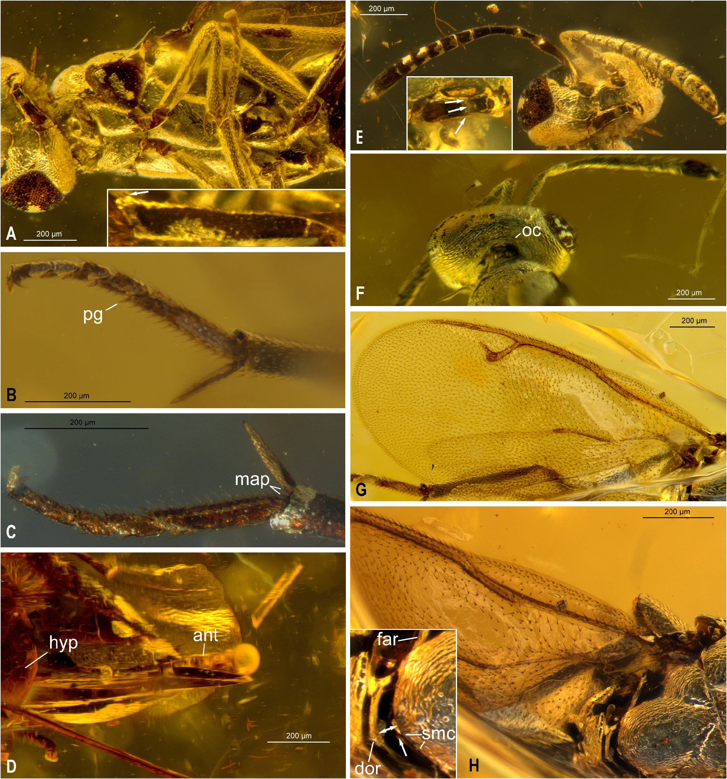
Description. Head with malar sulcus ( Figs 2G View FIGURE 2 , 3B View FIGURE 3 , 5E View FIGURE 5 ); clypeus with apical margin straight transverse and not delimited by a subapical transverse groove ( Figs 2G View FIGURE 2 , 5A, E View FIGURE 5 inset). Antenna 12- or 13-segmented with 7- ( Figs 2G View FIGURE 2 , 3B View FIGURE 3 ) or 8- ( Figs 4B, C View FIGURE 4 ) segmented funicle and 3-segmented clava; basal flagellomere lacking multiporous plate sensilla and the shortest funicular, subquadrate but not distinctly anelliform. Pronotum with strongly inclined neck abruptly merging into very short, transverse collar ( Figs 1A View FIGURE 1 , 4E, G View FIGURE 4 , 5F View FIGURE 5 ; Gibson 2008, figs 6‒8; Gibson 2009, figs 59, 60; Simutnik et al. 2020, figs 1C, D) and without a median line ( Figs 4E, G View FIGURE 4 ; Gibson 2008, fig. 4). Mesonotum with transscutal articulation straight, articulated along its entire width ( Figs 4E, G, H View FIGURE 4 ; Gibson 2008, figs 7, 8; Gibson 2009, fig. 59; Simutnik et al. 2020, fig. 1D); mesoscutum without parapsidal lines or sulcate notauli, but with notaular furrow extending from mesothoracic spiracle posterior of pronotum to transscutal articulation at anterolateral margin of axilla, the furrows delineating median and lateral mesoscutal lobes, with median lobe uniformly convex, not differentiated into anteromedian convex and posteromedian depressed region ( Figs 2H View FIGURE 2 , 4A, E‒G View FIGURE 4 ; Gibson 2008, figs 6, 7; Simutnik et al. 2020, fig. 1D); mesoscutellar-axillar complex with axillae triangular, their inner angles separated by narrowly truncate base of mesoscutellum when mesonotum not flexed; mesoscutellum without differentiated frenum or axillular carina. Mesosoma in lateral view with convex acropleuron extending posteriorly to metapleuron and ventrally at least to anterodorsal margin of mesocoxa, with variably large mesepisternal region ventral to acropleuron ( Figs 1A, B View FIGURE 1 , 5A View FIGURE 5 ). Mesopectus in ventral view with sulcate discrimen but apparently without transepisternal line ( Figs 2B View FIGURE 2 , 5A View FIGURE 5 ; Simutnik et al. 2020, fig. 2A); mesopectus with posterior margin abutting bases of mesocoxae ( Figs 2B, C View FIGURE 2 , 5A View FIGURE 5 ), without external membranous region between mesopectus and each mesocoxa so that mesocoxae unable to rotate anteriorly entirely out of their combined fossa. Prepectus bare; in lateral view anteriorly rounded to angulate and protuberant, extending to or slightly overlapping posterolateral margin of pronotum, with dorsal margin virtually straight and extending posteriorly distinctly beyond base of tegula ( Figs 1A, B View FIGURE 1 : arrows, 5A) and forming almost a right-angle relative to straight or only slightly curved posterior, truncate margin along anterior margin of acropleuron ( Figs 1A, B View FIGURE 1 , 5A View FIGURE 5 ); in dorsal view prepectus thin ( Fig. 4F View FIGURE 4 : pre) to slightly convex ( Fig. 2H View FIGURE 2 : pre) but not conspicuously bulbous. Fore wing with typical venation consisting of submarginal, marginal, postmarginal and stigmal veins, and stigmal vein with distinct uncus ( Figs 4A, D View FIGURE 4 , 5G View FIGURE 5 ; Gibson 2008, fig. 16); disc with broad, oblique bare band beyond basal fold either contiguous with ( Gibson 2008, fig. 16; Simutnik et al. 2020, fig. 1H) or separated from parastigma by setae ( Figs 5G, H View FIGURE 5 ; Gibson 2009, fig. 62). Legs with mesocoxa inserted near metacoxa, distant from procoxa, in lateral view mesocoxa with anterior margin distinctly posterior to midline of acropleuron ( Figs 1A, B View FIGURE 1 , 3A View FIGURE 3 ); tarsi 5- segmented; protibia with bifurcate, curved tibial spur ( Gibson 2008, fig. 21; Simutnik et al. 2020, fig. 1I), without dorsal spicules but with dorsoapical spicule ( Fig. 5A View FIGURE 5 insert: arrow; Gibson 2008, fig. 21: pas); mesotibia with apical pegs arranged in 1 or 2 rows ( Fig. 5C View FIGURE 5 map; Gibson 2008, figs 18, 19); mesotarsus ventrally with spine- to peg-like setae along both margins ( Figs 5B, C View FIGURE 5 ; Gibson 2008, figs 18, 19; Gibson 2009, figs 64, 65); metatibia with two spurs ( Simutnik et al. 2020, fig. 2C). Gaster sessile; Gt 7 and Gt 8 fused into syntergum, though at least sometimes with an oblique groove posterior to cercus distinguishing presumptive Gt 7 and Gt 8 ( Gibson 2008, fig. 17); syntergum with cercus not advanced and cercal setae not kinked; ovipositor sheaths exerted for short distance beyond apex of gaster ( Fig. 5D View FIGURE 5 ; Gibson 2008, fig. 17; Gibson 2009, fig. 63).
Remarks. Gibson (2009) described the mesoscutum of N. masneri as uniformly convex, without distinct notauli except for a very short furrow anteriorly at the level of the lateral margin of the pronotum mesal to the spiracle visible in lateral view ( Gibson 2009, fig. 60). He also stated that minute air bubbles reduced clarity of observation of the inclusion and that the mostly slightly lanceolate- or spatulate-appearing setae, as seen in dorsal view, likely resulted from the setae being surrounded by a thin layer of air because in lateral view the pronotal and mesoscutal setae appear more normal, hair-like. With the discovery of the new species of Neanaperiallus , which clearly shows complete, furrow-like notauli ( Figs 4A, E‒G View FIGURE 4 ) similar to those of L. janzeni ( Fig. 2H View FIGURE 2 ), the apparent absence of complete notauli for N. masneri ( Fig. 4H View FIGURE 4 ; Gibson 2009, fig. 59) likely reflects an artefact of the furrows not being visible except in lateral view immediately behind the pronotum. Different views and states of preservation of different individuals of L. janzeni imaged by Gibson (2008) also show different forms and relative visibility of the notaular furrows ( Gibson 2008, cf. figs 4, 6‒8), the furrows being least visible in direct dorsal view ( Gibson 2008, figs 4, 8). We therefore interpret complete notaular furrows as a feature of N. masneri and describe this feature for the family.
Both available inclusions of Neanaperiallus have an unflexed mesonotum in which the axillae are separated medially ( Figs 4E‒H View FIGURE 4 ). Because of this, it is unknown whether the basally truncate mesoscutellum separating the inner angles of the axillae results from the posterior margin of the mesoscutum overlapping the axillae medially, as for L. janzeni ( Gibson 2008, cf. figs 7, 8), or whether the mesonotum articulates, hinge-like, along the transscutal articulation and the inner angles of the axillae are separated by an anteriorly truncate mesoscutellum. Consequently, our family description includes description of relative mesoscutellar-axillar structure when the mesonotum is not flexed, but it remains uncertain whether the mesonotal articulatory structure in Neanaperiallus is the same as in Leptoomus .


No known copyright restrictions apply. See Agosti, D., Egloff, W., 2009. Taxonomic information exchange and copyright: the Plazi approach. BMC Research Notes 2009, 2:53 for further explanation.
|
Kingdom |
|
|
Phylum |
|
|
Class |
|
|
Order |
|
|
SuperFamily |
Chalcidoidea |
|
Family |