
Pochytoides, Berland & Millot, 1941
|
publication ID |
https://doi.org/10.5852/ejt.2018.418 |
|
publication LSID |
lsid:zoobank.org:pub:2526B156-673C-4693-B499-95387C7F0C78 |
|
DOI |
https://doi.org/10.5281/zenodo.14587571 |
|
persistent identifier |
https://treatment.plazi.org/id/03C987FA-3370-871E-FF31-F8E80619F9A1 |
|
treatment provided by |
Plazi |
|
scientific name |
Pochytoides |
| status |
|
A key to the species of Pochytoides View in CoL
Males
1. Pedipalp with short apophysis on the patella ( Fig. 7C View Fig ) ...................................... P. patellaris sp. nov.
– Pedipalp without patellar apophysis ................................................................................................. 2
2. Embolic division serrated distally ( Fig. 3A View Fig ) ...................................................... P. monticola sp. nov.
– Embolic division smooth distally ..................................................................................................... 3
3. Embolus almost straight, with accompanying membrane ( Fig. 9E View Fig ) ................................................... ................................................................................ P. poissoni ( Berland & Millot, 1941) comb. nov.
– Embolus bent, without conspicuous membrane ............................................................................... 4
4. Anterior lobe of bulb expanding distally, axe-shaped ( Fig. 11E View Fig ) ........................... P. securis sp. nov.
– Anterior lobe of bulb tapering distally .............................................................................................. 5
5. Embolic division curved toward prolateral side of the palp ( Fig. 5A View Fig ) ..................... P. obstipa sp. nov.
– Embolic division sigmoid, its tip curved toward retrolateral side .................................................... 6
6. Prolateral edge of anterior lobe of bulb with spine at base ( Fig. 13C View Fig ) ................. P. spiniger sp. nov.
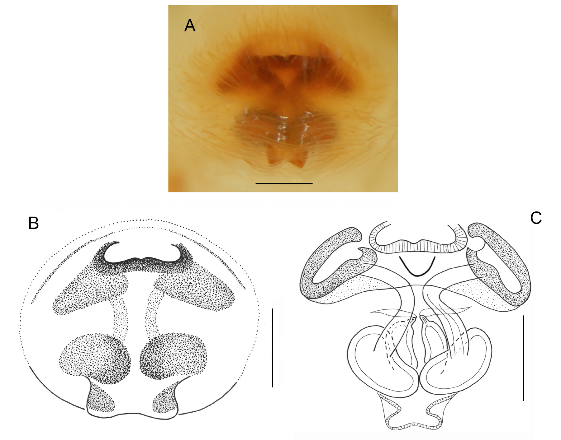
– Prolateral edge of anterior lobe of bulb strongly sclerotized, but without spine ( Fig. 1B View Fig ) ................ ............................................................................................................................... P. lamottei sp. nov.
Females
1. Epigyne without pockets ................................................................................................................... 2
– Epigyne with paired pockets at epigastric furrow ............................................................................ 3
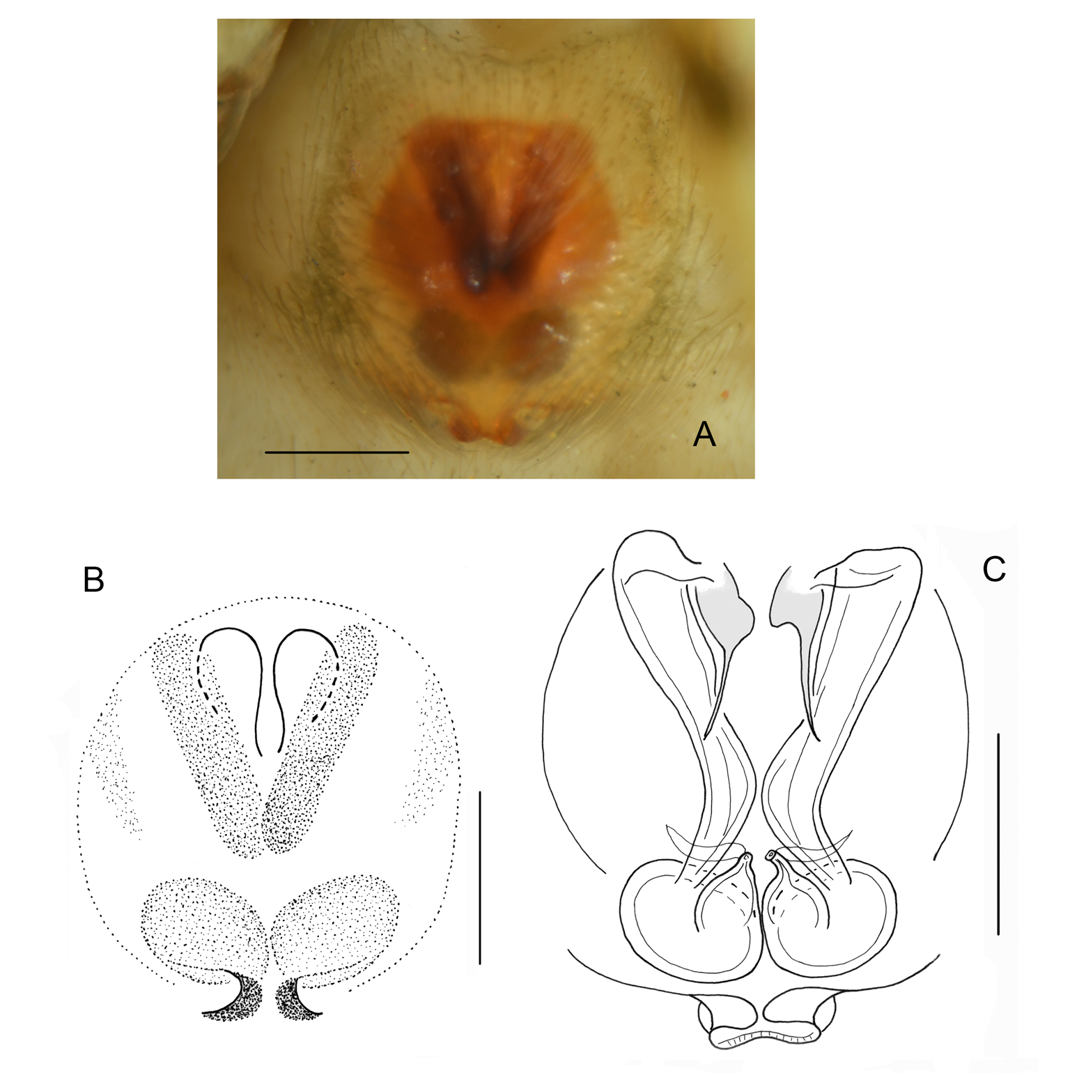
2. Area occupied by atria broader than spermathecae area ( Fig. 8D View Fig. 8 ) ..................................................... .................................................................................... P. perezi ( Berland & Millot, 1941) comb. nov.
– Area occupied by atria narrower than spermathecae area ( Fig. 15C View Fig. 15 ) ................... P. spiniger sp. nov.
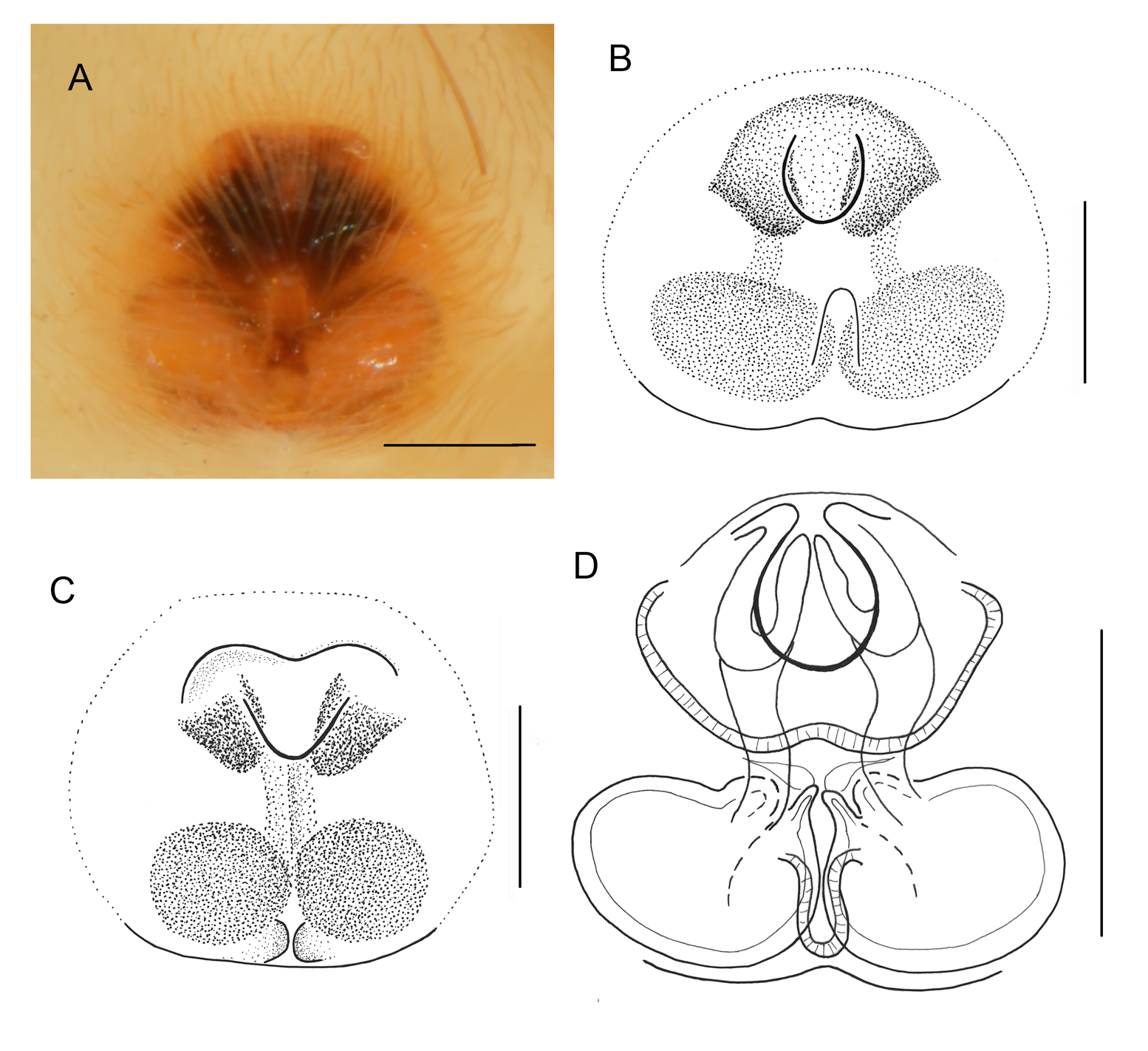
3. Atria weakly sclerotized ( Fig. 12C View Fig. 12 ) ........................................................................ P. securis sp. nov.
– Atria strongly sclerotized .................................................................................................................. 4
4. Area occupied by atria narrower than spermathecae area ( Fig. 2C View Fig. 2 ) ..................... P. lamottei sp. nov.
– Area occupied by atria broader than spermathecae area ................................................................... 5
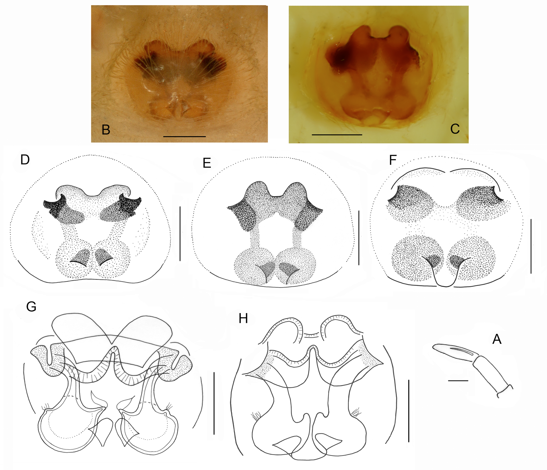
5. Two semicircular hoods in front of atria, spermathecae spherical ( Fig. 10D View Fig ) .................................... ................................................................................ P. poissoni ( Berland & Millot, 1941) comb. nov.
– Shallow depression in front of atria, spermathecae oval ................................................................... 6
6. Posterior edge of epigynal depression straight, pockets placed posteriorly to spermathecae ( Fig. 4B View Fig. 4 ) ............................................................................................................... P. monticola sp. nov.
– Posterior edge of epigynal depression arched, pockets placed at spermathecae level ( Fig. 6C View Fig. 6 ) .......... .................................................................................................................................... P. obstipa sp. nov.


No known copyright restrictions apply. See Agosti, D., Egloff, W., 2009. Taxonomic information exchange and copyright: the Plazi approach. BMC Research Notes 2009, 2:53 for further explanation.
|
Kingdom |
|
|
Phylum |
|
|
Class |
|
|
Order |
|
|
Family |
|
|
SubFamily |
Salticinae |
|
Tribe |
Aelurillini |