
Axelrodichthys megadromos, Lionel Cavin, Xavier Valentin & Géraldine Garcia, 2016
|
publication ID |
https://doi.org/ 10.1016/j.cretres.2016.02.002 |
|
DOI |
https://doi.org/10.5281/zenodo.6074328 |
|
persistent identifier |
https://treatment.plazi.org/id/03CA87FB-FF99-FF8A-FFF3-FE2DA07AF818 |
|
treatment provided by |
Plazi |
|
scientific name |
Axelrodichthys megadromos |
| status |
sp. nov. |
Axelrodichthys megadromos sp. nov.
( Figs. 2 View Fig. 2 and 3 View Fig. 3 )
Etymology. From the greek MέƳaς, megas, large and dRόM Ο ς, dromos,
driveway. The specific epithet emphasizes the arrival of this taxon from Gondwana probably by dispersal during the Cretaceous and refers to the building of a new highway in the vicinity of the type locality in the present time.
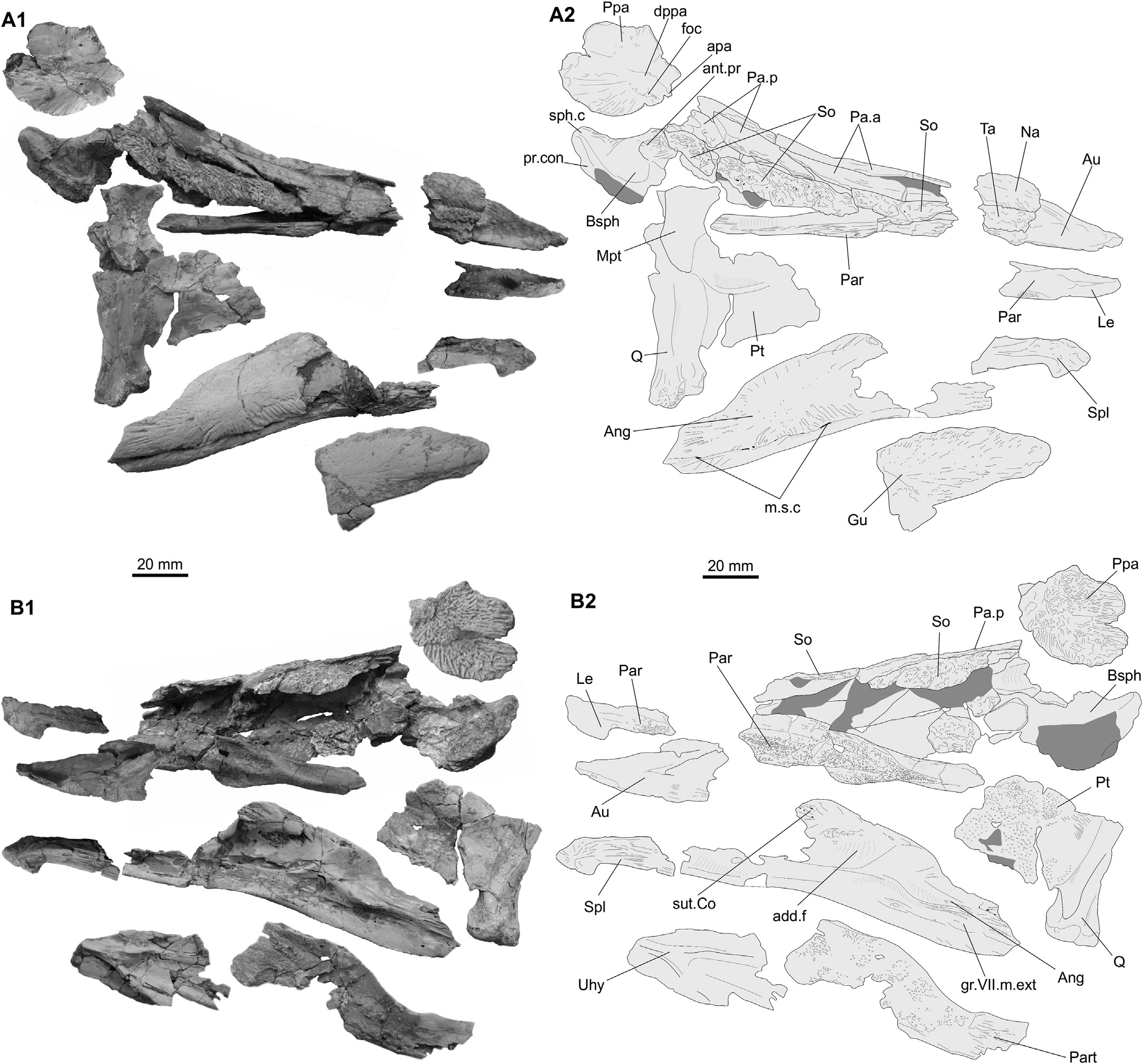
Holotype. MDE F-61 (X. Valentin), holotype, a partial skull comprising the ethmosphenoid portion, elements of the suspensorium, of the lower jaw and some other components ( Figs. 2 View Fig. 2 and 3 View Fig. 3 ).
Type locality and horizon. MDE F-61 was collected from a locality near the village of Ventabren (Bouches-du-Rhône Department, Southeastern France) in a lower Campanian “Valdo-Fuvelian” facies composed of lignitic marls interbedded with lacustrine limestones. Diagnosis. A species of Axelrodichthys with an ethmosphenoid moiety approximately 3.5 times longer than wide in its narrowest part; very elongate penultimate posterior supraorbital; posterior extremity of the parasphenoid tooth plate heart-shaped and forming frontwards lateral wings separated by a medial ridge; parietals and nasals with no ornamentation, except faint ridges along their lateral margin; supraorbitals and postparietals with strong reticulated ornamentation.
4. Description
The ethmosphenoid portion consists of a basisphenoid, most of the skull roof, the parasphenoid with no posterior extremity, and the right lateral ethmoid. The ethmosphenoid portion is not complete and a gap is present, but estimation of the total length based on the preserved parts indicates that this portion is approximately 3.5 times longer than wide in its narrowest part.
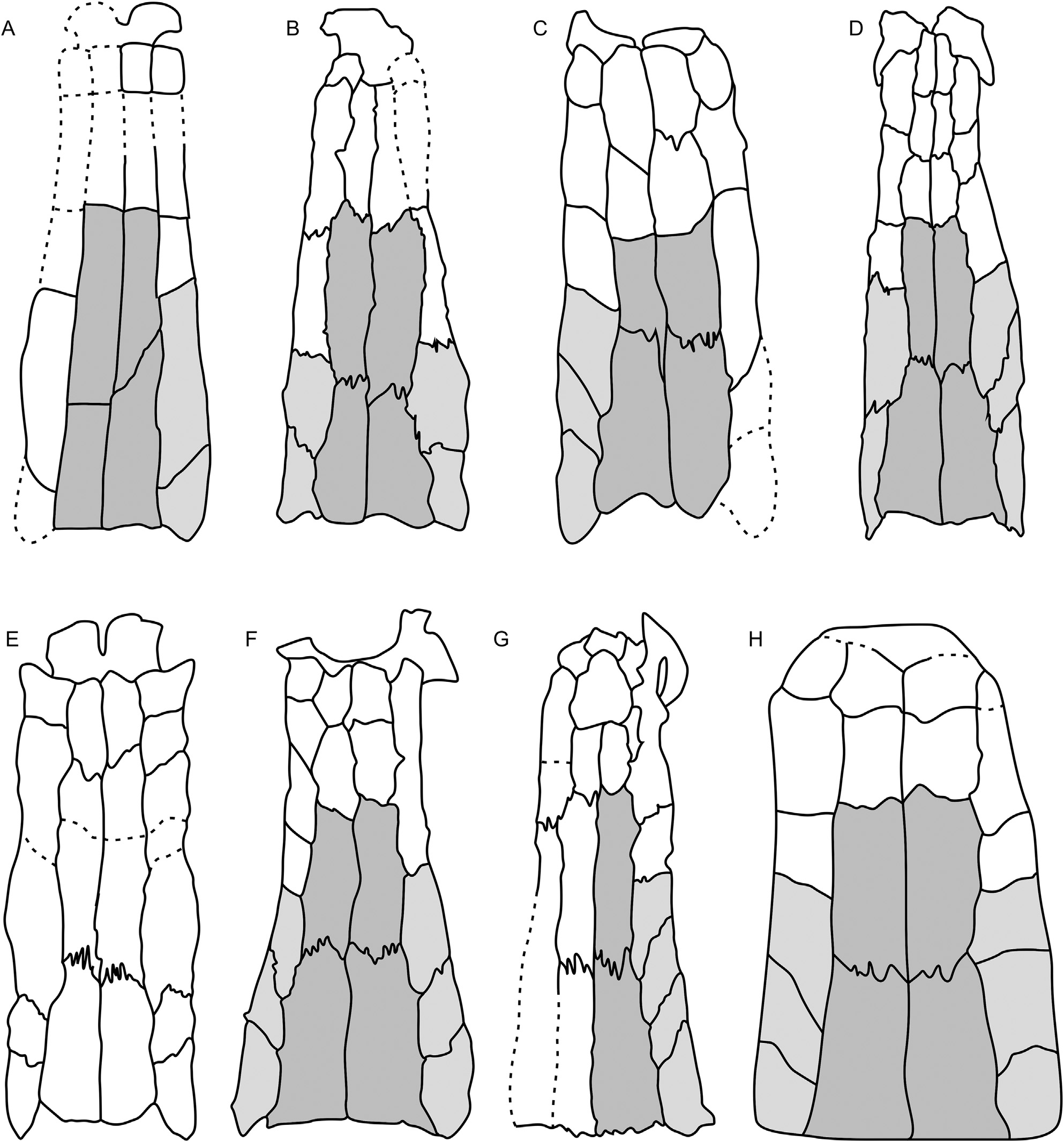
Skull roof. The preserved part of the supraorbital series in A. megadromos indicates that the ethmosphenoid portion has concave margins in dorsal view ( Fig. 4 View Fig. 4 A), as in Axelrodichthys araripensis and A. maiseyi , and to the contrary of Mawsonia , where it is slightly convex ( Wenz, 1975; Maisey, 1986). The medial series of paired bones of the skull roof is composed of two pairs of parietals and at least two pairs of nasals. These ossifications bear no ornamentation, except faint ridges along their lateral margin. The anterior and posterior pairs of parietals are elongate and approximately 3 times longer than wide. The suture between both right parietals is oblique and the posterior parietal extends beneath the anterior one, as in most coelacanths. The suture between both left parietals is almost perpendicular to the skull roof axis. Anterior and posterior parietals are approximately equal in length. In M. lavocati ( Figs. 4 View Fig. 4 D, E) and Mawsonia cf. gigas ( Fig. 4 View Fig. 4 F) the anterior parietals are slightly shorter ( Cavin and Forey, 2004 and Maisey, 1986, respectively) and longer in M. brasiliensis ( Yabumoto, 2002, Fig. 4 View Fig. 4 G) than the posterior parietals. In A. maiseyi ( Fig. 4 View Fig. 4 C) the anterior parietals are slightly shorter than the posterior ones ( De Carvalho et al., 2013), while the situation is reverse in Axelrodichthys araripensis ( Maisey, 1986, Fig. 4 View Fig. 4 B). Pairs of parietals of similar lengths were reported in M. tegamensis ( Wenz, 1975, Fig. 4 View Fig. 4 H) and Parnaibaia ( Yabumoto, 2008) . In A. megadromos , the ventral processes of the posterior parietal are poorly preserved, but apparently relatively small and sutured to the antotic process of the basisphenoid. The lateral series of bones is composed of supraorbitals and tectals, which are as wide as the parietals, at least in the central part of the ethmosphenoid portion. A wide lateral bones series is a mawsoniid feature according to Wenz (1981). These ossifications bear a strong ornamentation composed of reticulate ridges. Although incompletely preserved and fractured, we identified on the right series a short supraorbital lining the posterior half of the posterior parietal, an elongate supraorbital spanning the rest of the posterior parietal and the posterior half of the anterior parietal, two relatively short supraorbitals and, on a separate piece, one element regarded as a tectal. On the left side, an elongate supraorbital corresponding to penultimate posterior right supraorbital is present and a single supraorbital is preserved more anteriorly. Although the number of supraorbitals plus tectals is not definitely identified, they are low in number, less than 8. In mawsoniids, supraorbitals may have variable shape and size between individuals of the same species, and even on both sides of a single specimen. However, the proportionally elongate penultimate posterior supraorbital present here is unknown in other mawsoniids. A large pore for the supraorbital canal opens posteriorly above the ventral process of the posterior parietal, between the latter ossification and the supraorbital. The canal runs anteriorly between the two series of bones, as usual in most coelacanths, and open to the exterior through small pores in the supraorbitals, which are difficult to observe because of the coarse ornamentation of these bones.
Basisphenoid and parasphenoid. On the basisphenoid, the antotic process protrudes laterally and the sphenoid condyles are close, moderately developed, and present a squarish outline in dorsal view. As in M. lavocati and A. araripensis , the ventral side of the antotic process is grooved for the anterior division of the adductor mandibulae ( Maisey, 1986; Cavin and Forey, 2004; Yabumoto and Uyeno, 2005). The processus connectens is elongate and arched in lateral view. In posterior view, the basisphenoid is similar to those of M. cf. gigas , A. arapipensis and M. lavocati (see Maisey [1986: figs 2B and 19B] and Cavin and Forey [2004: fig. 6B]). The anterior three-fourths of the parasphenoid is preserved, with its anterior tip on a separate fragment but separated from the rest of the ossification by a gap. The posterior-most preserved portion of the bone has parallel margins then the ossification broadens in its mid-length and forms lateral wings separated by a medial ridge, which also broadens frontwards. The medial ridge and posteriormost part of the lateral wings are covered with fine shagreen of teeth. There is no mention of such a longitudinally grooved toothed surface in A. araripensis , but the arrangement is similar in the specimen of M. lavocati described by Cavin & Forey (2004), in which the grooves are interpreted as the site of insertion for the anterior end of the basicranial muscle. The posterior extremity of the parasphenoid tooth plate draws a narrow spine, as opposed to that of M. lavocati ( Wenz, 1981; Yabumoto and Uyeno, 2005) ( Fig. 5 View Fig. 5 ). The parasphenoid teeth show typical mawsoniid features, i.e. they are small, hemispherical and marked by fine striations that radiate from the apex. Such morphology is present in palatal, coronoid, prearticular and parasphenoid teeth of Mawsonia and Axelrodichthys , but also in some non-mawsoniid coelacanths ( Cavin and Forey, 2004). The right lateral ethmoid (‘ectethmoid’ of Maisey [1986]) is preserved attached to the anterior tip of the parasphenoid, but the suture line between both ossifications is not visible. The base of the dorsal limb contacting the tectal is preserved: it is posteriorly very inclined indicating that the tip of the snout was shallow. The anterior lateral process has a posterior margin forming a right angle with the long axis of the bone, but it is regularly curved anteriorly.
Otico-occipital portion. From this portion of the skull, only a left postparietal has been identified. The almost complete bone bears a strong ornamentation of coalescent ridges, which are as marked as on the supraorbital. The general outline is oval, but the thin margin of the bone is poorly preserved: The original contour was probably more angled, as in other mawsoniids. The anterior apophysis for articulation with the descending process of the posterior parietal is not as strongly developed as in other mawsoniids and located almost in the middle of the anterior margin, as in M. lavocati ( Yabumoto & Uyeno, 2005) . The otic sensory canal opens in a fossa situated just posterolaterally to the articular process.
Suspensorium. A portion of the right palatoquadrate is preserved. The quadrate bears a large double and asymmetrical condyle for articulation with the lower jaw, a typical feature in most coelacanths. The pterygoid is fragmentary but its ventral margin is preserved and straight, without the ventral swelling characteristic of the latimeriids ( Dutel et al., 2012). The ossification overlaps the vertical branch of the quadrate medially and bears tiny teeth similar in shape to the parasphenoid teeth. The metapterygoid is partly preserved. It bears dorsally an elongate antotic articular facet and the bone forms an expended anterior process in lateral view comparable to the pattern present in Mawsonia and in Axelrodichthys . The bone is proportionally deeper than in M. cf. gigas ( Maisey, 1986) , but reminiscent of the arrangement observed in A. araripensis and M. lavocati ( Maisey, 1986; Yabumoto and Uyeno, 2005). The autopalatine is visible as a triangular bone stuck un- der the nasal and tectal bones.
Lower jaw. A large angular, well preserved except its anterior extremity, is very similar to the isolated angular described from the locality of Cruzy ( Cavin et al., 2005). On the lateral side, four slit-like openings for the mandibular sensory canal are present along the posterior half of the ventral margin of the bone. The ornamentation consists of faint grooves and ridges radiating from the posterior third of the bone. In internal view, the groove for the external mandibular ramus of the facial nerve, the adductor fossa and the sutural area with the principal coronoid are well-marked. An incomplete bone showing a straight shaft and an internally curved extremity is identified as a right splenial by comparison with this bone in M. gigas ( Carvalho & Maisey, 2008) . With the exception of a smooth area along the medial margin of the shaft, the external face is ornamented with irregular ridges, and bears five openings for the sensory canals, three being concentrated along the lateral margin of the curved anterior extremity. More pores were possibly present on the unpreserved posterior part. An incomplete right prearticular is preserved as a thin blade of bone, with its dorsal margin fitting approximately the outline of the right angular. The posterior shallow portion, corresponding to the level of the articular facet is smooth while the deeper portion is covered with the typical striated hemispherical teeth.
Cheek bones and operculo-gular region. Two fragments of possible cheek ossifications are present and connected by matrix to the ethmosphenoid portion of the braincase and to the metapterygoid. They are reminiscent of postorbital and/or squamosal bones. Two plate-like bones are identified as fragments of gulars. The external face is ornamented with diverging faint grooves, which are more marked on the periphery of the bone. A fragmentary platelike bone bearing a Y-shaped crest on one side is identified as the urohyal. There is no evidence of the posterior fork, which is straight and deep in Megalocoelacanthus and more V-shaped in Axelrodichthys (Dutel et al., 2 0 1 2).
5. Discussion
5.1. Affinities
The relatively coarse ornamentation of the skull roof, in particular of the suborbital bones and of the postparietal, allows referring MDE F-61 to a mawsoniid coelacanth ( Forey, 1998). Another mawsoniid character, first noticed by Wenz (1981) in Mawsonia but observed later in Axelrodichthys and present on our specimen is the width of the supraorbital series at its mid-length, which is nearly equal to the width of the anterior parietal. In other coelacanths, the lateral series of bones (the supraorbital series) is narrower than the mediolateral series (the anterior parietals). The angular of A. megadromos is similar to the isolated angular described by Cavin et al. (2005) from Cruzy, which was referred to the Mawsonia- Axelrodichthys complex based on the following characters: ornamentation consisting of ridges radiating from a point in the posterior third of the bone, inflated lateral surface and few slit-like ventral openings of the sensory canal. Only the medial contact surface of the angular with the prearticular, which is the wellmarked in the Cruzy specimen and in the Mawsonia-Axelrodichthys complex, is slightly less developed in MDE F-61. This minor difference is regarded as intraspecific variation, and we refer the specimen form Cruzy to A. megadromos . Dutel et al. (2015) pointed out other mawsoniid characters on the basisphenoid, which are observed in MDE F-61: The dorsum sella is narrow and the close sphenoid condyles are separated by a marked notch.
Issues about the taxonomical status of the South American and African species of Mawsonia , e.g. the specific position of M. cf. gigas from the Santana Formation and the validity of the African Mawsonia species based on fragmentary remains only (M. Lybica, M. lavocati ), are beyond the scope of this article. The following discussion rests on comparisons with species from the Mawsonia - Axelrodichthys complex, when the material allows comparison, regardless the taxonomic statuses of these species. On the basis of the original diagnosis of Axelrodichthys ( Maisey, 1986) , together with subsequent emended diagnoses of this genus and of Mawsonia ( Maisey, 1991a, b; Forey, 1998), the single discriminant generic character preserved on our material rests on the proportions of the parietonasal shield: It is at least three times longer than broad in Axelrodichthys and between two and two and a half times longer than broad in Mawsonia . Based on this character, the new species belongs to the genus Axelrodichthys . It should be noticed here that on the basis of this character, the specimen of M. lavocati described by Yabumoto & Uyeno (2005) and the reconstruction by Cavin and Forey (2004) of the ethmosphenoid portion based on two specimens, all from the Cenomanian of Morocco, reveal proportions closer to those of Axelrodichthys than to those of Mawsonia , ca 3 times longer than broad. Although slightly distorted, the profile of the skull roof of the ethmosphenoid portion of MDE F-61 was likely concave in lateral view on the living fish, a character regarded as diagnostic for Axelrodichthys by Carvalho et al. (2013). Another feature discussed by Carvalho and Maisey (2008) is the size of the posterior parietals relatively to the supraorbitals. In most coelacanths, the posterior parietal sutures laterally with more than three supraorbitals, while in most mawsoniids ( Diplurus , Chinlea , Mawsonia ), the posterior parietal sutures with three supraorbitals. In A. araripensis and MDE F-13 only, the posterior parietal sutures with two supraorbitals. In A. maiseyi , the pattern is similar to Mawsonia , i.e. suture with three supraorbitals. According to De Carvalho et al. (2013), the occurrence of few large pores for the mandibular sensory canal, as observed in MDE F-13, is regarded as a character of Axelrodichthys (contra several small pores in Mawsonia ). Other characters distinguishing Mawsonia from Axelrodichthys present on the braincase, such as proportion between the parietonasal and otico-occipital moieties, presence/absence of a median extracapsular, development of the descending process of the supratemporal, shape and arrangement of the cheek ossifications (shape of the postorbital, squamosal and lachrymojugal) and of the lower jaw (overlap surface of the angular with the dentary) are not observable on our specimen.
The shared characters of MDE F-61 and Axelrodichthys , in particular with the type species A. araripensis , justify its inclusion into the genus Axelrodichthys . A. megadromos can be separated from A. araripensis by the very elongate penultimate posterior supraorbital, by the weaker ornamentation of the parietals and by the posterior extremity of the parasphenoid tooth plate, which is heartshaped and forms frontwards lateral wings separated by a medial ridge. The first two characters also separate A. megadromos from A. maiseyi .
5.2. Palaeobiogeography
Most Triassic ( Diplurus , Chinlea ) and Jurassic ( Parnaibaia ) mawsoniids are found in freshwater deposits ( Forey, 1998; Yabumoto, 2008). Trachymetopon , resolved as the sister-genus to the Mawsonia - Axelrodichthys complex ( Dutel et al., 2015), was found in marine deposits in the Lower and Upper Jurassic of Europe ( Dutel et al., 2014, 2015). During the Cretaceous, most of the mawsoniid occurrences are from freshwater deposits, or deposits with strong freshwater inputs, with the possible exception of Mawsonia and Axelrodichthys from the Santana Formation in Brazil. Although probably marine, the palaeoenvironment of the Santana Formation, however, did certainly not correspond to an open marine habitat. Based on the absence of open marine forms, Cretaceous mawsoniids are putative good indicators of continental connections, although narrow marine environments were possibly not impassable barriers for them.
The Late Cretaceous biogeography of the European Archipelago was much discussed. The first palaeobiogeographical studies of Late Cretaceous tetrapods from southern Europe indicated Gondwanan affinities with Africa and South America ( Buffetaut, 1989; Le Loeuff, 1991). More recent studies ( Pereda-Suberbiola, 2009; Pereda- Suberbiola et al., 2015) based on an updated compilation of tetrapod families indicate that most of families have affinities with northern land masses (Palaeolaurasia, Euramerica, Asia or endemic to Europe) with only a few elements showing Gondwanan affinities. This pattern is possibly made more complex by the presence of a spatial barrier, which separated the European Archipelago between a Tethyan area in the east and a southwestern area ( Weishampel et al., 2010). This separation is exemplified by the eastern most islands showing Asian affinities ( Csiki et al., 2010; Ösi et al., 2010) and the western most islands showing more North American affinities in the Late Maastrichtian ( Martin et al., 2005; Puértolas et al., 2011). On the western part of the Archipelago, the Campanian-Maastrichtian time interval witnessed an important temporal turnovers ( Le Loeuff et al., 1994; Blain et al., 2010), while on the eastern part, the composition of the vertebrate assemblage appeared to have been more stable ( Csiki-Sava et al., 2016).
Because all Cretaceous mawsoniid occurrences known so far are from Western Gondwana (South America and Africa) plus Madagascar, the occurrence of A. megadromos in the lower Campanian of the Ibero-Armorican Island of the European archipelago, associated with the observation that it is absent so far in the Early and early Late Cretaceous of that region, is a strong indication that fishes from this lineage dispersed from southern continental masses during the Late Cretaceous. Dispersal events between Western Gondwana and Europe in the Late Cretaceous have already been recorded in the ginglymodian and characiform freshwater or brackish lineages ( Cavin et al., 1996 and Otero et al., 2008, respectively). The close relationship between A. araripensis and A. megadromos might indicate closer connections of the southern European Archipelago with South America than with Africa. However, the systematic statuses of several African species, in particular the possible affiliation of M. lavocati with the genus Axelrodichthys as discussed above, makes premature any conclusive statement.
6. Conclusion
The occurrence of A. megadromos in the Campanian- Maastrichtian of southern France represents one of the latest fossil occurrences of coelacanths worldwide, together with the North American Megacoelacanthus . Moreover, it represents the latest mawsoniid occurrence after a long gap in the fossil record of this family, the preceding one being from the lower Upper Cretaceous of South America and Africa, i.e. 30 myr earlier. This finding indicates that the Ibero-Armorican Island may have acted as a refugium, as it was already suggested for some dinosaurs of the ‘Haţeg Island’ ( Weishampel et al., 2010). But the long temporal range of the genus Axelrodichthys , extending from the Albian to the Campanian, i.e. ca 40 myr, together with the similar long range of its sister genus Mawsonia , which stretches along the whole Early Cretaceous up to the Cenomanian, indicates rather that morphological transformation in both genera of this lineage was slow. This observation was already made for the post-Devonian actinistian lineage in a whole ( Schaeffer, 1952; Cloutier, 1991; Forey, 1998; Schultze, 2004; Friedman and Coates, 2006; Cavin and Guinot, 2014). The belated occurrence of a long-range mawsoniid genus in the Upper Cretaceous of Europe is regarded here as the consequence of physiological and/or genetic features present in this lineage, which make these organisms evolving morphologically slowly, rather that the result of insular environmental factors causing a refugium effect. Moreover, this occurrence is the evidence than on the Ibero- Armorican Island at least, some continental faunal elements had their origin in southern land masses and reached the region by dispersal events.


| MDE |
MDE |
No known copyright restrictions apply. See Agosti, D., Egloff, W., 2009. Taxonomic information exchange and copyright: the Plazi approach. BMC Research Notes 2009, 2:53 for further explanation.