
Centromeriana Melichar, 1912
|
publication ID |
https://doi.org/ 10.5852/ejt.2017.278 |
|
publication LSID |
lsid:zoobank.org:pub:1D3B6980-ED06-4A6F-BA16-AAECAF1CDBFF |
|
DOI |
https://doi.org/10.5281/zenodo.3845951 |
|
persistent identifier |
https://treatment.plazi.org/id/03CB0C7F-FFA1-FFCF-9D77-C06DA135F90A |
|
treatment provided by |
Carolina |
|
scientific name |
Centromeriana Melichar, 1912 |
| status |
|
Genus Centromeriana Melichar, 1912 View in CoL
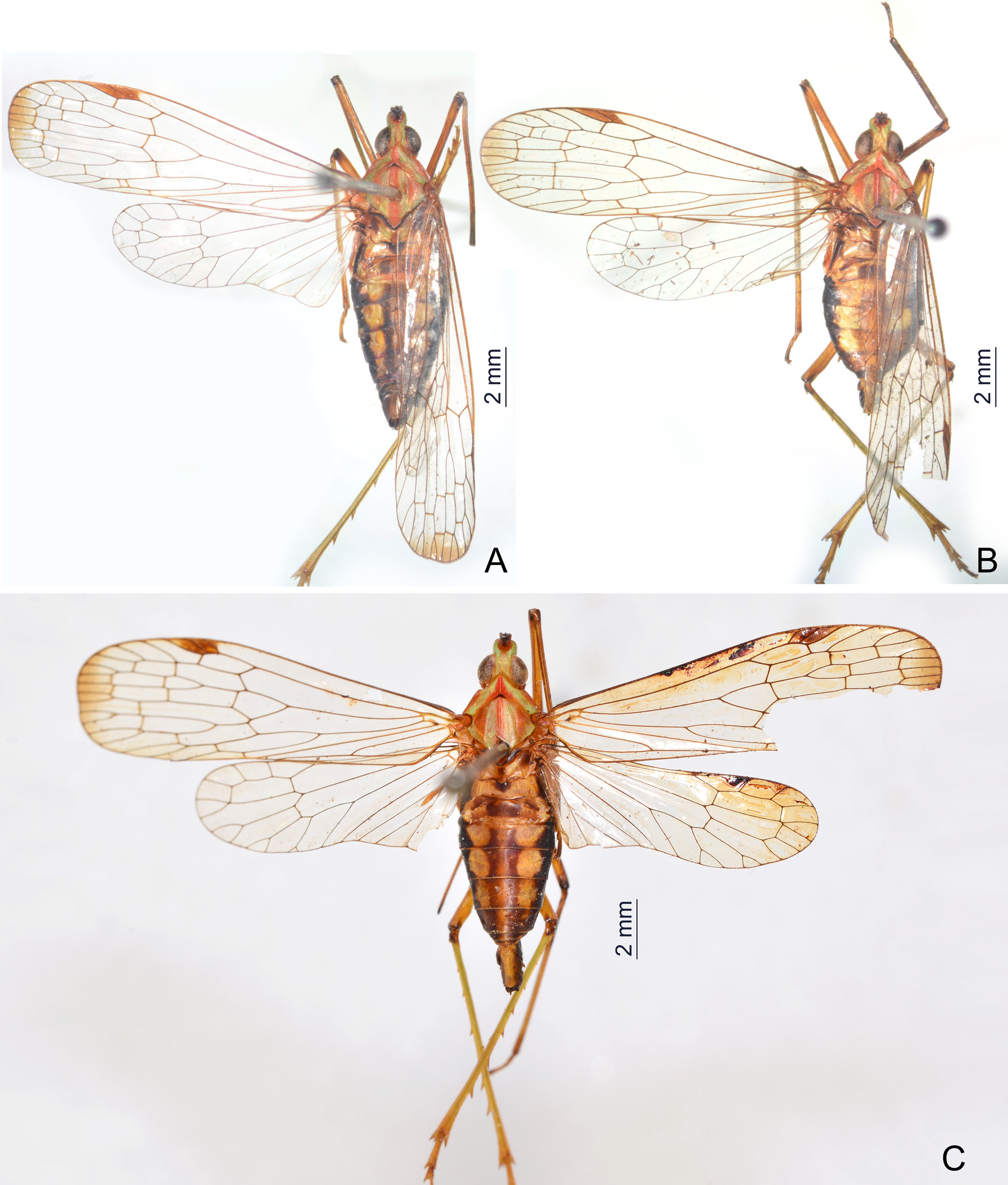
Figs 1–8 View Fig View Fig View Fig View Fig View Fig View Fig View Fig View Fig
Centromeriana Melichar, 1912: 45 View in CoL .
Centromeriana View in CoL – Schmidt 1915: 349 (in list of material). — Metcalf 1946: 39 (in catalogue). — Fennah 1958a: 52 (in key).
Type species
Dictyophara jocosa Gerstaecker, 1895 View in CoL (original designation).
Emended diagnosis
The genus can be distinguished by the following combination of characters: cephalic process moderately long and slender, conical and strongly curved upward; vertex with lateral carinae abruptly constricted and strongly upturned in front of eyes, then gradually convergent anteriad and acuminate at apex; frons with median carina robust and strongly convex, intermediate carinae distinctly expanded outward in apical third, their apical portion being distinctly visible in posterodorsal view; genae with a longitudinal carina above eyes; pronotum with median carina sharp and high, intermediate carinae absent; forewings with sparse transverse veins, stigmal area small and quadrangular, with two or three cells; legs elongate and slender, fore femur not flattened and dilated, without spine; hind tibia with eight apical teeth; apical spines of tarsomeres with long setae; female abdominal sternite VII with a pair of large horn-shaped spines strongly produced ventrad near anterior margin and a pair of blunt triangular lobes on posterior margin; gonocoxae VIII with a pair of small triangular sclerotized plates on posterolateral margin of endogonocoxal lobes.
Redescription
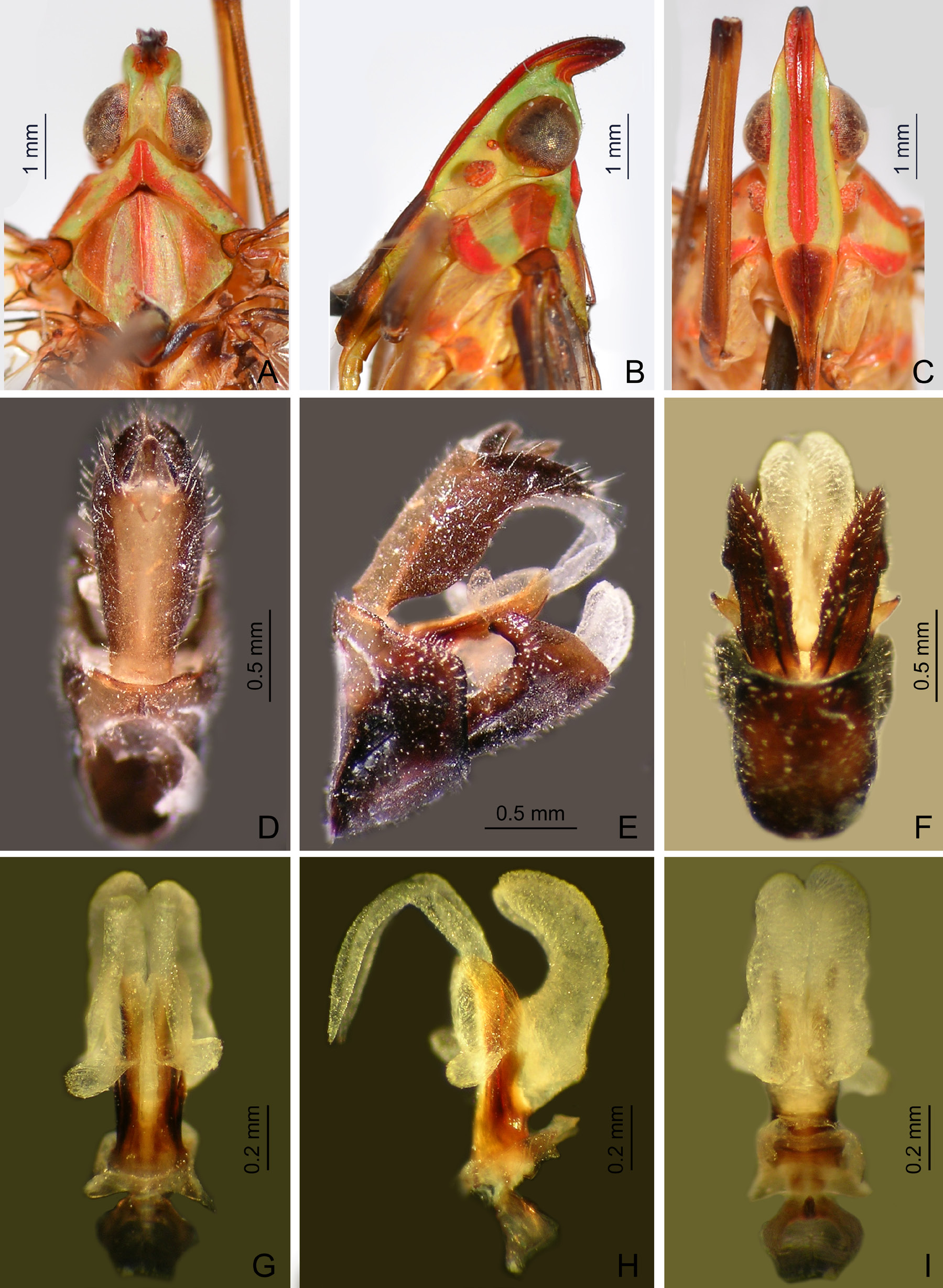
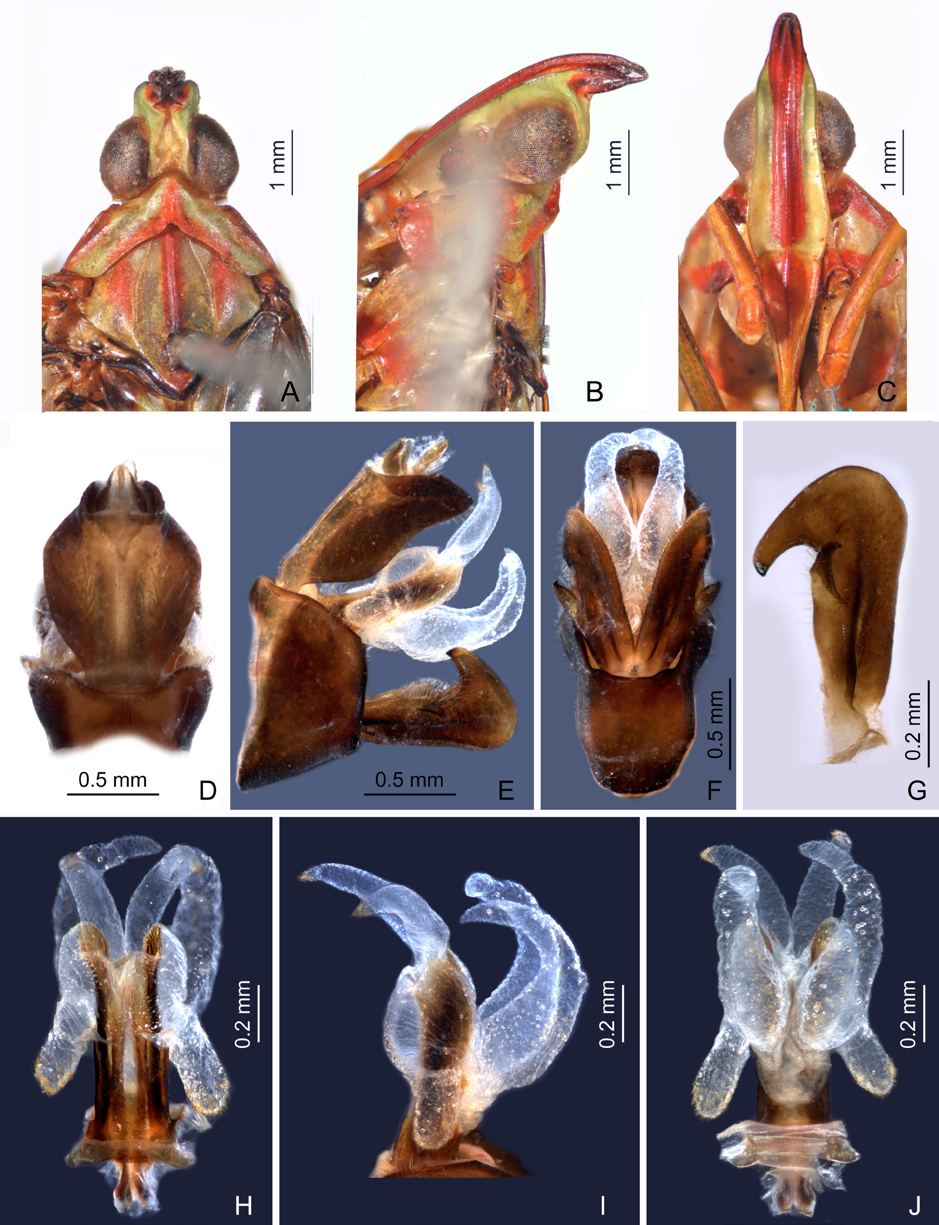
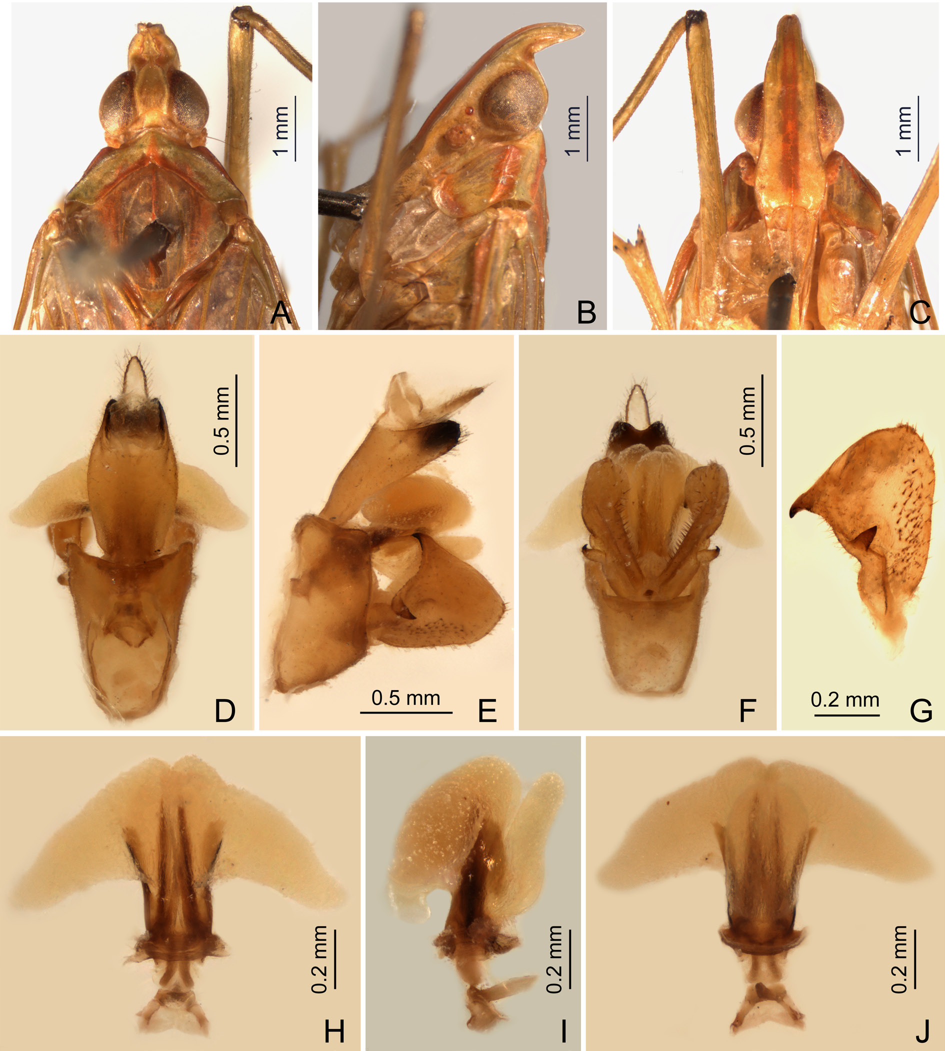
HEAD ( Figs 4 View Fig A–C, 6A–C, 7A–C, 8A–C). Produced into a moderately long and slender cephalic process. Cephalic process ( Figs 4B View Fig , 6B View Fig , 7B View Fig , 8B View Fig ) conical, strongly curved upward, and gradually narrowed toward apex. Vertex ( Figs 4A View Fig , 6A View Fig , 7A View Fig , 8A View Fig ) broadest at base, basal width narrower than transverse diameter of eyes, posterior plane elevated above pronotum; lateral carinae strongly ridged, foliaceous, and sub-parallel in basal third, abruptly constricted and strongly upturned in front of eyes, then gradually convergent anteriad, and acuminate at apex; posterior margin of vertex ridged and angularly concave at about 90–100°; median carina distinct on a bulge between eyes. Frons ( Figs 4C View Fig , 6C View Fig , 7C View Fig , 8C View Fig ) with lateral carinae ridged, nearly parallel, slightly expanded outward below antennae; intermediate carinae slightly converging posteriorly and nearly approaching to frontoclypeal suture, but only sharp and distinct in apical third where they are distinctly expanded outward in ventral view and curved anterodorsad in lateral view, so their apical portion is distinctly visible in posterodorsal view; median carina robust and strongly convex in lateral view. Postclypeus and anteclypeus ( Figs 4C View Fig , 6C View Fig , 7C View Fig , 8C View Fig ) convex medially, with distinct median carina. Rostrum very long, surpassing middle of hind femora; basal segment distinctly longer than distal one. Genae ( Figs 4B View Fig , 6B View Fig , 7B View Fig , 8B View Fig ) with a longitudinal carina above eyes, appearing as bifurcated from lateral carinae of vertex in dorsal view ( Figs 4A View Fig , 6A View Fig , 7A View Fig , 8A View Fig ). Compound eyes ( Figs 4 View Fig A–C, 6A–C, 7A–C, 8A–C) large and globose. Ocelli ( Figs 4B View Fig , 6B View Fig , 7B View Fig , 8B View Fig ) relatively large, reddish. Antenna ( Figs 4B View Fig , 6B View Fig , 7B View Fig , 8B View Fig ) with very small scape; pedicel large and subglobose, with more than 50 distinct sensory plaque organs distributed over entire surface; flagellum long, setuliform.
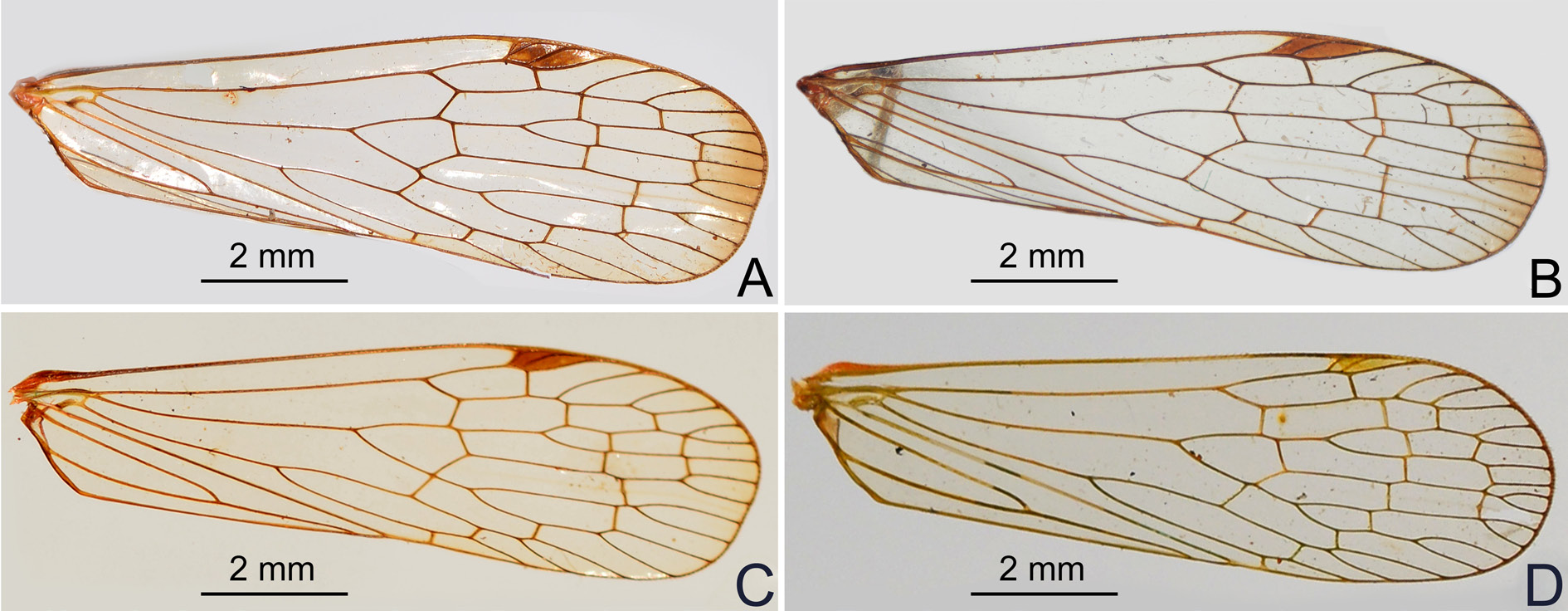
THOARAX. Pronotum ( Figs 4A View Fig , 6A View Fig , 7A View Fig , 8A View Fig ) distinctly shorter than mesonotum medially, narrow anteriorly, broad posteriorly; anterior margin pointed medially, forming a sharp angle, lateral marginal areas straight and sloping with two long longitudinal carinae on each side between eyes and tegulae, lower lateral carinae expanded and visible in dorsal view; posterior margin angularly concave at about 100–110°; median carina sharp and high, with a lateral pit on each side, intermediate carinae absent. Mesonotum ( Figs 4A View Fig , 6A View Fig , 7A View Fig , 8A View Fig ) tricarinate, lateral carinae incurved anteriorly toward median carina. Forewings ( Fig. 3 View Fig A–D) hyaline, much longer than abdomen, with ratio of length to width about 3:1; veins with short setae on ventral side; venation with sparse transverse veins; MP bifurcating MP 1+2 and MP 3+4 near middle and beyond CuA; number of apical cells between R and CuA equal to 14; stigmal area small and quadrangular, with two or three cells. Legs elongate and slender, fore and middle femora distinctly elongate, fore femur not flattened and dilated, without spine near apex; hind tibia with 5–7 lateral spines and eight apical teeth; hind tarsomeres I and II each with 7–8 apical spines; apical spines of tarsomeres with long setae instead of platellae.
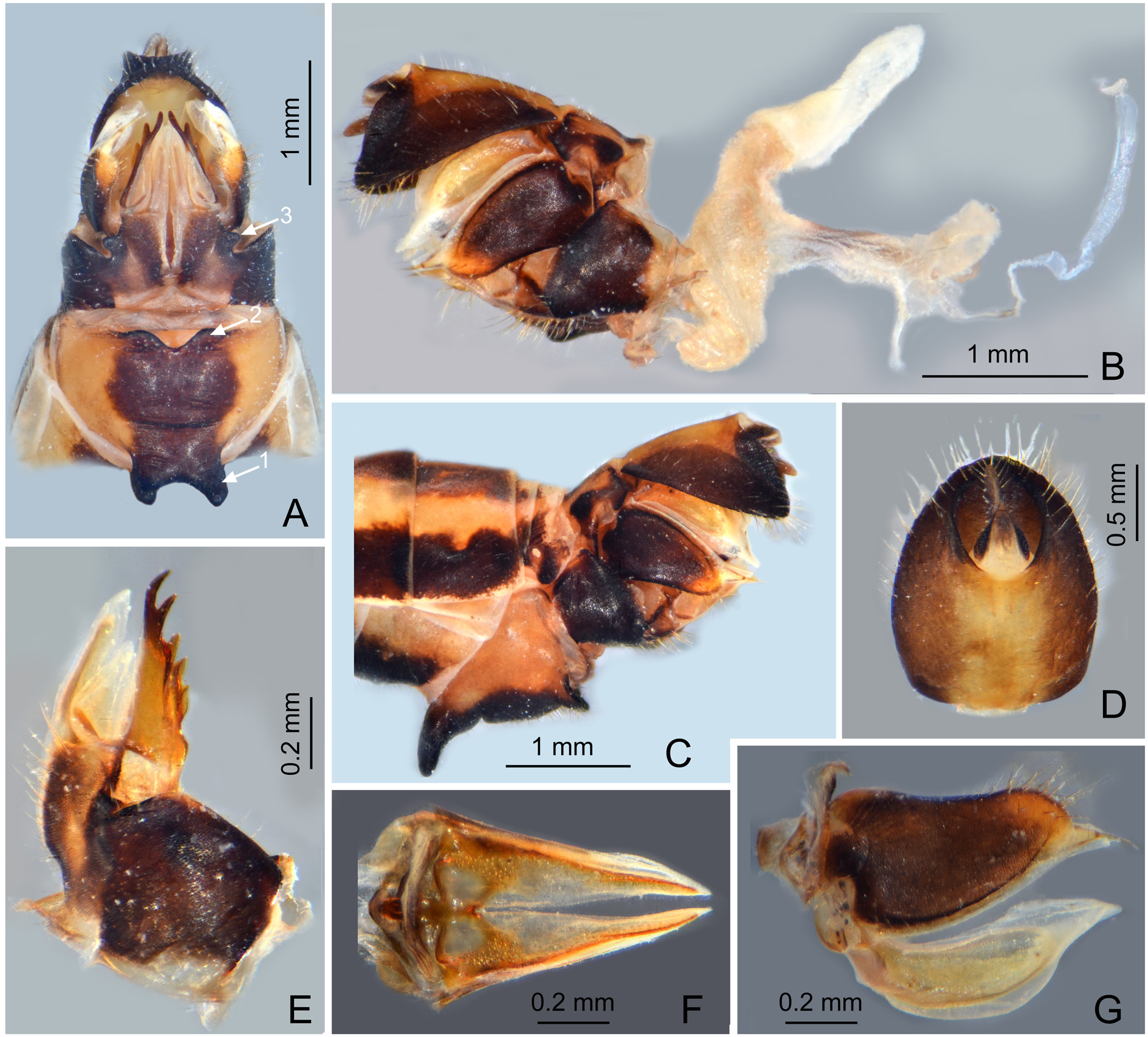
ABDOMEN. With pregenital segments elongate and broad, without distinct median and intermediate carinae dorsally. Female abdominal sternite VII with a pair of large horn-shaped spines directed ventrad near anterior margin and a pair of blunt triangular lobes on posterior margin ( Fig. 5A View Fig ).
MALE GENITALIA. Pygofer ( Figs 4 View Fig D–F, 6D–F, 8D–F) in lateral view distinctly wider ventrally than dorsally, dorsal margin slightly excavated to accommodate segment X, dorsoposterior margins angular. Gonostyles ( Figs 4E View Fig , 6G View Fig , 8G View Fig ) symmetrical, base narrow, expanded toward apex, broadest subapically; dorsal margin with a claw-like, sclerotised process at apex directed dorsad, outer dorsal edge with a hook-like sclerotised process near middle directed ventrad. Aedeagus ( Figs 4 View Fig G–I, 6H–J, 8H–J) with one pair of long endosomal processes extended from phallotheca or lacking such processes; phallobase sclerotised basally and membranous and inflated apically, with paired lobes covered with numerous minute superficial spines ( Figs 4 View Fig G–I, 6H–J) or without spines ( Fig. 8 View Fig H–J). Segment X ( Figs 4D View Fig , 6D View Fig , 8D View Fig ) large, in dorsal view with apex deeply excavated to accommodate anal style; anal style elongate and large.
FEMALE GENITALIA ( Fig. 5 View Fig A–G). Gonocoxae VIII ( Fig. 5E View Fig ) with two membranous and flattened endogonocoxal processes (Gxp) on endogonocoxal lobe: Gxp1 large and elongate, with a long sclerotized plate in it; Gxp2 smaller and shorter. A pair of small triangular sclerotized plates on posterolateral margin of endogonocoxal lobes ( Fig. 5A View Fig ). Gonapophyses VIII ( Fig. 5E View Fig ) with anterior connective lamina large and sclerotized, with seven teeth of varying sizes and shapes. Gonapophyses IX ( Fig. 5F View Fig ) with posterior connective lamina triangular, symmetrical, fused with the intergonocoxal plate at base; intergonocoxal plate extended cephalad into genital cavity, forming wall of gonospiculum. Gonoplacs ( Fig. 5G View Fig ) with two lobes homologous; lateral lobe large and moderately sclerotized, with long setae at apex; the posterior lobe membranous, containing long sclerotized plate. Segment X ( Fig. 5D View Fig ) large and broad in dorsal view, apex deeply excavated to accommodate anal style; anal style large and elongate. Female ectodermal genital ducts ditrysian. Bursa copulatrix ( Fig. 5B View Fig ) superficially membranous, regularly gridded, without sclerotized ornamentations. A pair of large digitiform glands ( Fig. 5B View Fig ) branched at anterior extremity of the anterior vagina on each side of the spermatheca. Spermatheca ( Fig. 5B View Fig ) divided clearly into five parts: orificium receptaculi, ductus receptaculi, diverticulum ductus, pars intermedialis, and glandula apicalis.
Diversity and distribution
Centromeriana is comprised of four species being endemic to the Congolian region of the western tropical Africa as defined by Linder et al. (2012) and closely matching to the Guineo-Congolian region of White (1979, 1983) including the ‘ Dahomey gap’.
Ecology and economic importance
Unknown. Heinrichs & Barrion (2004) listed Centromeriana sp. among insects occurring on rice in Gambia. However, based on morphological characters they specify in their identification key (p. 155) and a schematical drawing of the head and thorax (fig. 355: 157), this is a misidentification; the record probably refers to some other dictyopharid genus. Oke et al. (2015) reported adult Centromeriana spp. as minor pests of leaves of Amaranthus spp. in Nigeria but the identification is uncertain.
Key to species of Centromeriana View in CoL
1. Forewing membrane with a fuscous macula apically ( Fig. 3 View Fig A–B) .................................................. 2
– Forewing membrane with apex clear ( Fig. 3 View Fig C–D) ........................................................................... 3
2. Phallobase with ventral lobes robust and thumb-like, broad apically, confluent medially ( Fig. 4 View Fig H–I). Male segment X, in dorsal view, narrow, ratio of length to width near middle about 2.2:1 ( Fig. 4D View Fig ); in lateral view, with narrow and sharp, hook-like apical lobes, strongly projecting ventrad ( Fig. 4E View Fig ) ............................................................................................... C. jocosa ( Gerstaecker, 1895) View in CoL
– Phallobase with ventral lobes more slender, convergent and tapering apically, but divergent medially ( Fig. 6I, J View Fig ). Male segment X, in dorsal view, broad, ratio of length to width near middle about 1.3:1 ( Fig. 6D View Fig ); in lateral view, with broad and blunt apical lobes, weakly projecting ventrad ( Fig. 6E View Fig ) ........................................................................................................... C. lindbergae View in CoL sp. nov.
3. Abdomen dorsally and ventrally dark brown and black, with a longitudinal row of yellowishochraceous spots on each side ( Fig. 2A View Fig ). Cephalic process relatively short, curved upward and slightly backward in more than 90° ( Fig. 7B View Fig ) ................................................. C. rhinoceros View in CoL sp. nov.
– Abdomen dorsally and ventrally greenish-ochraceous ( Fig. 2B View Fig ). Cephalic process relatively long, curved upward in nearly 90° ( Fig. 8B View Fig ). Male genitalia as in Fig. 8 View Fig D–J ... C. simplex Melichar, 1912 View in CoL


No known copyright restrictions apply. See Agosti, D., Egloff, W., 2009. Taxonomic information exchange and copyright: the Plazi approach. BMC Research Notes 2009, 2:53 for further explanation.
|
Kingdom |
|
|
Phylum |
|
|
Class |
|
|
Order |
|
|
InfraOrder |
Fulgoromorpha |
|
Family |
|
|
Tribe |
Orthopagini |
Centromeriana Melichar, 1912
| Song, Zhi-Shun, Malenovský, Igor & Liang, Ai-Ping 2017 |
Centromeriana
| Fennah R. G. 1958: 52 |
| Metcalf Z. P. 1946: 39 |
| Schmidt E. 1915: 349 |
Centromeriana
| Melichar L. 1912: 45 |