
Epistylis smalli, Utz, Laura R. P., Santos, Mariana Silva Dos & Araujo, Gabriella Oliveira De, 2015
|
publication ID |
https://doi.org/10.11646/zootaxa.4040.4.10 |
|
publication LSID |
lsid:zoobank.org:pub:828BD39A-0921-4E9F-BBD8-F2D5B18C7B08 |
|
DOI |
https://doi.org/10.5281/zenodo.5630370 |
|
persistent identifier |
https://treatment.plazi.org/id/03CB1F58-4711-FD68-D3CB-FD4AFC19C20F |
|
treatment provided by |
Plazi |
|
scientific name |
Epistylis smalli |
| status |
sp. nov. |
Epistylis smalli n. sp.
Diagnosis. Freshwater Epistylis with a dichotomously branched colony bearing elongate zooids measuring on average 173 µm in length, and 49 µm in width. A C-shaped macronucleus lies transversally on the oral half of the cell, close to the infundibulum. All infundibular polykinetids are composed of three rows of kinetosomes; Oral polykinetid 1 (OPK1) has rows of equal lengths, OPK2 ends at the adstomal curvature of PK1 and presents rows of different lengths, and PK3 has all rows of the same length and terminates at the adstomal end of OPK1.
Type locality. Guaíba Lake ( 30º6'38"S, 51º15'38"W) located in Porto Alegre municipality, Rio Grande do Sul state, Brazil.
Etymology. This species name is dedicated to Eugene B. Small for his relevant contributions to ciliate taxonomy.
Deposition of Slide. One slide with protargol stained specimens was deposited in the Protist Collection of the Museum of Science and Technology of the Pontificia Universidade Católica do Rio Grande do Sul under the number MCTP3.
Morphology of live specimens. Colonies of E. smalli were dichotomously branched with secondary branches terminating at the same level ( Fig 1 View FIGURE 1 A and 2A). As characteristic of the genus Epistylis the basal or secondary stalks did not present spasmoneme ( Fig 1 View FIGURE 1 A and 2B). The main basal stalk was considerably long ( Table 1 View TABLE 1 ) and presented smooth longitudinal ridges ( Fig 1 View FIGURE 1 H). Live colonies had up to 34 zooids, but generally presented 6-8 zooids that were similar in size and shape. Zooids were elongate and ranged from 145 to 200 µm in length and from 45 to 80 µm in width ( Table 1 View TABLE 1 and Fig 1 View FIGURE 1 B and 2B). The peristomial lip was thick and wider than the body ( Table 1 View TABLE 1 and Fig 1 View FIGURE 1 B and 2B). The epistomial disk was elevated ( Fig 1 View FIGURE 1 B) and about ½ as wide as the peristomial lip ( Table 1 View TABLE 1 ). A Cshape macronucleus was located on the first half of the cell close to the infundibulum ( Fig 1 View FIGURE 1 C and 2B).
Morphology of stained specimens. The infraciliature and the nuclear apparatus of Epistylis smalli was easily recognized in protargol staining. The size of stained zooid ranged from 92.5 to 117.5 µm in length and from 42.5 to 57.5 µm in width ( Table 2 View TABLE 2 ). A total of 26 somatic myonemes extend from the scopula to the epistomial disk ( Fig 1 View FIGURE 1 D). A C-shaped macronucleus lies on the first half of the cell ( Fig 1 View FIGURE 1 D and 2B). The oral infraciliature was typical of peritrich ciliates with an outer haplokinety and an inner polykinety that performs approximately 1 ½ turns before entering the infundibulum ( Fig 1 View FIGURE 1 E and 2C). Inside the infundibulum the oral polykinetid 1 (OPK1) is accompanied by the infundibular polikinetids 2 (OPK2), and 3 (OPK3). All infundibular polykinetids present three rows of kinetosomes. The middle row of OPK1 is longer than the other two ( Fig 1 View FIGURE 1 E and 2C). Rows in OPK2 are of different lengths: the row closer to OPK1 is shorter than the others ( Fig 1 View FIGURE 1 F and 2C). All three rows of OPK2 terminate at the adstomal curvature of OPK1. OPK3 have three rows of kinetosomes of equal length that terminate above the adstomal end of OPK1 ( Fig 1 View FIGURE 1 G and 2C). The trochal band in the aboral half of the cell is composed by a single row of staggered kinetosomes ( Fig 1 View FIGURE 1 I).
Species in the genus Epistylis Ehremberg, 1830 are colonial having contractile zooids on top of non-contractile stalks. Morphologically species of Epistylis are identified by the size and shape of the zooid, overall shape of the colony, and arrangement of the oral polykinetids revealed by protargol impregnation. The species collected from the Guaiba Lake in the present study was identified as Epistylis smalli n. sp. after being compared with several species in the genus Epistylis . These species presented similar zooid and colony shape, and sometimes a similar zooid size, but differed in other relevant morphologhical characters.
For example, Epistylis constricta Kellicott, 1855 presents an elevated epistomial disk, and a contractile vacuole below the peristomial border similar to E. smalli , but has a smaller size, presents transversal folds when the zooid is contracted and generally is observed as epibiont ( Kahl, 1935; See Table 1 View TABLE 1 for comparisons). E. rotans Sveç, 1897 has a slender zooid, but differs from E. smalli in the organization of the colony, the ability to swim and in the epibiotic way of life ( Kahl, 1935). Epistylis niagarae Kellicott, 1883 has an prominent epistomial disk similar to the studied species, but differs from it in the size of the zooid, in the presence of a strongly projected snout when contracting, and in the epibiotic way of life ( Kahl, 1935).
Epistylis chrysemydis Bishop & Jahn, 1941 is the described species most similar to E. smalli . The zooids of E. chrysemydis are slender measuring 120 – 220 µm in length and 60 – 110 µm in width, which is in the range found for E. smalli ( Table 1 View TABLE 1 ). In addition, the macronucleus of E. chrysemydis is C-shaped and surrounds the infundibulum as observed for the species described here ( Fig 1 View FIGURE 1 C and 2B). On the other hand, the arrangement of the oral polykinetids of E. chrysemydis is different from the observed for E. smalli . The oral polykinetid 1 (OPK1) of E.chrysemydis has three rows of kinetosomes of equal length, while in E. smalli the middle row is longer than the other two ( Fig 1 View FIGURE 1 E and 2C). The rows in OPK3 of E. chrysemydis terminate at the same level of OPK1 and OPK2, while in E. smalli OPK3 terminates above the adstomal end of OPK1. OPK2 is similar in both species.
The arrangement of kinetosomal rows in oral polykinetids has been used by several authors to distinguish species in peritrich ciliates (e.g. Esteban & Fernandez-Galiano, 1989; Leitner & Foissner, 1997; Clamp, 2005; Sun et al., 2006; Utz et al., 2014). The relative length of rows and the points they terminate is definitive for species identification ( Sun et al., 2006; Gentekaki & Lynn, 2010). Despite the similarities in the size and shape of the live zooid and the overall shape of the colonies observed between E. chrysemydis and E. smalli , differences were pointed out in the pattern of kinetosomal rows in the oral polykinetids. These differences are strong enough to consider a species-level distinction between E. chrysemydis and E. smalli .
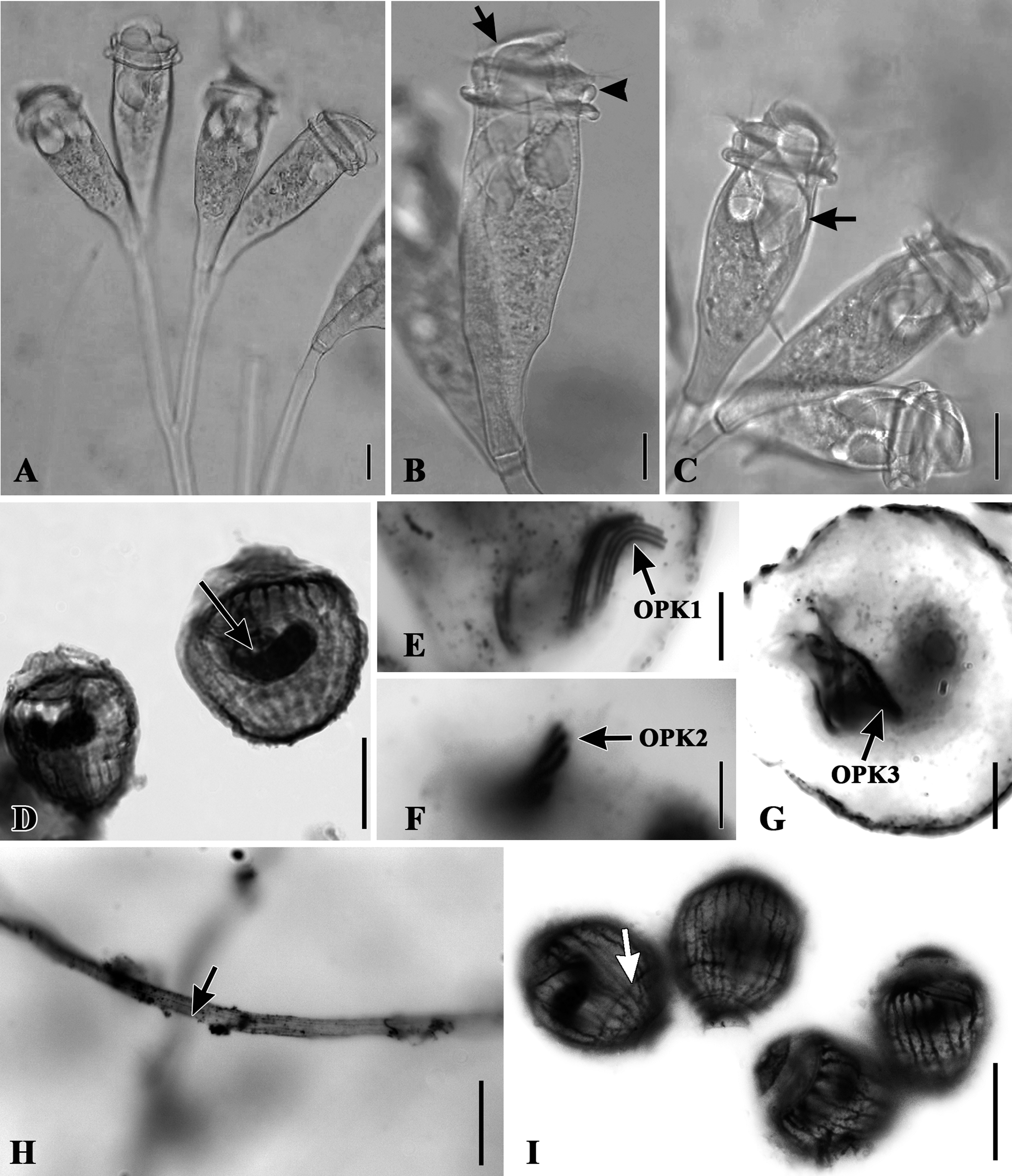
TABLE 1. Measurements of live colonies of Epistylis smalli attached to slides collected from Guaiba lake, Southern Brazil. A total number of 20 zooids were measured for each character.
| Character | Mean (µm) | SD* (µm) | Mode | CV** (%) | Range |
|---|---|---|---|---|---|
| Length of the Body from epistomial disk to aboral end | 173.1 | 16.4 | 187.5 | 9.5 | 145–200 |
| Length of the body from peristomial lip to aboral end | 137.5 | 15 | 137.5 | 10.9 | 112.5–165 |
| Width of the Body below the peristomial lip | 49.6 | 6.7 | 45 | 13.5 | 42.5–65 |
| Width of the body at midpoint between oral and aboral ends | 55.5 | 10.4 | 57.5 | 18.7 | 45–80 |
| Width of peristomial Lip | 63.4 | 5.1 | 62.5 | 8.1 | 57.5–72.5 |
| Width of epistomial disk | 36.6 | 4.9 | 37.5 | 13.3 | 27.5–47.5 |
| Thickness of peristomial Lip | 24.5 | 2.2 | 25 | 9.1 | 20- 30 |
| Width of scopula | 18.1 | 8.4 | 17.5 | 46.2 | 2.5–20 |
| Length of basal stalk | 371.7 | 153.9 | 300 | 41.4 | 152.5–770 |
| Width of basal stalk | 15.1 | 4.0 | 17.5 | 26.5 | 10–17.5 |
| * Standard Deviation; ** Coefficient of Variation. |
TABLE 2. Measurements of Protargol stained colonies of Epistylis smalli collected from Guaiba Lake, Southern Brazil. A total of 16 zooids were measured for each character.
| Character | Mean (µm) | SD* (µm) | Mode CV** (%) | Range |
|---|---|---|---|---|
| Total length of the body from epistomial disk to aboral end | 104.5 | 7.0 | 100 6.7 | 92.5 – 117.5 |
| Width of the body at midpoint between oral and aboral ends | 47.6 | 4.0 | 47.5 8.4 | 42.5 – 57.5 |
| Distance between Trochal Band and Scopula | 9.7 | 3.1 | 10 3.4 | 10 - 15 |
| Length of the Macronucleus | 39.5 | 9.2 | 40 23.2 | 15 - 50 |
| Width of Macronucleus at Midpoint | 6.2 | 1.3 | 5.0 20.7 | 5.0 – 7.5 |
| * Standard Deviation; ** Coefficient of Variation. | ||||
| Discussion |
No known copyright restrictions apply. See Agosti, D., Egloff, W., 2009. Taxonomic information exchange and copyright: the Plazi approach. BMC Research Notes 2009, 2:53 for further explanation.
|
Kingdom |
|
|
Phylum |
|
|
Class |
|
|
Order |
|
|
Family |
|
|
Genus |