
Microzercon opiparus ( Błaszak, 1984 ), Ujvári, 2013
|
publication ID |
https://doi.org/10.5281/zenodo.5736238 |
|
persistent identifier |
https://treatment.plazi.org/id/03CB878E-653B-5124-FE1E-FD4F8611BD62 |
|
treatment provided by |
Felipe |
|
scientific name |
Microzercon opiparus ( Błaszak, 1984 ) |
| status |
comb. nov. |
Microzercon opiparus ( Błaszak, 1984) comb. n.
( Figs 38–48 View Fig View Figs 39–41 View Figs 42–48 )
Bakerasopiparus BŁaSzak, 1984: 588.
Typelocalities. USA, Virgnia, GraysonCounty, WhitetopMt., 4000 ft. a.s.l. USA, West
Virginia, MercerCounty, CampCreek, St. Forest.
Materialexamined. USA, Tennessee, Cumberlandcounty , 07.07.1956, leg. Bohnsack, K ( HNHM E-Am-007 – 3 females, 6 males, 1 deutonymph); USA, North Carolina, Burke Co., LinvilleFallsgorgearea, BlueRidgePkwy. 975 ma.s.l., frommixeddeciduous & white pinelitter, 11.08.1986, leg. Lindquist, E. E. (slideCNCAZ0547 – 1 female) ; USA, WestVir- ginia, PocahontasCo., HillsCreekFallsarea, 27 kmE. Richwood, 915 ma.s.l., ex. deciduous litter, rottenwood, moss & substrate, 03.08.1986, leg. Lindquist, E. E. (slideCNCAZ0636 – 1 male; slide CNCAZ 0637 – 1 female; slide CNCAZ 0638 – 1 female; slide CNCAZ 0640 – 1 female; slide CNCAZ 0657 – 1 female; slide CNCAZ 0660 – 1 female; slide CNCAZ 0708 – 1 female; slide CNCAZ 0709 – 1 female); USA, Alabama, De Kalb Co., De Soto State Park , Little River Canyon, ex. Rhododendron litter on NW facing bank, 28.09.1992, leg. Behan, V. (slide CNCAZ 0781 – 1 female) .
Diagnosisoffemale. Dorsalsetaepiloseexceptj4–5. EachJ-setareaching basesofthefollowingoneoftheseries. Eachmarginalsetasimilarinappear- ance, denselypilose, brush-like. GlandsPo2 situatedinposition gdS2, online connectingS2 andS3, nearS3, Po3 situatedinposition gdJ2, posterolateralto J2. Anteriorsurfaceofpodonotalshieldcoveredbyscalespossessinglacypos- teriormargin. Opisthonotalsurfacesmooth. Posterodorsalcavitiesuniform, round, weaklydeveloped. Posterolateraltipsofperitrematalshieldsreaching levelofR3, freeorfusedwithlateralpartsofventrianalshield.
Descriptionoffemale. Lengthofidiosoma: 382 μm (360–398 μm); width: 327 μm (296–349 μm) (n = 10).
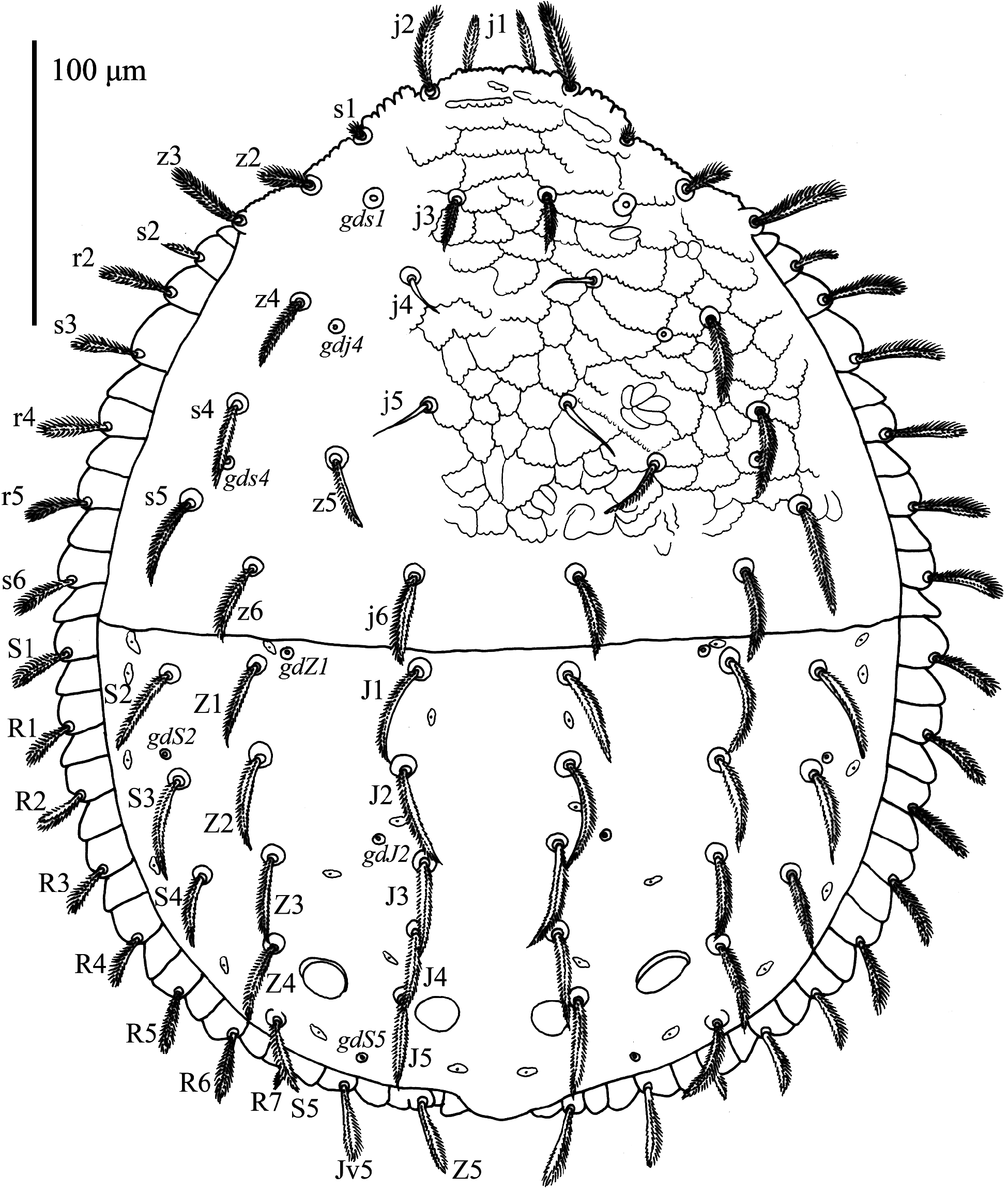
Dorsal side ( Fig. 38 View Fig ). Podonotum with 20 pairs of setae, j1–6, z2–6, s1–6, r2 and r4–5 inserteddorsally, r1 andr3 insertedventrally, onperitrematalshields. Dorsalpodonotal setadenselypilose, exceptj4–5 whichsmoothandneedle-like. Marginalpodonotalsetae brush-like, submarginalandcentralsetaepointed. Setaes5 situatedbetweenlevelofz5 and z6. Glands gds1 (po1) situated on line connecting j3 and z2; gdj4 (po2) situated on line connecting j5 and z4, near z4; gds4 (po3) slightly medial to line connecting s4 and s5. The surfaceinfrontofthelineconnectingsetaes5 coveredbyscalespossessinglacyposterior margin, theposteriormostpodonotalsurfacesmooth.
Opisthonotumwith 22 pairsofsetae: J1–5, Z1–5 andS1–5, marginalR-serieswith sevenpairsofsetae. SetaeJ1–5, Z1–4 andS2–5 similarinappearance, relativelylong, point- ed, denselypilose ( Fig. 42 View Figs 42–48 ). SetaeJ1–5 andZ1–4 constitutenearlyparallellines. SetaeZ4 situated on line connecting Z3 and S5. Setae S2 situated anterior to S3. Setae S2–4 not reaching edgesofopisthonotum, thetipsofS5 expandingbeyondmarginsoftheshield. SetaeZ5, S1 andR1–7 denselypilose, apicallybroadening, brush-like. R7 significantlyshorterthan therestofR-setae. Lengthofopisthonotalsetaeanddistancesbetweentheirinsertionsas inTable 5. Glands gdZ1 (Po1) situatedanteromedialtoinsertionsofZ1; gdS2 (Po2) situated on line connecting S2 and S3, near S3; gdJ2 (Po3) posterolateral to J2; gdS5 (Po4) on line connectingZ5 andS5, anteromedialtoinsertionsofJv5. Marginalserrationdeepandobtuse. Opisthonotalsurfacesmooth. Posterodorsalcavitiesuniform, round, weaklydeveloped. Lateralpairofcavitiessituatedanteriortocentralpair, betweeninsertionsofJ5 andZ4.
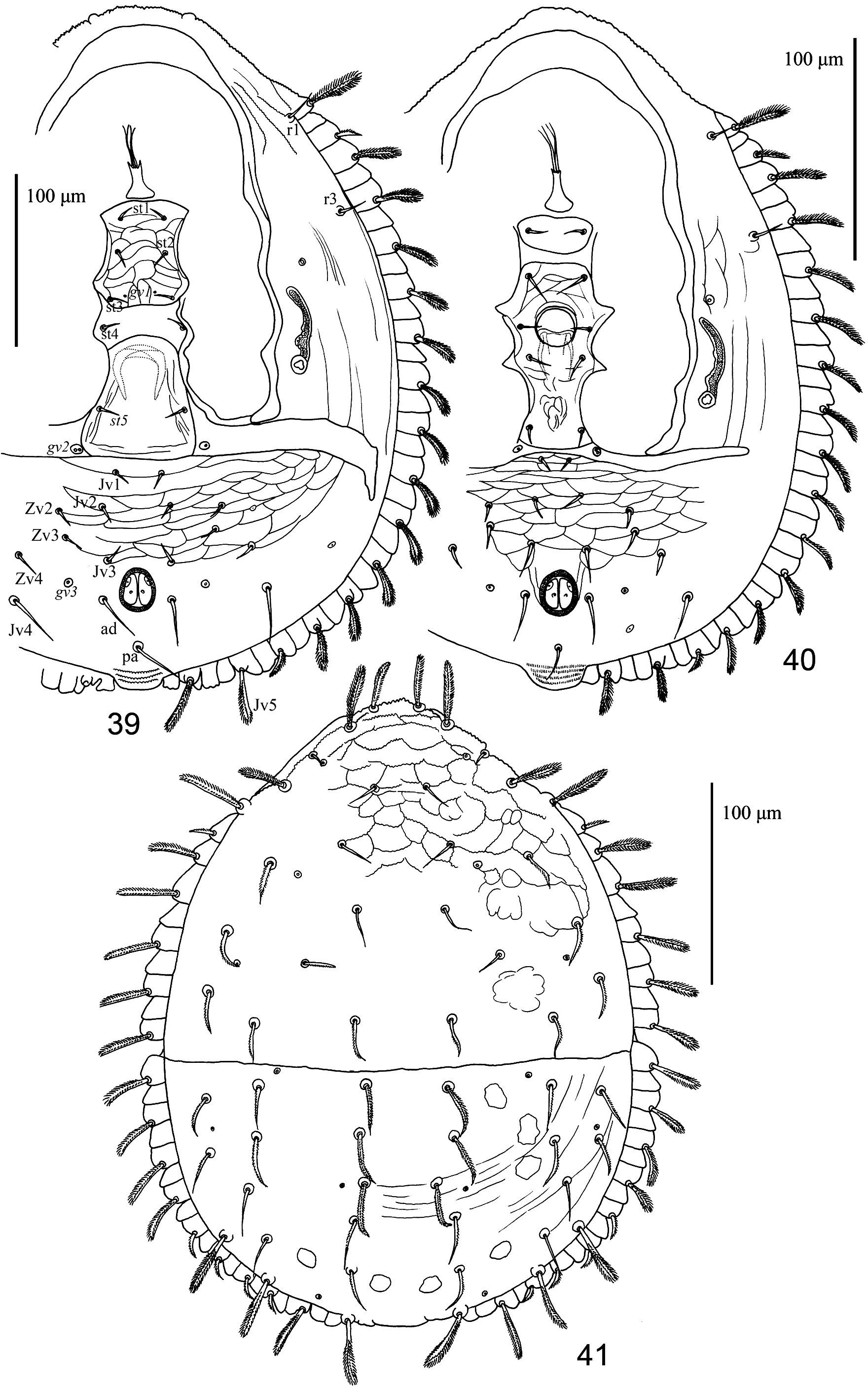
Ventralside ( Fig. 39 View Figs 39–41 ). Theslitbetweenperitrematalshieldsandbodymarginsnarrow. Posterolateraltipsofperitrematalshieldsfreeorfusedwithlateralpartsofventrianalshield onlevelofsetaeR3. Peritrematalshieldsornamentedbyfinereticulationoflongitudinal lines. Peritremesfinelycurved, with 1–3 smalldilations. Peritrematalsetaer1 andr3 short, smoothandneedle-like. Tritosternumwithtwoslender, apicallybifurcate, marginallypilose laciniae, tritosternalbasevase-like. Sternalshield 51 μmlongand 37 μmwideatthelevelof setaest2, withnearlystraightposteriormargin, withreticulateornamentation. Sternalsetae smoothandneedle-like. Glands gv1 situatedanteromedialtosetaest3. Genitalshieldtypi- calforthefamily, withoutornamentation, genitalsetaesmoothandneedle-like. Glands gv2 present, withsingleordoubleopeningssurroundedbysmalladgenitalsclerites. Ventrianal shieldwithsmoothandneedle-likesetae, setaeZv1 absent. Adanalsetae, Jv4 andpostanal seta 2 timeslongerthanotherpreanalsetae. SetaeJv5 moderatelylong, brush-like, densely pilose. Analvalveswithvestigialeuanalsetae. Glands gv3 situatedanterolateraltoadanal setae. Anteriorsurfaceofventrianalshieldcoveredbytile-likepatterntolevelofJv3-Zv4.
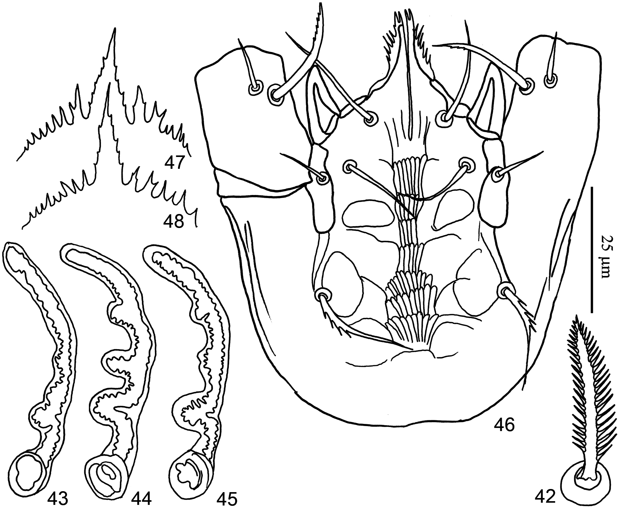
Gnathosoma ( Fig. 46 View Figs 42–48 ). Situationofhypostomalandsubcapitularsetaetypicalforthe family. Setae h1 elongate, apically tapering, smooth. Setae h2 shorter than h1, h3 shorter than h2, each smooth. Setae h4 slightly longer than h2, proximally serrate. Corniculi hornlike, internalmalaewithapairofbifurcateanterocentralbranchesandwithserratemar- gins. Cheliceraerelativelyslender, fixeddigitwith 6 teeth, movabledigitwith 4 teeth. Epistome of Prozercon - type ( Figs 47–48 View Figs 42–48 ).
Descriptionofmale ( Fig. 40 View Figs 39–41 ). Lengthofidiosoma: 312 μm (296–333 μm); width: 265 μm (247–296 μm) (n = 7). Chaetotaxy, adenotaxy and sculptural pattern of dorsal, ventri- analandperitrematalshieldsbasicallysimilartothoseoffemale, exceptshapeofj3 which smoothinmale. Lengthofopisthonotalsetaeanddistancesbetweentheirinsertionsas inTable 5. Peritrematalshieldsfusedwithanterolateralpartsofventrianalshield, onlya pairofhorizontalincisionscanbeobservedrunningtowardssetaeR1. Sternigenitalshield divided, thefirstpairofsternalsetae (st1) sittingonaseparatepieceofsclerotizedarea, therestofsternalsetaeandgenitalopeningsituatedontheposteriorsclerotizedareaofthe sternigenitalregion, whichendsonlevelofposteriormarginofcoxaeIV. Setaest5 present. Sternalsetaest3 situatedonlevelofcentralpartofgenitalopening. Analvalveswithves- tigialeuanalsetae. Eachcharactersofgnathosomasimilartothoseoffemale, butterminal partoffixeddigitofcheliceraebifurcate.
Descriptionofdeutonymph ( Fig. 41 View Figs 39–41 ). Lengthofidiosoma: 311 μm; width: 258 μm (n = 1). Dorsalchaetotaxy, adenotaxyandappearanceofposterodorsalcavitiesbasicallysimilarto thoseofadults, exceptshapeofcentralandsubmarginalsetaeofdorsalshields, whichless pilosethanthoseofadults, setaes1, j3–5 andZ3–4 appearstobesmoothorbarelypilose. LengthofopisthonotalsetaeanddistancesbetweentheirinsertionsasinTable 5. Dorsal ornamentationsimilartothatofadults, butlessexpressed. Eachcharactersofgnathosoma similartothoseofadultfemale.
Remarks. Similarlytothatof Macrozerconpraecipuus, somedifferencescan beobservedbetweenthespecimensoftheCNCandHNHMcollections. The
ventral view of gnathosoma, female, 47–48 = epistomes of female.
CNCspecimenscorrespondwiththetypespecimensdescribedbyBŁaSzak (1984), theyarerelativelylarge (376–398 μm), posterolateraltipsoftheirperi- trematalshieldsendfreely, andtheirperitremespossessextratubercules ( Figs 44–45 View Figs 42–48 ). Incontrast, theHNHMspecimensaresmaller (360–370 μm), posterolateraltipsoftheirperitrematalshieldsarefusedwiththeventrianalshield, andtheirperitremeshaveonlyasingletubercule ( Fig. 43 View Figs 42–48 ).
| HNHM |
Hungarian Natural History Museum (Termeszettudomanyi Muzeum) |
| V |
Royal British Columbia Museum - Herbarium |
No known copyright restrictions apply. See Agosti, D., Egloff, W., 2009. Taxonomic information exchange and copyright: the Plazi approach. BMC Research Notes 2009, 2:53 for further explanation.