
Pelogenia kinbergi ( Hansen, 1882 )
|
publication ID |
https://doi.org/10.5852/ejt.2022.807.1717 |
|
publication LSID |
lsid:zoobank.org:pub:67DAF0D7-9D9F-4F0F-8004-9CCE0071BF33 |
|
DOI |
https://doi.org/10.5281/zenodo.6421721 |
|
persistent identifier |
https://treatment.plazi.org/id/03CB87CF-C679-FF86-B803-FCC0B2DBF7ED |
|
treatment provided by |
Felipe |
|
scientific name |
Pelogenia kinbergi ( Hansen, 1882 ) |
| status |
|
Pelogenia kinbergi ( Hansen, 1882) View in CoL
Figs 2C–D, I–J View Fig , 17–18 View Fig View Fig
Psammolyce kinbergi Hansen, 1882: 5 View in CoL , pl. 1 figs 10–13.
Pelogenia kinbergi View in CoL – Pettibone 1997: 56, figs 41–42 (syn., comb. nov.).
Material examined
GUYANA • 1 incomplete spec.; off Georgetown ; 08°28′ N, 58°12′ W; R/V Pillsbury, Stn 0694; depth 80 m; 15 Jul. 1968; UMML 6806-0694 View Materials GoogleMaps .
Description
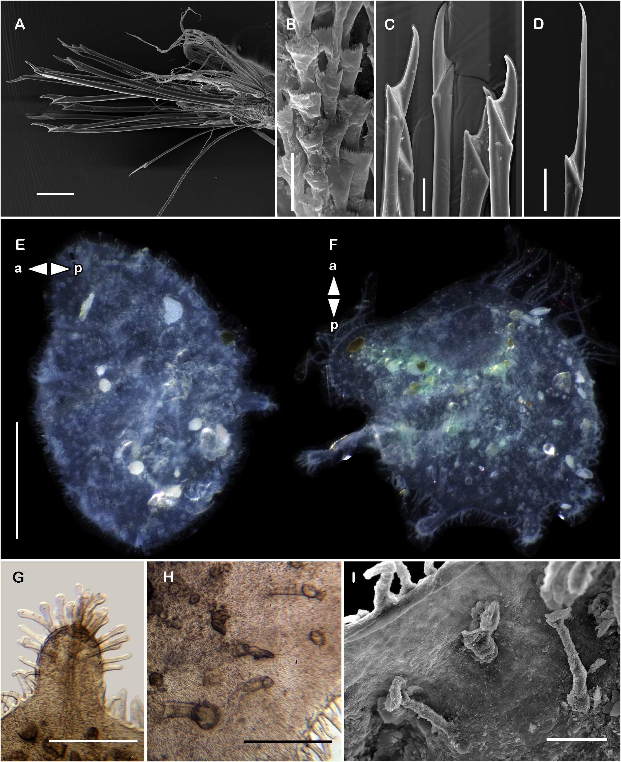
BODY. Pale yellow, long, broad ( Fig. 17A View Fig ); 67 segments, 4.5 cm long, 1.5 cm to segment 30, 0.5 cm wide. Middorsal line covered with coarse sand attached to adhesive papillae ( Fig. 17B View Fig ). Venter partially covered with short globular and long dendritic papillae ( Fig. 17C View Fig ).
PROSTOMIUM. Spherical. Two pairs of eyes, anterior eyes slightly larger and inserted anteroventrally ( Fig. 17D View Fig ). Lateral antennae long, slender; ceratophores longer than style, dorsally fused with tentacular segment, completely covered by median antennal ceratophore ( Fig. 17E View Fig ). Median antenna with bulbous ceratophore,twice as long as prostomium,with transverse ridges; style long, slightly shorter than ceratophore ( Fig. 17D View Fig ). Middorsal lobe of segment II absent. First segment directed anteriorly; fused with tentacular segment (left and right parapodia fused anteriorly); biramous, chaetae simple verticillate. Dorsal tentacular cirrus longer than neuropodia including chaetae, ventral tentacular cirrus as long as dorsal tentacular one, longer than neuropodia; palps short, reaching segment five, with inner palpal sheaths ( Fig. 17B View Fig ).
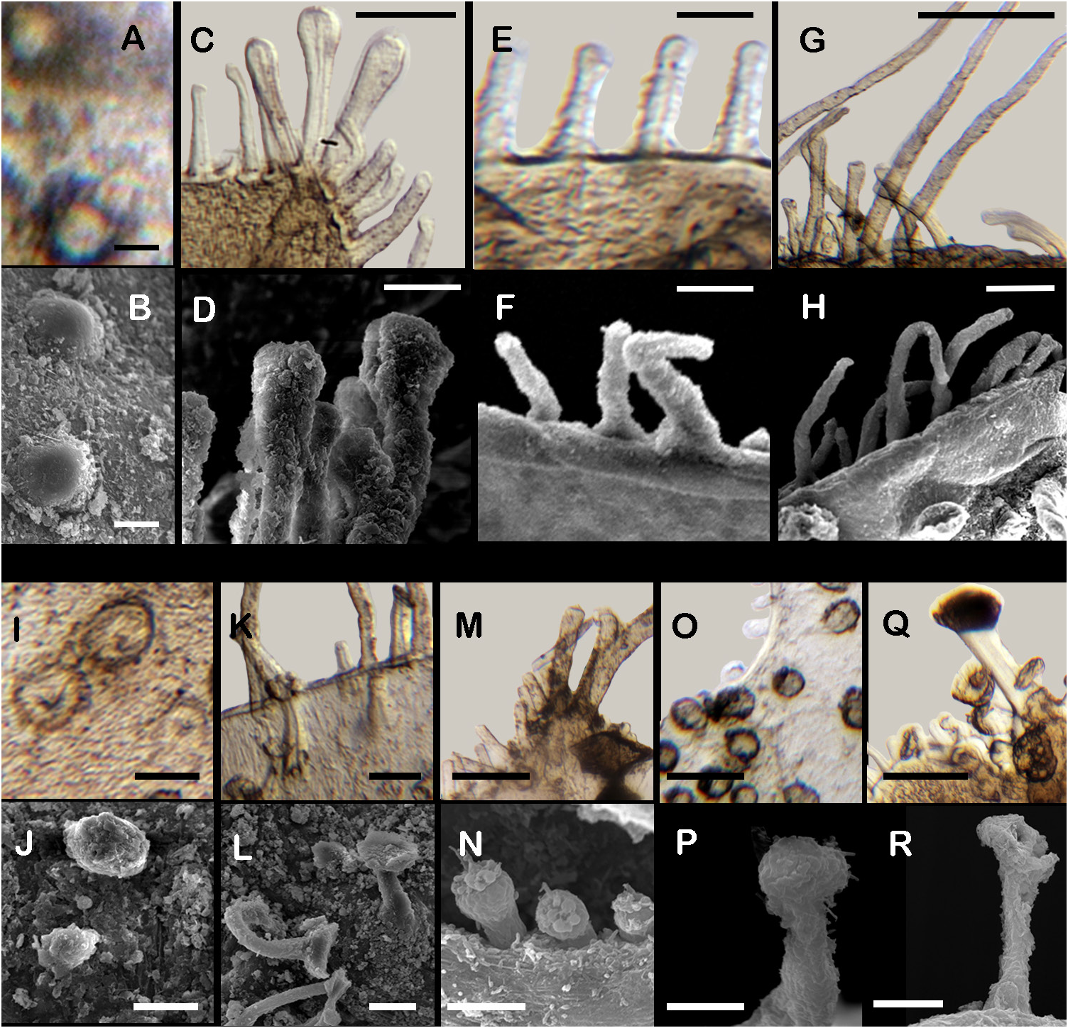
ELYTRA. First right elytron oval, with one large medial process, with two kinds of papillae ( Fig. 18E View Fig ): surface with flat papillae, elytral margin with pedunculate papillae with puffed tips ( Fig. 18G View Fig ). Posterior elytra oval notched, with two short medial processes, and four posterior enlarged processes, each distally expanded ( Fig. 18F View Fig ); four kinds of papillae: on the surface flat papillae, and pedunculate papillae with flat tips, concentrated along the largest process; elytral margin with pedunculate with puffed tips, and long dendritic papillae ( Figs 2C–D, I–J View Fig , 18H–I View Fig ).
RIGHT PARAPODIUM FROM SEGMENT II ( Fig. 17G View Fig ). Notopodia conical, papillate, as large as neuropodia; notopodial flange round, papillate. With up to 80 simple verticillate chaetae, tips hooked ( Fig. 17H View Fig ), shortest ones as long as notopodia, longest ones twice as long. Neuropodia truncated, short, papillate; with a truncated appendage ( Fig. 17G View Fig ). Neurochaetae only falcigers; all blades entire, slightly falcate: unit A, six falcigers with handles slender, with 19–21 transverse rows of spines, blades long, 12–13× as
.
long as wide ( Fig. 17I View Fig ); unit B, five falcigers with handles slender, with 29–31 transverse rows of spines, blades long, 13–14× as long as wide ( Fig. 17J View Fig ); units C and D undifferentiated, two falcigers with handles slender with 11–13 transverse rows of spines, blades long, 14–15× as long as wide ( Fig. 17K View Fig ).
RIGHT PARAPODIUM FROM SEGMENT III ( Fig. 17L View Fig ). Dorsal cirrophore as long as cirrostyle ( Fig. 17F View Fig ). Notopodia conical, short, smooth (non-papillate), half as long as neuropodia; notopodial flange conical, non-papillate. With up to 60 simple verticillate notochaetae, tips hooked, shortest ones as long as notopodia, longest ones twice as long ( Fig. 17M View Fig ). Neuropodia larger, conical, papillate. Neurochaetae only falcigers; units A and B with entire tips; units C and D with bifid tips: unit A, two falcigers with handles thick with 12–13 transverse rows of spines, blades medium-sized, 8 × as long as wide ( Fig. 17N View Fig ); unit B, four falcigers with handles thick with 8–12 transverse rows of spines, blades long, 9–10 × as long as wide ( Fig. 17O View Fig ); unit C, four falcigers with handles slender with 7–8 transverse rows of spines, blades long, 19–20 × as long as wide ( Fig. 17P View Fig ); unit D, two falcigers with handles slender with barely perceptible 8 transverse rows of denticles, blades long, 15 × as long as wide ( Fig. 17Q View Fig ).
RIGHT PARAPODIUM FROM SEGMENTS 21 AND 25 (MIDDLE SEGMENT) ( Figs 17R View Fig , 18A View Fig ). Notopodia conical, short, smooth (non-papillate), half as long as neuropodia; notopodial flange large, rounded. With up to 120 simple verticillate notochaetae, shortest ones twice as long as notopodia, longest ones 3 × as long ( Figs 17S View Fig , 18B View Fig ). Neuropodia larger, conical, papillate. Neurochaetae only falcigers; units A–subunit 1 with blades unidentate, falcate; unit D with bifid tips: unit A, two falcigers with handles slender with 2 rows of spines, and two subdistal rows of denticles, blades medium-sized, 4–5 × as long as ( Figs 17T View Fig , 18C View Fig ); unit B, three falcigers with handles thick with barely perceptible transverse rows of denticles, blades short, 2× as long as wide ( Figs 17U View Fig , 18C View Fig ); unit C, six falcigers with handles slender with transverse rows of denticles, blades short, 2–3 × as long as wide ( Fig. 17V View Fig ); subunit 1, one falciger with handle thick with one transverse row of spines and subdistal rows of denticles, blade massive mediumsized, 2× as long longer than wide ( Fig. 17W View Fig ); unit D, six falcigers with handles slender with transverse rows of denticles, blades long, 12–13 × as long as wide ( Figs 17X View Fig , 18D View Fig ).
POSTERIOR REGION. Lost.
Remarks
Pettibone (1997) redescribed P. kinbergi using Hansen’s (1882) description and topotype specimens. Moreover, through examination of the holotype and the original description of Eupholoe nuda Treadwell, 1936 , described from Bermuda, she concluded that this species should be regarded as its junior synonym. The specimens here examined agree with the description by Pettibone (1997), except for the proportion of the cirrophore and the style of the dorsal cirri from segment III. The specimen illustrated by Pettibone (1997: 57, fig. 41d) shows the dorsal cirri with a long cirrophore, and slightly shorter style, while the specimen here examined present the cirrophore slightly shorter than the style. The distortion of the cirrophore might be caused by fixation.
Also, intraspecific differences were noted in the elytra: the specimen here examined has a welldeveloped medial process on the first right elytron while the specimen examined by Pettibone has a barely expanded region at the same site ( Pettibone 1997: 58, fig. 42a); the examined specimen has posterior elytra with four medial processes, while Pettibone (1997: 58, fig. 42c) indicated only three. According to Pettibone (1997: 56) there is no existing type material of the species. However, Augener (1934: 123–125) redescribed the type material, originally deposited in Leiden, The Netherlands, but it might have been reidentified and placed elsewhere. Although the specific status is solved, it would be useful to confirm whether the type material is not lost.
Distribution
Grand Caribbean Region, from Florida to off João Pessoa, Brazil.

No known copyright restrictions apply. See Agosti, D., Egloff, W., 2009. Taxonomic information exchange and copyright: the Plazi approach. BMC Research Notes 2009, 2:53 for further explanation.
|
Kingdom |
|
|
Phylum |
|
|
Class |
|
|
Order |
|
|
Family |
|
|
SubFamily |
Pelogeniinae |
|
Genus |
Pelogenia kinbergi ( Hansen, 1882 )
| Cruz-Gómez, Christopher 2022 |
Pelogenia kinbergi
| Pettibone M. H. 1997: 56 |
Psammolyce kinbergi
| Hansen G. A. 1882: 5 |