
Ameletus longulus Sinichenkova, 1981
|
publication ID |
https://doi.org/ 10.11646/zootaxa.3630.3.7 |
|
publication LSID |
lsid:zoobank.org:pub:CD62DF54-8E58-478F-BAD9-F0AA8D9CAEC3 |
|
DOI |
https://doi.org/10.5281/zenodo.6155238 |
|
persistent identifier |
https://treatment.plazi.org/id/03CC87C4-3D33-5209-58A4-FC693CD5FBE9 |
|
treatment provided by |
Plazi |
|
scientific name |
Ameletus longulus Sinichenkova, 1981 |
| status |
|
Ameletus longulus Sinichenkova, 1981
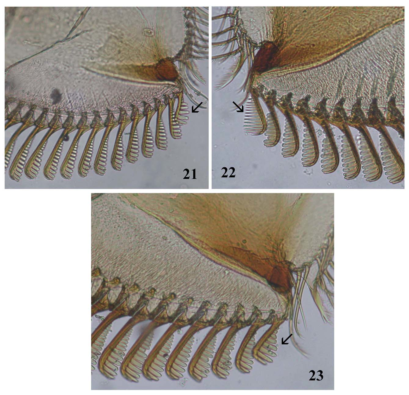
( Figs. 22 View FIGURES 21 – 23 , 27–49 View FIGURES 27 – 35 View FIGURES 36 – 46 View FIGURES 47 – 49 )
Sinitshenkova & Tshernova 1976: 16, figs. 10–17 ( Ameletus costalis ); Sinitshenkova 1977: 78, fig. 3, l–m ( Ameletus costalis ); Sinichenkova 1981: 77–78, fig. 3 ( Ameletus longulus ); Tshernova et al. 1986: 126, figs. 57:1–2 ( Ameletus costalis ); Tiunova & Potikha 2005 ( Ameletus costalis ); Kluge 2007:257 ( Ameletus costalis syn. nov.).
Material examined. Paratype larvae (No 2971a, collected together with the holotype), Russia, Primorskiy Kray, Terneiskii region, Sikhote-Alin State Nature Biosphere Reserve, Poltkov Stream, 17.VI.1974, Nadezdina.
Other material examined. Russia, Primorskiy Kray, Terneiskii region, Sikhote-Alin State Nature Biosphere Reserve: Mashinukovskiy Stream, 4–6.VI.1995, E Poticha, 4 male imagines; Zolotoi Stream, 21.VI.1995, E Poticha, 1 male subimago (reared), 2 female imagines; Serebrynka River, 11.VI.2002, E Poticha, 3 male imagines; Lazovskiy Nature Reserve, Proselochnaya Bay, Proselochniy Stream, 9.VI.2005, Y Sundukov, 1 male imago; Khabarovskiy Kray, Ulchskii region, Severnaya Bay, Tatarka River, about 200 m above mouth, 19.VI.2005, T Tiunova, 1 male imago (reared); Tatarka River, about 100 m above mouth, 22.VI.2007, I Tiunov, 1 male, 2 female imagines, 3 larvae; same place, 19.VI.2005, I Tiunov, 8 mature larvae and 2 larval exuvia; Bolshoi Somon River, 22.VI.2008, I Tiunov, 1 male imago.
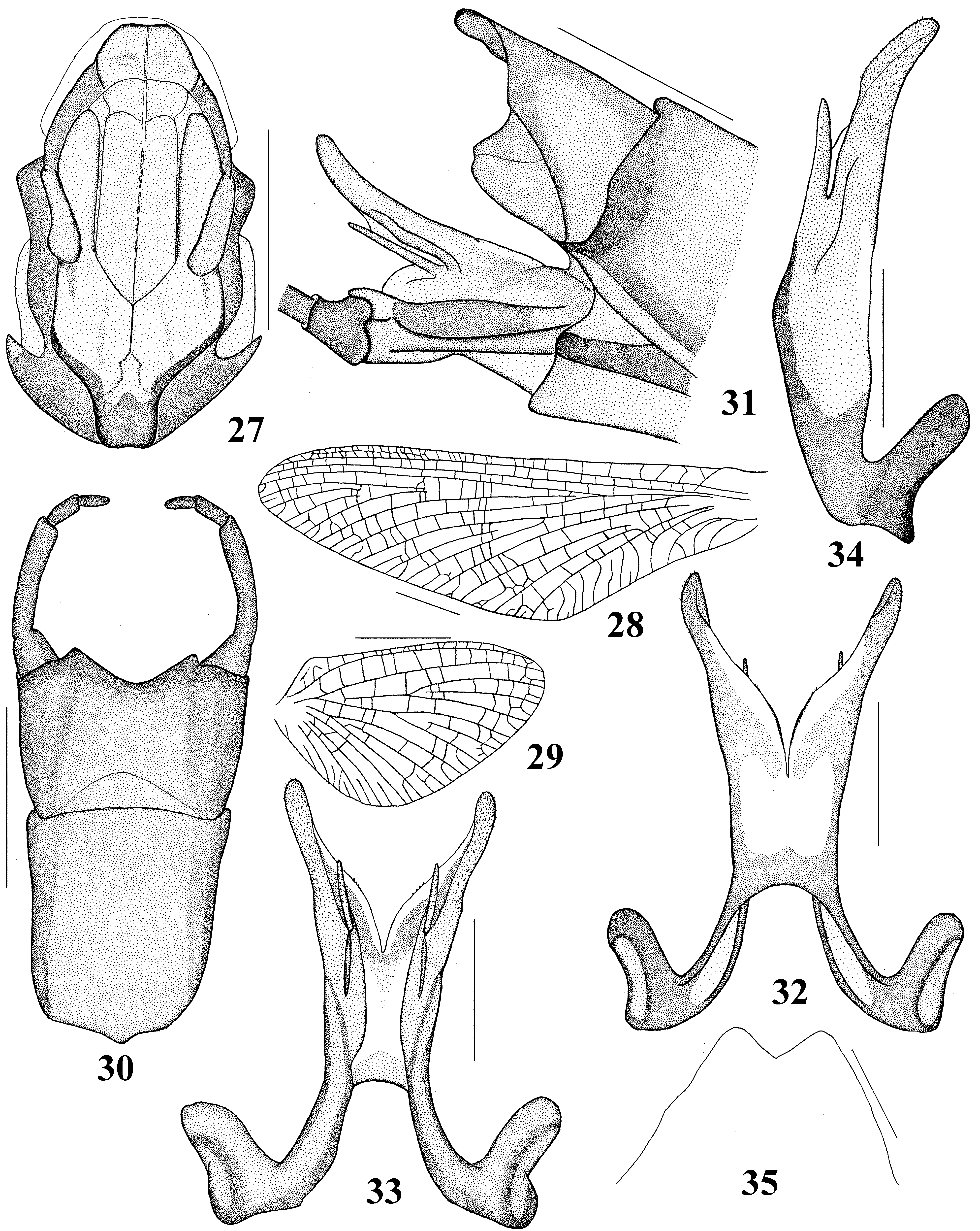
Description. Male imago (in alcohol). Length (mm): body 14.0–16.8; forewings 13.8–15.3; cerci 20.0–23.0. Head: upper portion of eyes gray; lower portion dark gray. Medial and lateral ocelli brown. Antennae brown. Thorax: Medioscutum and submedioscutum light brown; medial longitudinal suture narrow, brown. Sublateroscutum dark brown. Prelateroscutum yellowish. Medioparapsidal suture brown. Scutellum light brown; scuto-scutellar impression pale. Posterior scutal protuberance light brown or dirty yellowish; parascutellum dark ( Fig. 27 View FIGURES 27 – 35 ). Forelegs brown; middle and hind legs yellowish; tarsal segments brownish; dorsal surface of the fore tibia without spinules. Length (mm) of foreleg segments: femora 2.8–3.0; tibia 2.7–2.9; tarsal segments 0.7–1.1, 1.7–1.9, 1.5–1.6, 1.1–1.2, and 0.5–0.6. Wings brownish, hyaline; all veins brown ( Figs. 28–29 View FIGURES 27 – 35 ), crossveins of forewings between C and R veins brown, more contrasting than others. Abdomen: terga brownish, opaque; lateral and posterior margins brown; tergum I dark brown; terga II–IX in middle portion with pair of brown oblique stripes along the length; tergum IX–X darker. Sterna whitish or brownish; sternum I brown; sterna II–VII in the middle closer to the anterior margin with a dark triangular spot and two brownish oblique stripes; sternum IX dirty brown with darkish lateral margins. Styliger light brown, lateral sides darker; forceps dark brown ( Fig. 30 View FIGURES 27 – 35 ). Penis lobes light brown on inner sides and brown or dark brown on external sides; lateral penis lobes with rounded and divergent tips ( Figs. 32–33 View FIGURES 27 – 35 ); lateral penis lobes densely covered with small spines; each ventral plate bears a single long and pointed denticle ( Figs. 31, 34 View FIGURES 27 – 35 ); distance between base of the lateral penis lobe and denticle ¼ of maximum width of denticle ( Fig. 34 View FIGURES 27 – 35 ). Cerci brown with yellowish tips.
Female imago. Length (mm): body 16.4–20.7; forewings 16.6–18.0; cerci 24.0. Overall color light brown. Thorax: Mesonotum brown. Forelegs brown; middle and hind legs light brown, tarsal segments brown, joints darker. Color of wings as in male. Abdomen: tergum I dark brown; terga II–IX brown (abdomen with eggs). Color of sterna same as terga. Subgenital plate with deep depression ( Fig. 35 View FIGURES 27 – 35 ). Cerci dark brown with light brown or yellowish tips.
Male subimago. Overall color brown. Wings whitish and translucent. All crossveins of fore- and hind wings bordered by brown. Terga brown, monotonous; terga VII–X darker. Sterna brown. Forelegs brown; middle and hind legs lighter; joints dark brown. Cerci dark brown.
Mature larva. Length (mm): body 13.5–19.0; cerci 6.0–7.5. Head: dark brown. Antennae light brown, basal segments brown. Labrum (length 0.8–0.93 of width) with a deep and wide apical incision ( Fig. 36 View FIGURES 36 – 46 ). Incisor of left mandible with four denticles ( Fig. 37 View FIGURES 36 – 46 ). Incisor of right mandible with three denticles, the third somewhat larger than the first ( Fig. 38 View FIGURES 36 – 46 ). Labium dirty whitish; paraglossae length exceeds glossae length; distal margin of the glossae and part of the paraglossae brownish; third segments of labial palpi brownish ( Fig. 39 View FIGURES 36 – 46 ). First comb-shaped seta of maxilla with narrow and pointed denticles, their number more than ten ( Fig. 22 View FIGURES 21 – 23 ). Thorax: pronotum and mesonotum brown with pale median stripe. Pronotum with light oblique stripes on the sides; lateral margins darker. Legs brown; forelegs darker than middle and hind legs; distal part of femur of all legs light brown; a group of six or seven setae on the dorsal surface at the base of fore femora ( Fig. 40 View FIGURES 36 – 46 ). Foreleg: 2.1–2.3; tibia 1.2–1.3; and tarsus 1.4–1.5. Middle leg: femur 2.0–2.3; tibia 1.1–1.3; and tarsus 1.1–1.3. Hind leg: femur 2.1–2.5; tibia 1.1–1.3; and tarsus 1.1–1.4. Abdomen: tergum I dark brown; antero-lateral corners pale; terga II–IX with dark brown posterior margins and light brown lateral corners; maculation on terga II–IX not distinct in the form of paired brown and light stripes; tergum X brown; all terga with almost regular row of spines, which alternate between large and small. Sterna dark brown; sternum I pale; sterna II–IX with a pair of small white spots on antero-lateral corners; sterna VI–VIII with a well-defined ganglionic marking. Gills white or pale; gill I and II small, rounded in distal part; gill I with small anal rib, width 0.57 of length ( Fig. 41 View FIGURES 36 – 46 ); gill II with anal rib, width 0.60 of length ( Fig. 42 View FIGURES 36 – 46 ); gills III–VI with anal rib far from anal margin; gills III–V with rounded distal margin, width 0.58 of length ( Figs. 43–44 View FIGURES 36 – 46 ); gill VI with straight distal margin, width 0.55 of length ( Fig. 45 View FIGURES 36 – 46 ); gill VII with a truncated inner distal margin, width 0.46 of length ( Fig. 46 View FIGURES 36 – 46 ). The basal quarter of the cerci is dark brown, more distally, brown, tips light brown or yellowish; joints of each segment lighter.
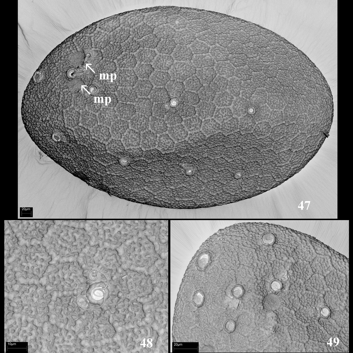
Eggs. General form of egg flattened ellipse with a length of 282–293 μm and a width of 176–187 μm ( Fig. 47 View FIGURES 47 – 49 ). Chorionic surface covered with large-mesh regular hexagonal cells; each cell with ribbed bottom; borders of cells narrow and very low; a few coiled threads, terminating in a small round knob, located in the middle of their borders, bordering higher hexagonal cells ( Fig. 48 View FIGURES 47 – 49 ); coiled threads terminated by a large round knob, located on one pole only ( Fig. 49 View FIGURES 47 – 49 ). Micropyles observed in the subequatorial area; sperm guide round ( Fig. 47 View FIGURES 47 – 49 ).
Distribution and biology. Russian Far East: Primorskiy and Khabarovskiy regions. Mature larvae and adults were collected in small foothill rivers.
Remarks. A. longulus was described based on young larvae, which, like all young larvae, have gills narrower than in mature larvae (Sinichenkova 1981). Kluge (2007) reviewed the genus Ameletus , and considered A. longulus as a junior synonym of A. costalis (Kluge 2007) . Recently, we collected adequate new material associating the male, female, eggs, and larvae. The identity of our material was confirmed by comparison with photos of the denticles in the mandible and maxilla the paratype (number of preparate 2971a) (kindly provided by N. Kluge). Herein, A. longulus is redescribed and new illustrations are presented, confirming that the larva illustrated by Sinichenkova (1981) is A. longulus .
Ameletus costalis (Matsumura 1931) ( Figs. 23 View FIGURES 21 – 23 , 50 View FIGURES 50 – 58 –71)
Matsumura 1931: 1474 ( Chimura costalis ); Imanishi 1932: 526–527, Pl 31 fig. 1, Pl 32 figs. 2–3 ( Ameletus sapporensis = Chimura costalis ); Imanishi 1933: 65–66, figs. 3,5 ( Ameletus costalis = Chimura costalis = Ameletus sapporensis ); Kluge 2007:257 ( Ameletus costalis = Ameletus longulus ).
Material examined. Japan, Nara Prefecture, Takami River, 23–25.III.1997, T Tiunova, 8 male, 4 female imagines (reared), 12 larval exuviae.
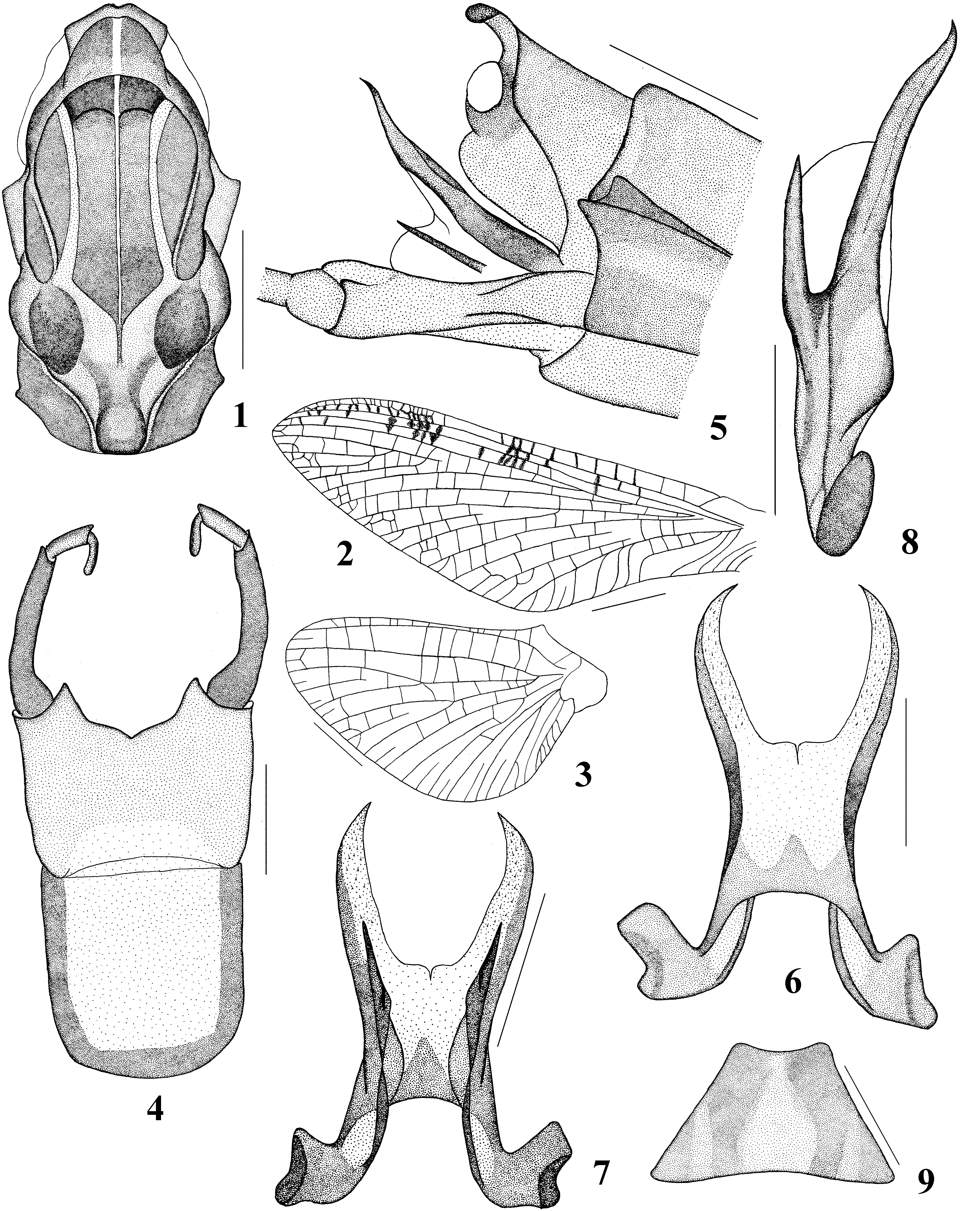
Description. Male imago (in alcohol). Length (mm): body 12.0–13.0; forewings 12.0–13.0; cerci 15.0–20.0. Head: upper portion of eyes gray; lower portion dark gray. Basal segments of antennae brown, tips pale. Thorax: Medioscutum, submedioscutum and anteronotal protuberance light brown; medial longitudinal suture narrow, brown. Anteronotal transverse impression brown. Sublateroscutum from dark brown. Prelateroscutum white with brown margins. Medioparapsidal suture brown. Scutellum light brown; scuto-scutellar impression brown with pair of whitish spots. Posterior scutal protuberance light brown; parascutellum brown ( Fig. 50 View FIGURES 50 – 58 ). Femur of forelegs brown, tibia light brown, dorsal surface with spinules ( Fig. 51 View FIGURES 50 – 58 ), first tarsal segments light brown, other brownish or yellowish; middle and hind legs yellowish, joint brown. Length (mm) of foreleg segments: femora 2.7–3.0; tibia 2.3–2.7; tarsal segments 0.5–0.7, 1.7–2.0, 1.5–1.8, 1.1–1.3, and 0.5–0.6. Wings hyaline; near the base brown; crossveins of forewings between C and R veins dark brown and bordered by brown; other veins from brown to brownish ( Fig. 52 View FIGURES 50 – 58 ). Hind wings brownish at the base, all veins yellowish or colorless. Abdomen: Styliger brownish, distal margin darker brownish. First segment of forceps extended distally, brown at the base; second and third segments pale yellowish ( Fig. 53 View FIGURES 50 – 58 ). Penis lobes pale on inner margin and brown on the external sides; lateral penis lobes rounded and bent medially ( Figs. 55–56 View FIGURES 50 – 58 ); each ventral plate bears a single long pointed denticle parallel to the lateral lobe; denticle tips curved inside ( Figs. 54 View FIGURES 50 – 58 ); the distance between bases of the lateral penis lobes and denticles 2.5 of the maximum width of the denticles ( Fig. 57 View FIGURES 50 – 58 ). Cerci brownish at the base and yellowish distally.
Female imago. Length (mm): body 16.5–18.5; forewings 16.5–17.4; cerci 20.0. Overall color light brown. Thorax: Mesonotum light brown. Forelegs brown; middle and hind legs light brown, tarsal segments darker. Color of forewings, as in the male; hindwings colorless. Abdomen: tergum I dark brown; terga II–IX light brown, posterior area darker (abdomen with eggs). Sterna brownish. Subgenital plate with almost smooth margin ( Fig. 58 View FIGURES 50 – 58 ). Cerci light brown, tips same but lighter.
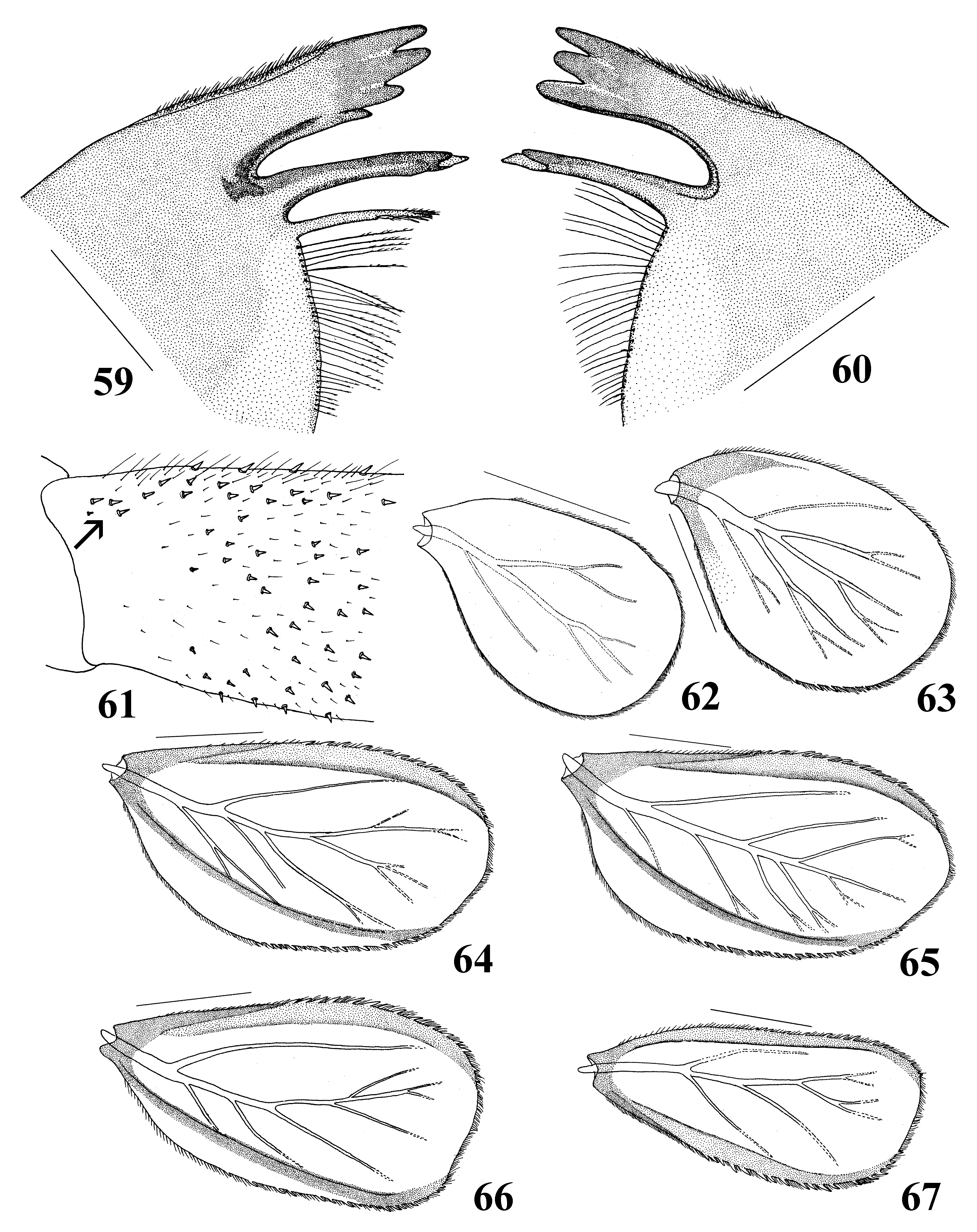
Mature larva. Length (mm): body 12.5–15.5; cerci 5.2–7.3. Head: Labrum (length 0.71–0.85 of width) with a shallow apical incision; first comb-shaped seta of maxilla with wide and rounded denticles, their number less than ten ( Fig. 23 View FIGURES 21 – 23 ). Incisor of the left mandible with four denticles; the first and second denticles of equal length, the fourth smallest ( Fig. 59 View FIGURES 59 – 67 ). Incisor of the right mandible with three denticles; the first length equal to the third, both almost equal to the second ( Fig. 60 View FIGURES 59 – 67 ). Thorax: Lengths (mm) of leg segments are as follows. Foreleg: 1.6–2.0; tibia 0.9–1.0; and tarsus 1.1–1.3. Middle leg: femur 1.6–2.1; tibia 0.9–1.3; and tarsus 1.1–1.2. Hind leg: femur 1.6–2.2; tibia 0.9–1.3; and tarsus 0.9–1.2. A dense group of four or five setae on the dorsal surface at the base of the fore femora ( Fig. 61 View FIGURES 59 – 67 ). Abdomen: All terga with irregular row of spines. Gill I and II small, rounded in distal part; gill I without anal ribs, width 0.65 of length ( Fig. 62 View FIGURES 59 – 67 ); gill II with small anal ribs, width 0.68 of length ( Fig. 63 View FIGURES 59 – 67 ); gill III–V with rounded distal margin, width 0.51–0.53 of length (64–65); gill VI with straight distal margin, width 0.53 of length ( Fig. 66 View FIGURES 59 – 67 ); gill VII with a truncated inner distal margin, width 0.48 of length ( Fig. 67 View FIGURES 59 – 67 ).
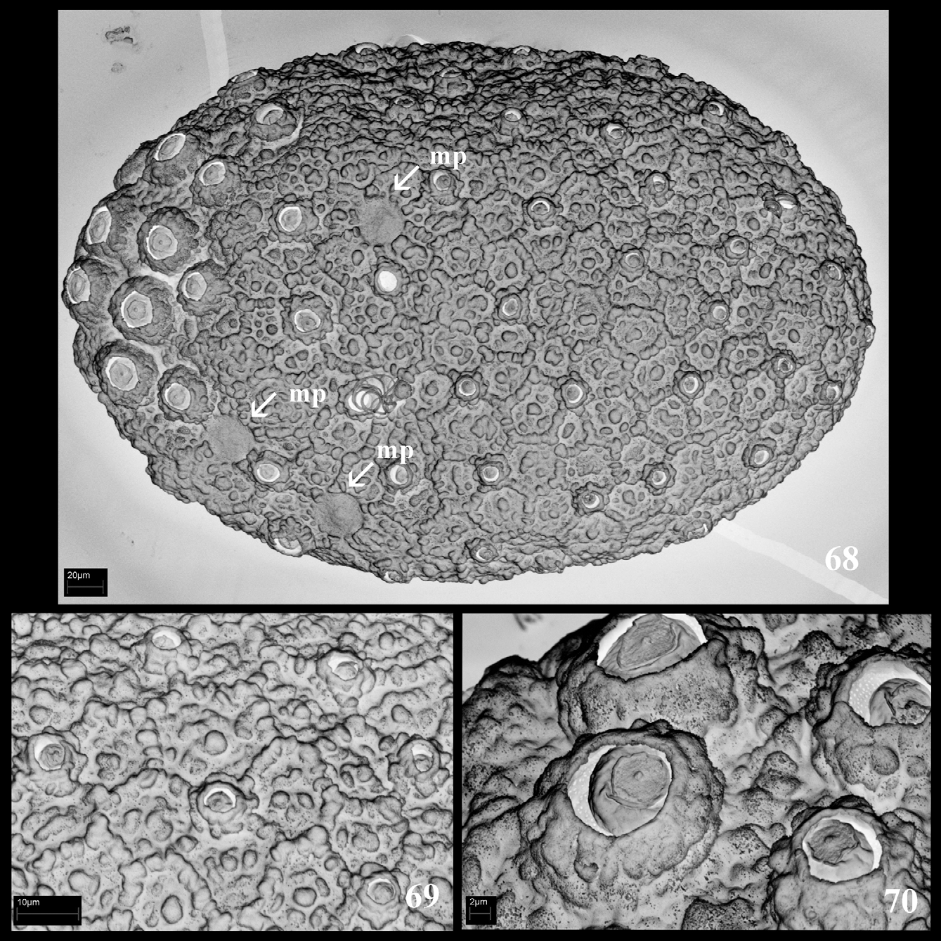
Eggs. General form of egg flattened ellipse with length 180–187 μm and a width 110–123 μm ( Fig. 68 View FIGURES 68 – 70 ). The chorionic surface covered by irregularly shaped coarse cells; each cell with a ribbed bottom and well-marked protuberances, one of which is the largest; borders of cells are in contact with tubercles, relatively high ( Fig. 69 View FIGURES 68 – 70 ); small round flower-like knob-terminated coiled threads, relatively rare and evenly distributed throughout the surface of the egg ( Fig. 69 View FIGURES 68 – 70 ); large and round knobs arranged on one pole only ( Figs. 68, 70 View FIGURES 68 – 70 ). Three micropiles visible in the subequatorial area; sperm guide round ( Fig. 68 View FIGURES 68 – 70 ); micropylar rim absent.
Distribution and biology. The larval exuviae and the subimago emerged from the larvae were collected in a foothill river in Japan at the end of March. The subimagos emerged on a warm sunny day. The mature larvae crawled, half-submerged to large stones protruding from the water and molted to subimago. According to Imanishi (1932), A. costalis is one of the earliest-flying Japanese species of the genus Ameletus .
Remarks. Until now it was thought that A. costalis inhabited streams of the Russian Far East and Japan. Study of a new material shows that the japan-continental species A. costalis represents a group of species, including the Japanese species A. costalis s. s. and two separate continental species. The new species herein described, A. khasanensis sp. nov. and the redescribed species A. longulus . Ameletus longulus was regarded to be a synonym of A. costalis , when A. costalis was accepted in wide sense, including the continental form (Sinitshenkova & Tshernova 1976; Tiunova & Potikha 2005; Tiunova 2006, 2007, 2009).
Discussion. The male imago of A. khasanensis sp. nov is similar to A. costalis by its forewings, in which the crossveins between the C and R veins are dark brown and are bordered by brown ( Figs. 2 View FIGURES 1 – 9 , 52 View FIGURES 50 – 58 ); however, the male imago can be distinguished from that of A. costalis by the absence of spinules on the dorsal surface of the fore tibia and the structure of the penis. The male imago of A. longulus can be distinguished from A. khasanensis sp. nov. and A. costalis by the color of forewings, which are brownish with all veins brown in A. longulus ( Figs. 28–29 View FIGURES 27 – 35 ). By the structure of the gills, A. khasanensis sp. nov., A. longulus , A. costalis , A. formosus Kang & Yang 1994, A. atratus Kang & Yang 1994 and A. montivagus Kang & Yang 1994 can be assigned to a group of species in which gills III–VI bear an anal rib far from the anal margin. The larvae of A. khasanensis sp. nov. differ from those of A. longulus , A. formosus and A. atratus in the structure of gills I. In A. khasanensis sp. nov., gill I has no vestigial anal ribs ( Fig. 15 View FIGURES 10 – 20 ), in contrast to A. formosus , A. atratus [ Fig.1, 3 View FIGURES 1 – 9 (Kang & Yang 1994)], and A. longulus ( Figs. 41–42 View FIGURES 36 – 46 ), in which gill I has vestigial anal ribs. The larvae of A. khasanensis sp. nov. differ from A. montivagus by the width/ length ratio of gill IV. In A. khasanensis sp. nov. gill IV width/length ratio is 0.54 ( Fig. 18 View FIGURES 10 – 20 ), in contrast to A. montivagus , in which this ratio is 0.55–0.58 [ Fig. 2 View FIGURES 1 – 9 (Kang & Yang 1994)]. The larvae of A. khasanensis sp. nov. differ from those of A. costalis in the length of the denticles on the right mandible ( Fig. 12 View FIGURES 10 – 20 ), in the absence of the group of setae on the dorsal surface at the base of the fore femora ( Fig. 14 View FIGURES 10 – 20 ) and the width/length ratio of gill VII ( Fig. 20 View FIGURES 10 – 20 ). Ameletus longulus is similar to A. formosus in the presence of vestigial anal ribs on gills I–II, but differ from this species based on the width/length ratio of gill IV. In A. longulus , gill IV width/length ratio is 0.55; in A. formosus it is 0.47– 0.49 (Kang & Yang 1994).


No known copyright restrictions apply. See Agosti, D., Egloff, W., 2009. Taxonomic information exchange and copyright: the Plazi approach. BMC Research Notes 2009, 2:53 for further explanation.