
Dolichodectes furcilobus, Mironov, Sergey, Literak, Ivan, Hung, Nguyen Manh & Capek, Miroslav, 2012
|
publication ID |
https://doi.org/10.5281/zenodo.282115 |
|
DOI |
https://doi.org/10.5281/zenodo.6170967 |
|
persistent identifier |
https://treatment.plazi.org/id/03CF4855-FF88-EE71-FF57-C2B5FA9AF998 |
|
treatment provided by |
Plazi |
|
scientific name |
Dolichodectes furcilobus |
| status |
sp. nov. |
Dolichodectes furcilobus sp. n.
( Figs. 16–18 View FIGURE 16 View FIGURE 17 View FIGURE 18 )
Type material. Male holotype ( ZISP 4777), 3 male and 3 female paratypes from Copsychus malabaricus (Scopoli) (Muscicapidae) , VIETNAM: Ninh Binh, Cuc Phuong National Park, 20°21' N 105°35' E, 1 February 2010, coll. I. Literak, Nguen Manh Hung and M. Capek.
Type depository. Holotype, 1 male and 1 female paratypes—ZISP, remaining paratypes UMMZ, IEBR.
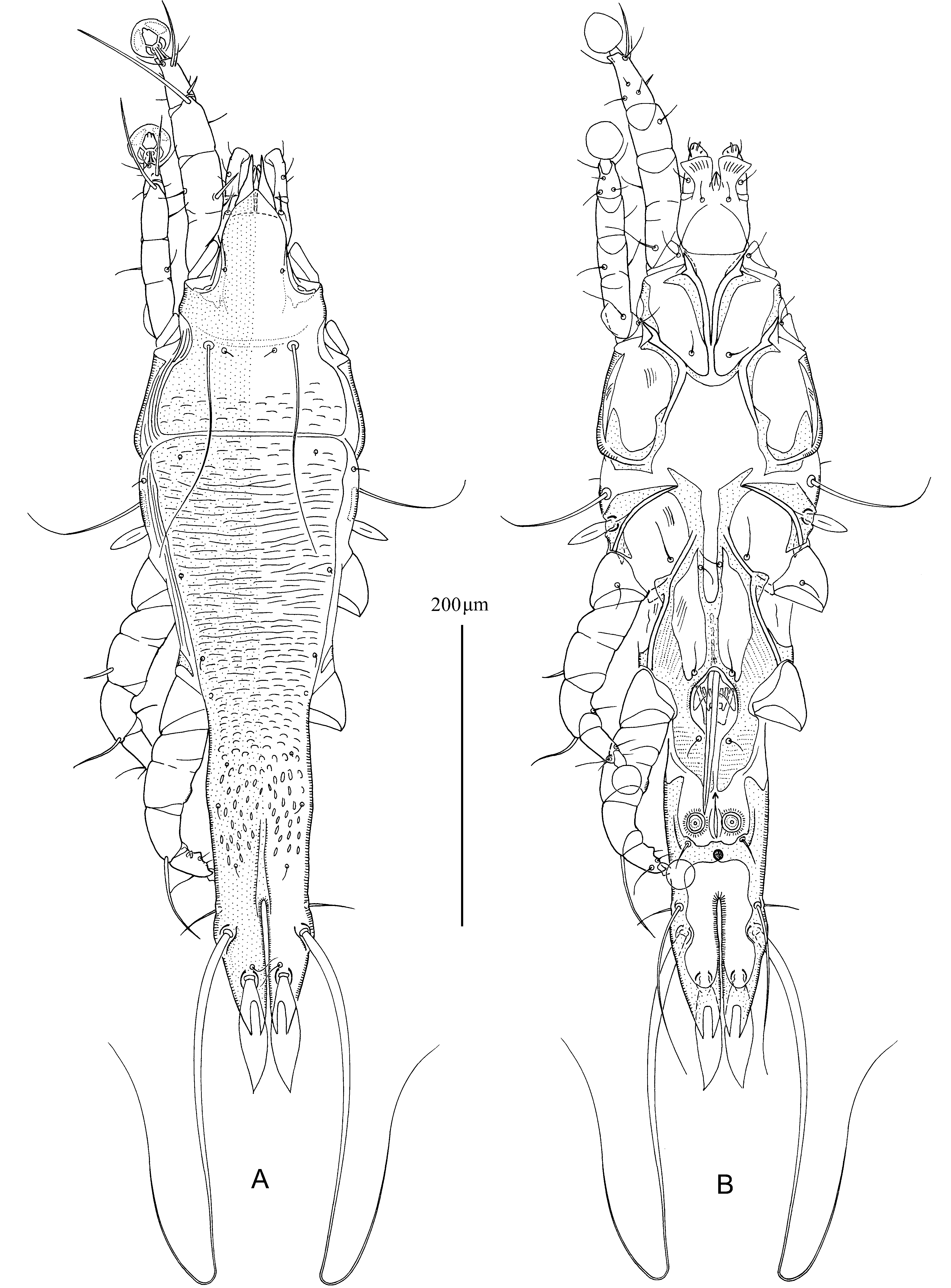
Description. MALE ( holotype, range for 3 paratypes). Length of idiosoma 456 (415–450), width 150 (145–155), length of hysterosoma 385 (360–380). Prodorsal shield: antero-lateral extensions widely connected to bases of epimerites Ia, lateral margins without incisions around scapular setae, posterior margins straight, length 155 (145–155), width at posterior margin 128 (120–128), posterior part with transverse dashes ( Fig. 16 View FIGURE 16 A). Setae ve represented by microsetae. Scapular setae se separated by 57 (55–58). Humeral shields present, poorly developed, separated from epimerites III, not encompassing setae cp. Setae c2 situated on soft tegument near anterior end of humeral shields. Setae c3 lanceolate, 29 (28–30) × 9 (8–9). Hysteronotal shield: greatest length 400 (375–395), width at anterior margin 120 (125–130), anterior margin straight, anterior angles rounded, anterior half of shield from margin to level of trochanters IV with transverse striae and dashes, area from level of trochanters IV to bases of opisthosomal lobes with small ovate lacunae. Opisthosomal lobes 3 times longer than wide, lateral margins at level of setae h2 noticeably convex, posterior end of each lobe with pair of acute and long extensions, length of apical extensions 22 (17–22) ( Fig. 18 View FIGURE 18 A). Setae h3 approximately equidistant from lobar apices and bases of setae h2. Terminal cleft a narrow parallel-sided slit, lateral margins almost touching, length 93 (84–94), width at level of setae h2 2–3. Supranal concavity a long and narrow median groove stretching from anterior end of terminal cleft to level of setae e2. Setae f2 and ps2 situated at same transverse level. Setae h1 slightly closer to level of setae f2 than to setae e2. Setae h3 lanceolate, with acute tips, length 75 (70–75), greatest width 17 (15–17); setae ps2 90 (80–85) long, extending to lobar apices; setae ps1 filiform, about 20 long, situated slightly anterior to bases of setae h3. Distance between bases of dorsal setae: c2:d2 128 (120–130), d2:e2 102 (90–102), e2:h2 90 (78–85), h2:h3 31 (30–32), d1:d2 55 (52–55), e1: e2 33 (26–33), h1:ps2 28 (26–28), ps1:h3 9 (8–10), h2:h2 51 (48–52), h3:h3 24 (22–24), ps2:ps2 55 (52–55).
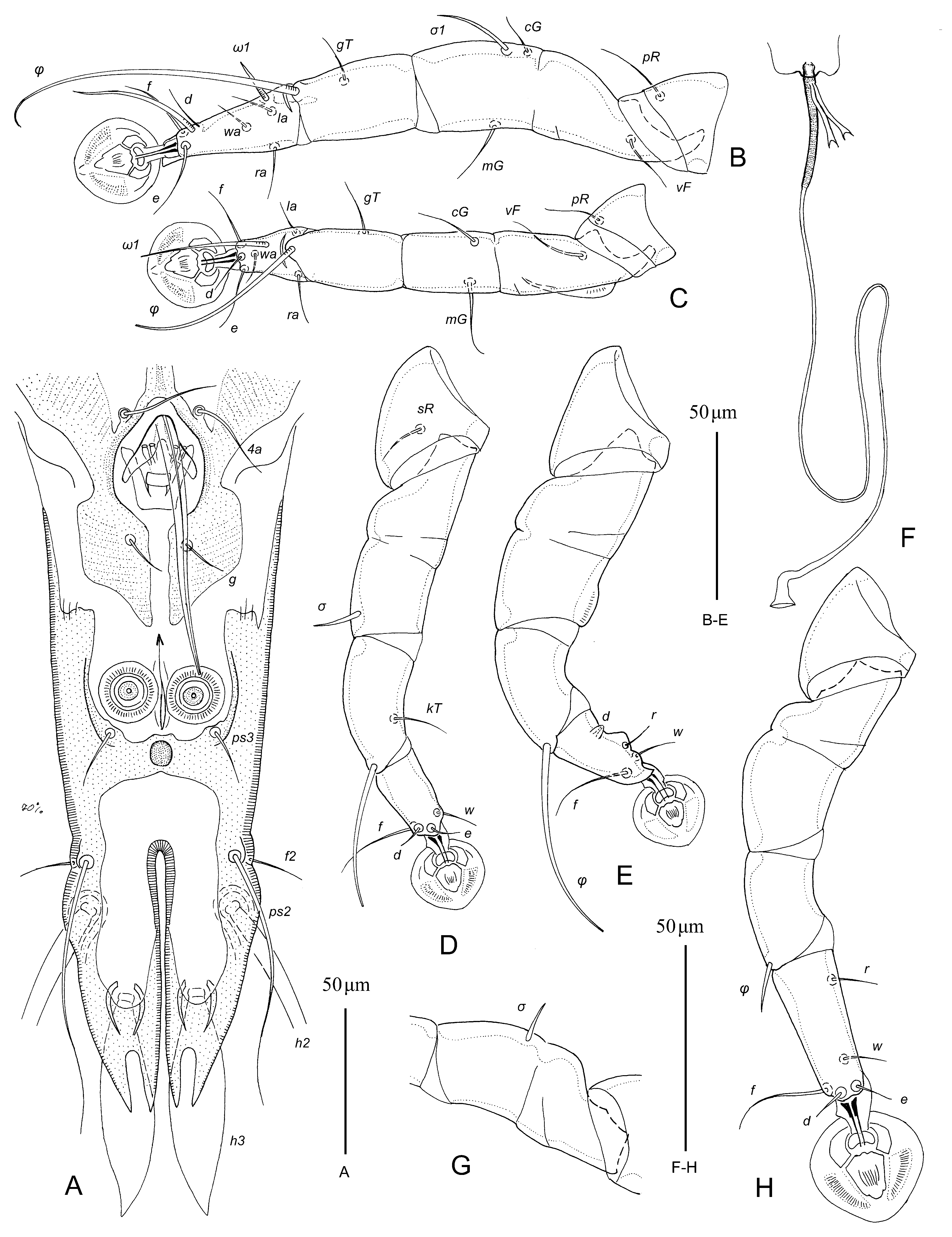
Epimerites I fused into a Y, sternum about 1/3rd of total length of epimerites, posterior end of sternum with transverse extensions connected with medial part of epimerites II ( Fig. 16 View FIGURE 16 B). Coxal fields I, II without large sclerotized areas. Coxal fields I–IV closed. Rudimentary sclerites rEpIIa absent. Coxal fields IV with large sclerotized areas at bases of trochanters IV. Genital arch of moderate size, 22 (20–22) long, 31 (27–32) wide; basal sclerite of genital apparatus large, shaped as inverted trapezium. Aedeagus 95 (90–95) long, extending to anterior margins of anal suckers. Genital papillae well distinct, situated at midlevel of genital arch, arranged in transverse row. Paragenital apodemes fused to each other by their medial parts, anterior branches of these apodemes fused with inner margins of epimerites IIIa and posterior branches fused with epimerites IV. Genital shields, epimerites IVa, posterior branches of paragenital apodemes and shield-like areas of coxal fields IV fused altogether to form almost complete sclerotized oval surrounding genital apparatus ( Fig. 18 View FIGURE 18 A). Genital shields elongate and not fused each other at midline of body. Setae 4b on anterior branches of paragenital apodemes, setae 4a on posterior branches of paragenital apodemes, setae g on inner margin of genital shields. Opisthoventral shields wide, fused together by wide transverse bridge immediately posterior to anal opening; anal field flanked from posterior and lateral sides by opisthoventral shields and transverse bridge. Anal suckers 15 (13–15) in diameter, corolla without indentations. Setae ps3 situated on anterior margin of transverse band connecting opisthoventral shields. Distance between ventral setae: 3a:4b 5 (5–7), 4b–4a 75 (66–75), 4a–g 44 (42–44), g–ps3 66 (64–66), ps3–ps3 37 (36–38), ps3:h3 93 (88–92).
Legs I longer and thicker than legs II, femora II with narrow ventral crests, tarsus I with small apicoventral claw–like extension, other segments of these legs without processes. Solenidion σ 1 of genu I 20 (18–20) long, situated in proximal part of segment; genual setae cG I, II, mG I, II filiform. Genu IV with narrow heavily sclerotized ventral crest. Solenidion ω 1 of tarsus II elongate, extending to distal margin of ambulacral disc; seta d of tarsus II half as long as corresponding seta f. Seta d of tarsus III much shorter than corresponding setae f. Tarsus IV 33 (26–33) long, with small apical claw-like process and blunt-angular ventral extension; seta d hemispherical, with thick walls, situated in proximal part of this segment; seta e indistinct. Solenidion φ of tibia IV extending to midlevel of ambulacral disc. Length of solenidia: ω 1 I 12 (11–12), ω 1 II 40 (30–38), φI 90 (84–90), φII 55 (48–54), φIII 35 (32–35), φIV 54 (46–52).
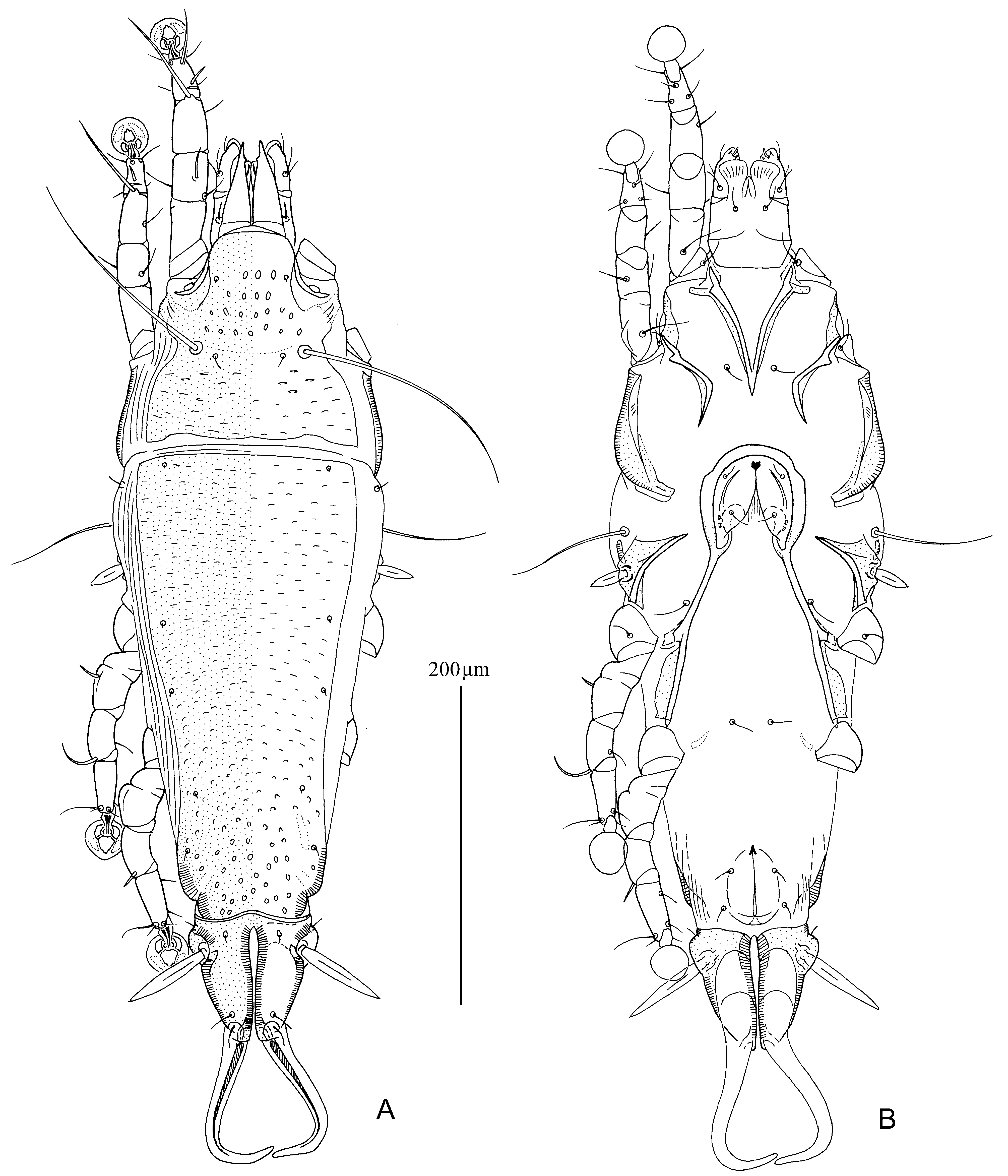
FEMALE (3 paratypes). Length of idiosoma 485–515, width 164–175, length of hysterosoma 342–360.
Prodorsal shield: antero–lateral extensions wide and connected to epimerites Ia, posterior margin sinuous, 128–134 long, 130–134 wide, anterior part with small ovate lacunae, posterior part with transverse dashes ( Fig. 17 View FIGURE 17 A). Setae ve represented by microsetae. Setae se separated by 66–70. Humeral shields represented by small longitudinal sclerite situated posterior to bases of setae cp. Setae c2 situated laterally on soft tegument. Setae c3 lanceolate, 22–24 × 7–8. Anterior and lobar parts of hysteronotal shields completely separated from each other by narrow transverse band of soft tegument. Hysteronotal shield noticeably enlarged in anterior part, anterior margin straight, posterior margin slightly concave, length 285–290, width at anterior margin 132–144; anterior two thirds of this shield with dash-like transverse striae, posterior one third with small ovate lacunae. Length of lobar region 75–78, width 76–82, anterior margin convex. Terminal cleft parallel-sided, narrow, with margins almost touching, 66–72 long, 4–5 wide in anterior part. Supranal concavity absent. Setae f2 present. Setae h1 situated on lobar shield near its anterior margin. Setae h2 spindle-like, 54–56 long, 7–9 wide. Setae ps1 approximately equidistant from inner and outer margins of opisthosomal lobes and close to lobar apices. Setae h3 filiform, 9–12 long, about 1/8th of terminal appendages. Distance between dorsal setae: c2:d2 130–132, d2: e2 92–100, e2:h2 62–71, h2:h3 48–51, d1:d2 40–44, e1: e2 35–38, h1:h 2 11–13, h2:ps1 37–44, h1:h1 32–33, h2:h2 57–62.
Epimerites I fused into a Y, sternum about 1/5th of total length of epimerites ( Fig. 17 View FIGURE 17 B). Lateral parts of coxal fields I, II without wide sclerotized areas. Epimerites IVa scarcely distinct. Translobar apodemes of opisthosomal lobes wide, not fused to each other anterior to terminal cleft. Copulatory opening ventral, situated immediately posterior to anal opening. Primary spermaduct with punctate enlargement in most proximal part, secondary spermaducts 10–11 long ( Fig. 18 View FIGURE 18 F). Distance between pseudanal setae: ps2:ps2 36–38, ps3:ps 3 22–25, ps2:ps 3 20–22.
Legs I slightly longer that legs II, femur II with narrow ventral crest, other segments of these legs without processes. Solenidion σ 1 of genu I 15–16 long, situated at midlevel of segment. Genual setae cG I, cG II, mG I, mG II filiform. Genu IV with small dorsal inflation. Setae d of tarsi II–IV much shorter than corresponding setae f. Solenidion φIV about 1/4th of corresponding tarsus ( Figs. 18 View FIGURE 18 G, H). Length of solenidia: ω 1 I 9–11, ω 1 II 10–11, φI 62–73, φII 54–56, φIII 25–27, φIV 12–15.
Differential diagnosis. Among previously known Dolichodectes species, the new species is most similar to D. myrmecocichlae Mironov and Fain, 2003 described from Myrmecocichla nigra (Vieillot) (Muscicapidae) by having the following features in males: the posterior part of hysteronotal shield is ornamented with small ovate lacunae, the genital shields is separated into two pieces, and the transverse bridge flanking the anal field is straight and not touching the anterior margin of terminal cleft. Dolichodectes furcilobus sp. n. clearly differs from that species by the following features: in males, the posterior ends of opisthosomal lobes have a pair of long spine–like extensions, sternum formed by epimerites I is connected to epimerites II by transverse bands, the median part of paragenital apodemes is entire, and solenidion ω 1 of tarsus II is strongly elongated and extends to the distal part of ambulacral disc; in females, the lobar shield is completely separated from the anterior part of hysteronotal shield, and the proximal part of the primary spermaduct is uniformly enlarged. In males of D. mirmecocichlae , the posterior ends of opisthosomal lobes is simply acute, the posterior end of sternum has short lateral extensions not reaching corresponding epimerites II, the median part of paragenital apodemes is split into several separate fragments, and solenidion ω 1 of tarsus II extends to distal end of the segment; in females, the lobar shield is not separated from the anterior part of hysteronotal shield, and the primary spermaduct has a short ampuliform enlargement separated from the head of spermatheca by 15–20 micrometers.
Etymology. The specific epithet refers to the bifurcate posterior ends of the opisthosomal lobes in males.
No known copyright restrictions apply. See Agosti, D., Egloff, W., 2009. Taxonomic information exchange and copyright: the Plazi approach. BMC Research Notes 2009, 2:53 for further explanation.
|
Kingdom |
|
|
Phylum |
|
|
Class |
|
|
Order |
|
|
Family |
|
|
Genus |