
Proterothrix alcippeae, Mironov, Sergey, Literak, Ivan, Hung, Nguyen Manh & Capek, Miroslav, 2012
|
publication ID |
https://doi.org/10.5281/zenodo.282115 |
|
DOI |
https://doi.org/10.5281/zenodo.6170971 |
|
persistent identifier |
https://treatment.plazi.org/id/03CF4855-FF8D-EE4C-FF57-C298FA71F835 |
|
treatment provided by |
Plazi |
|
scientific name |
Proterothrix alcippeae |
| status |
sp. nov. |
Proterothrix alcippeae sp. n.
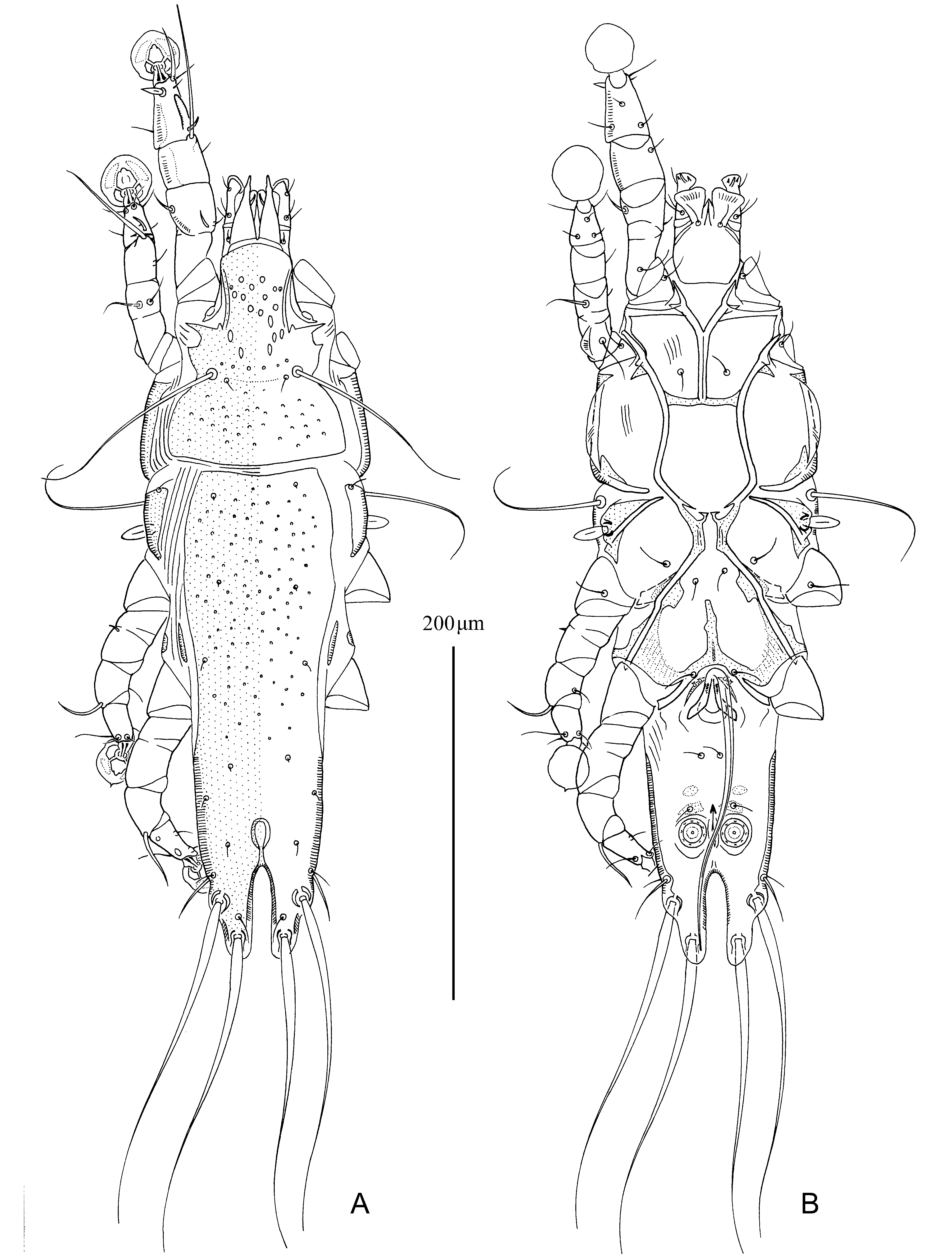
( Figs. 19–21 View FIGURE 19 View FIGURE 20 View FIGURE 21 )
Type material. Male holotype ( ZISP 4780), 6 male and 10 female paratypes from Alcippe rufogularis (Mandelli) (Pellorneidae) , VIETNAM: Ninh Binh, Cuc Phuong National Park, 20°21' N 105°35' E, 2 February 2010, coll. I. Literak, Nguen Manh Hung and M. Capek.
Type depository. Holotype, 4 male and 6 female paratypes—ZISP, remaining paratypes—UMMZ, IEBR.
Description. MALE ( holotype, range for 6 paratypes in parentheses). Length of idiosoma 400 (390–406), width 124 (120–130), length of hysterosoma 265 (260–270). Prodorsal shield: entire, antero-lateral extensions acute, lateral margin shallowly concave, posterior margin slightly convex, length 126 (122–130), width 90 (88–94), surface with circular and ovate lacunae in anterior part and with minute pit-like lacunae in posterior part ( Fig. 19 View FIGURE 19 A). Setae ve rudimentary, represented by alveoli. Scapular setae se separated by 51 (46–54). Scapular shields narrow. Humeral shields present, well developed, separated from epimerites III. Setae cp situated off humeral shields. Setae c2 situated on anterior end of humeral shield. Subhumeral setae c3 lanceolate, 15 (14–15) × 7 (6–7). Hysteronotal shield: length 272 (265–275), width in anterior margin 88 (82–90), anterior margin slightly concave, surface of anterior half with numerous mall pit-like lacunae. Lateral hysteronotal sclerites present, situated slightly anterior to level of trochanters IV.
Opisthosomal lobes elongated, nearly twice as long as wide at base, distal half about half as wide as basal part, posterior margin of lobar apices rounded and poorly sclerotized; setae h3 situated near lobar apices ( Fig. 21 View FIGURE 21 A). Terminal cleft narrowly U-shaped, 53 (48–55) in length, 11 (9–12) in width at midlevel; margins of terminal cleft without narrow membranes. Supranal concavity ovate, with clearly outlined margin. Setae f2 and ps2 situated at same transverse level. Setae h1 at level of supranal concavity. Setae ps1 at midlevel between setae h2 and h3, closer to inner margin of opisthosomal lobe than to outer margin. Setae h3 and h2 represented by macrosetae of subequal length, 150 (150–170) and 160 (145–170), respectively; setae ps2 setiform, 20 (20–25) long; setae ps1 filiform 20 (18–22) long. Distance between dorsal setae: c2:d2 100 (95–104), d2: e2 75 (70–78), e2:h3 81 (78–85), h2:h3 22 (20–25), d1:d2 46 (42–50), e1: e2 22 (12–22), h1:ps2 17 (13–18), ps1:h3 13 (10–14), h2:h2 48 (44–50), h3:h3 26 (25–27), ps2:ps2 57 (53–59).
Epimerites I fused into a long Y, posterior end of sternum connected with middle parts of epimerites II by transverse sclerotized bands. Epimerites II very long, extending to level of sejugal furrow and fused with anterior ends of elongated epimerites IIIa forming a pentagonal frame in median part of ventral propodosoma; frame unclosed in the very posterior end ( Fig. 19 View FIGURE 19 B). Inner tips of epimerites IIa and epimerites III almost touching posterior ends of epimerites II. Rudimentary sclerites rEpIIa absent. Coxal fields I closed, coxal fields II, III nearly closed, with narrow gaps between tips of corresponding epimerites. Coxal fields IV with triangle-shaped sclerotized area at bases of trochanters IV. Epimerites IVa present, well developed, their anterior tips fused with corresponding posterior ends of pregenital sclerites and sclerotized areas of coxal fields IV. Setae 4a situated on anterior end of epimerites IV. Pregenital sclerite shaped as an inverted Y, its anterior tip almost extending to level of trochanters III. Genital arch of moderate size, 27 (22–30) in length (from its base to bending of aedeagus backward), 26 (24–26) in width at base; basal sclerite of genital apparatus large and short; aedeagus 162 (162–165) long, almost extending to level of lobar apices ( Fig. 21 View FIGURE 21 A). Genital papillae situated on small narrow sclerites lateral to genital arch. Genital shields represented by two pairs of small roughly ovate sclerites situated anterior to anal field. Anal suckers 17 (15–17) in diameter, corolla with 9–10 indentations, surrounding membrane without striae. Opisthoventral shields not developed. Setae ps3 situated on posterior pair of adanal sclerites, distance between them subequal to distance between centers of anal suckers. Distance between ventral setae: 3a:4b 9 (8–10), 4b–4a 51 (50–55), 4a–g 46 (46–50), g–ps3 29 (28–31), ps3–ps3 24 (20–25), ps3:h3 73 (70–75).
Legs I longer and noticeably thicker than legs II; femur II with wide ventral crest; genu, tibia and tarsus I with lateral longitudinal heavily sclerotized crests, tarsus I with thumb-like dorsal process anterior to base of solenidion ω 1 ( Figs. 21 View FIGURE 21 B, C); other segments of legs I, II without processes. Solenidion σ 1 of genu I small spiculiform, 9 (7–9) long, situated at very base of segment; setae cG I, cG II filiform, setae mG I mG II strongly thickened but with filiform apex. Seta e of tarsus I narrowly lanceolate, 12 (11–12) long. Seta d of tarsus II slightly longer than corresponding seta f; seta d of tarsus III approximately half the length of corresponding seta f. Legs III, IV similar in size. Solenidion σ of genu III situated at base of segment ( Fig. 21 View FIGURE 21 D). Solenidion φ of tibia IV slightly extending beyond tarsal apex. Tarsus IV 24 (22–24) long, with a small apico-ventral extension bearing seta w; setae d, e button-like, situated in basal and apical parts of segment, respectively, seta d in diameter twice as large as seta e (Figs, 21E). Length of solenidia: ω 1 I 7 (6–7), ω 1 II 6 (5–6), φI 75 (73–78), φII 55 (49–55), φIII 26 (24–26), φIV 24 (22–26).
FEMALE (10 paratypes). Length of idiosoma 460–485, width 140–155, length of hysterosoma 323–355. Prodorsal shield: entire, antero-lateral extensions acute, lateral margins without incisions, posterior angles not expressed, posterior margin convex, length 128–135, width 100–108, surface with numerous small circular lacunae, lacunae in anterior part noticeably larger than in posterior one ( Fig. 20 View FIGURE 20 A). Setae ve rudimentary, represented by alveoly. Setae se separated by 59–69. Scapular shields narrow. Humeral shields narrow, separated from epimerites III; setae cp situated on soft tegument near ventral margin of humeral shields. Setae c2 situated on anterior ends of humeral shields. Setae c3 lanceolate, 17–18 x 7–8. Anterior and lobar pieces of hysteronotal shield separated dorsally by narrow transverse band but remain connected ventro-laterally by narrow bands. Anterior hysteronotal shield roughly rectangular, anterior margin slightly concave, greatest length 235–255, width at anterior margin 104–115, surface with numerous very small circular lacunae as on posterior part of prodorsal shield. Length of lobar region 100–110, width 72–80, anterior marging concave. Terminal cleft parallel-sided, narrow; length 62–66, width at midlevel 2–5. Supranal concavity well developed, circular. Setae h1 on lobar shield, slightly anterior to supranal concavity. Setae h2 spindle-like, with short terminal filaments, length including filaments 60–68, width 7–8. Setae ps1 near inner margins of opisthosomal lobes. Setae h 3 10–15 long, about 1/8th–1/10 th the length of terminal appendages. Distance between dorsal setae: c2:d2 135–150, d2: e2 77–82, e2:h2 62–64, h2:h3 47–52, d1:d2 84–93, e1: e2 40–44, h1:h2 33–42, h2:ps 1 15–17, h1:h1 36–42, h2:h2 60–62.
Epimerites I fused as a short-stemmed Y, sternum shorter than 1/5th of total length of epimerites, with very short lateral extensions. Lateral parts of coxal fields I, II without heavily sclerotized areas ( Fig. 20 View FIGURE 20 B). Epimerites IVa present, poorly sclerotized. Translobar apodemes of opisthosomal lobes present, wide, fused to each other anterior to terminal cleft. Epigynum horseshoe-shaped, greatest width 58–64. Copulatory opening situated ventrally at anterior margin of fused translobar apodemes. Distal half of primary spermaduct enlarged, approximately 3 times wider than proximal half; secondary spermaducts 5–6 long ( Fig. 21 View FIGURE 21 F). Distance between pseudanal setae: ps2:ps2 44–51, ps3:ps 3 17–20, ps2:ps 3 20–22; setae ps2 situated at level of posterior end of anal opening.
Legs I slightly longer and thicker than legs II; femur II with wide ventral crest; other segments of legs I, II without processes. Solenidion σ 1 of genu I, 8–9 long, situated slightly close to distal margin of this segment. Genual setae cG I, cG II filiform, mG I spiculiform, mG II spiculiform with filiform terminal part. Seta e of tarsus I strongly enlarged in basal part, apical part filiform ( Fig. 21 View FIGURE 21 G). Setae d, e, f, of tarsus II subequal in length, setae d of tarsi III, IV about 3 times shorted than respective setae f. Genu IV slightly inflated dorsally, with narrow longitudinal crest, genu III not modified ( Figs. 21 View FIGURE 21 H, I). Solenidion φ of tibia IV much shorter than that on tibia III. Length of solenidia: ω 1 I 11–14, ω 1 II 7–8, φI 70–77, φII 47–53, φIII 21–24, φIV 6–7.
Differential diagnosis. Within the wolffi species group, Proterothrix alcippeae sp. n. obviously belongs to the paradoxornis species complex ( Mironov & Proctor 2009) by having setae e of tarsi I lanceolate in males. Among three previously known species of this complex, the new species is most similar to P. sarabushae Mironov and Proctor, 2009 described from Paradoxornis verreauxi (Sharpe) (Sylviidae) (in other classifications, the genus Paradoxornis Gould is included in Timaliidae or constitute a separate family Paradoxornithidae ; see Robson, 2007) by the following features in males: tarsus I has a thumb-like dorsal extension ( Fig. 21 View FIGURE 21 B), a pregenital sclerite is present and fused with the inner tips of epimerites IV, lateral hysteronotal sclerites are present. Proterothrix alcippeae differs from P. sarabushae by these features: in both sexes, setae mG of genua I, II are distinctly thickened but have filiform apex; in males, the pentagonal sclerotized frame in the postero–median part of propodosoma remains open posteriorly, the inner tips of epimerites IIa are separated from epimerites II, the posterior part of hysteronotal shield is without lacunae; in females, spindle–shaped setae h2 have short filiform apices, the posterior margin of anterior hysteronotal shield lack incisions. In both sexes of P. sarabushae , setae mG of genua I, II are lanceolate; in males, the pentagonal sclerotized structure is completely closed, the inner tips of epimerites IIa are fused with the posterior ends of epimerites II, almost all surface of the hysteronotal shield with exception of opisthosomal lobes is covered with small circular lacunae; in females, setae h2 are simple spindle–like, the posterior margin of anterior hysteronotal shield has a pair of deep lateral incisions extending beyond bases of setae e2.
Etymology. The specific epithet derives from the generic name of the host and is a noun in the genitive case.


| ZISP |
Zoological Institute, Russian Academy of Sciences |
No known copyright restrictions apply. See Agosti, D., Egloff, W., 2009. Taxonomic information exchange and copyright: the Plazi approach. BMC Research Notes 2009, 2:53 for further explanation.
|
Kingdom |
|
|
Phylum |
|
|
Class |
|
|
Order |
|
|
Family |
|
|
Genus |