
Montsauria pellornei, Mironov, Sergey, Literak, Ivan, Hung, Nguyen Manh & Capek, Miroslav, 2012
|
publication ID |
https://doi.org/10.5281/zenodo.282115 |
|
DOI |
https://doi.org/10.5281/zenodo.6170959 |
|
persistent identifier |
https://treatment.plazi.org/id/03CF4855-FF9F-EE7E-FF57-C3B8FA6BFE58 |
|
treatment provided by |
Plazi |
|
scientific name |
Montsauria pellornei |
| status |
sp. nov. |
Montsauria pellornei sp. n.
( Figs. 7–9 View FIGURE 7 View FIGURE 8 View FIGURE 9 )
Type material. Male holotype ( ZISP 4743), 9 male and 10 female paratypes from Pellorneum ruficeps Swainson (Pellorneidae) , VIETNAM: Ninh Binh, Cuc Phuong National Park, 20°21' N 105°35' E, 1 February 2010, coll. I. Literak, Nguen Manh Hung and M. Capek.
Type depository. Holotype, 7 male and 8 female paratypes—ZISP, remaining paratypes—UMMZ, IEBR.
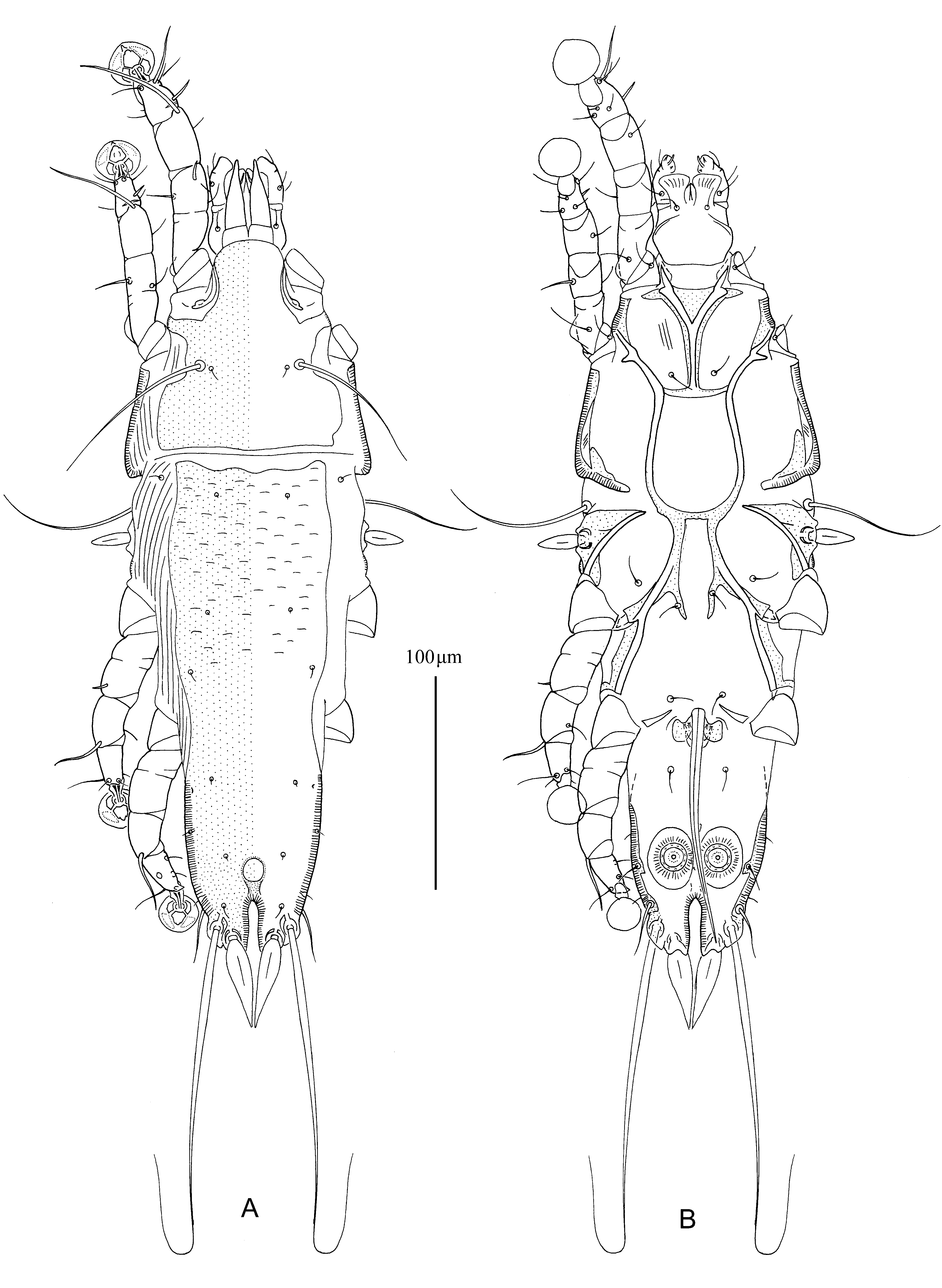
Description. MALE ( holotype, range for 9 paratypes). Length of idiosoma 340 (335–355), width 115 (113–123), length of hysterosoma 218 (210–235). Prodorsal shield: entire, antero-lateral extensions narrow, connected with bases of epimerites Ia, lateral margins with shallow incisions not extending to bases of setae se, posterior margin almost straight, length of shield 82 (90–98), width 88 (86–03), surface without ornamentation ( Fig. 7 View FIGURE 7 A). Setae ve absent. Scapular setae se separated by 48 (48–52). Scapular shields narrow. Humeral shields absent. Setae cp and c2 situated on soft tegument. Subhumeral setae c3 lanceolate, 20 (19–21) × 7.5 (7–8). Hysteronotal shield: length 223 (220–245), width at anterior margin 75 (73–78), anterior margin slightly sinuous, anterior half with sparcely disposed dash-like transverse striae.
Opisthosomal lobes short, slightly longer than wide, straight, posterolateral margin rounded; lobar apex with pair of blunt teeth at bases of setae h3 ( Fig. 9 View FIGURE 9 A). Terminal cleft narrow, almost parallel-sided, length 24 (22–26), greatest width 6 (5–7). Supranal concavity circular, well outlined, 13 (12–13) in diameter. Setae f2 anterior to bases of setae ps2. Setae h1 situated at level of anterior margin of supranal concavity. Setae ps1 situated at level of opisthosomal lobe bases, equidistant from outer and inner margins of these lobes. Setae h3 lanceolate, 45 (42–46) long, 10 (10–11) wide; setae ps2 28 (26–30) long. Distance between dorsal setae: c2:d2 88 (88–94), d2: e2 79 (78–84), e2:h3 53 (52–62), d1:d2 28 (28– 33), e1: e2 20 (20–24), h1:ps2 27 (25–28), ps1:h3 16 (16–18) h2:h2 37 (36–40), h3:h3 20 (20–22), ps2:ps2 44 (44–47).
Epimerites I fused into a Y, sternum half as long as total length of epimerites, posterior end of sternum connected to middle parts of epimerites II by transverse branches. Epimerites II strongly elongate, extending to level of sejugal furrow; posterior ends of epimerites II fused with anterior ends of epimerites IIIa, which are in turn connected to each other by transverse sclerite. Posterior end of sternum, posterior parts of epimerites II, and anterior ends of epimerites IIIa form large completely closed frame in median part of ventral propodosoma ( Fig. 7 View FIGURE 7 B). Medial part of epimerites IIIa with extensions directed backward and bearing bases of setae 4b. Rudimentary sclerites rEpIIa absent. Coxal fields I closed, coxal fields II, III almost closed. Coxal fields IV without sclerotized area at bases of trochanters IV. Epimerites IVa present, short. Genital arch small, with short lateral extensions, 17 (15–18) in length, 22 (22–25) in width including extensions; basal sclerite of genital apparatus semicircular, small; aedeagus 113 (110–115) long, extending to level of lobar apices ( Fig. 9 View FIGURE 9 A). Genital papillae not connected by their bases, arranged in transverse row. Anal suckers 13 (11–13) in diameter, corolla with 11–13 indentations. Opisthoventral shields narrow, with short and acute extension bearing setae ps3 at level of anal suckers. Distance between ventral setae: 3a:4b 7 (4–10), 4b–4a 47 (46–51), 4a–g 36 (35–38), g–ps3 46 (45–48), ps3–ps3 51 (50–54), ps3:h3 36 (36–40).
Legs I slightly longer and thicker than legs II; femora I, II with ventral crest, other segments of legs I, II without processes. Solenidion σ 1 of genu I 6 (5.5–7) long, situated in distal half of segment; setae cG I, cG II, mG I, mG II filiform. Setae d of tarsi II, III shorter than corresponding setae f. Legs III, IV similar in size. Solenidion φ of tibia IV extending to midlevel of ambulacral disc. Tarsus IV 22 (22–24) long, with short apical claw-like process; setae d button-like, situated at midlevel of segment, seta e indistinct ( Figs. 9 View FIGURE 9 B–E). Length of solenidia: ω 1 I 10 (10–11), ω 1 II 7 (7–9), φI 60 (60–65), φII 45 (45–49), φIII 22 (22–24), φIV 27 (27–30).
FEMALE (10 paratypes). Length of idiosoma 427–442, width 130–140, length of hysterosoma 295–315. Prodorsal shield: entire, antero-lateral extensions acute, lateral margins with incusion extending to bases of setae se, posterior margin sinuous, length 120–130, width 102–110, surface without ornamentation ( Fig. 8 View FIGURE 8 A). Setae ve absent. Setae se separated by 52–55. Scapular shields not developed dorsally. Humeral shields absent; setae cp and c2 situated on soft tegument. Setae c3 lanceolate, 17–18 × 6–7. Anterior and lobar pieces of hysteronotal shield separated dorsally by wide transverse band of soft tegument but remain connected ventro-laterally. Anterior hysteronotal part of shield roughly rectangular, anterior margin straight, length 215–225, width at anterior margin 93–108, anterior part can bear small sparcely disopised transverse dashes. Length of lobar region 77–84, width 75–77, anterior margin shallowly concave. Terminal cleft parallel-sided, narrow, with margins almost touching, length 52–57, width at midlevel 2–5. Supranal concavity circular, well outlined. Setae h1 on anterior margin of lobar shield. Setae h2 spindle-like, 45–47 × 8–9. Setae ps1 approximately equidistant from inner and outer margins of opisthosomal lobes. Setae h3 short, 22–24 in length, about 1/5th the length of terminal appendages. Distance between dorsal setae: c2:d2 103–110, d2: e2 90–95, e2:h2 35–42, h2:h3 50–55, d1:d2 42–46, e1: e2 32–34, h1:h 2 4–11, h2:ps 1 22–27, h1:h 1 24–31, h2:h2 55–57.
Epimerites I fused into a Y with very short and acute stem. Lateral parts of coxal fields I, II without heavily sclerotized areas ( Fig. 8 View FIGURE 8 B). Epimerites IVa absent. Translobar apodemes of opisthosomal lobes present, wide and well sclerotized, fused to each other anterior to terminal cleft. Epigynum horseshoe-shaped, outer margins smooth, greatest width 57–60. Copulatory opening situated ventrally at anterior margin of fused translobar apodemes. Proximal part of primary spermaduct near head of spermatheca slightly thickened, distal 1/5th of primary spermaduct enlarged forming bursa copulatrix, head of spermatheca shaped as small cup with sinuous margin, secondary spermaducts 13–15 long ( Fig. 9 View FIGURE 9 F). Distance between pseudanal setae: ps2:ps2 43–45, ps3:ps 3 20–22, ps2:ps 3 16–18.
Legs I slightly thicker than legs II; femur II with angular ventral crest; other segments of legs I, II without processes. Solenidion σ 1 of genu I 6–7 long. Genual setae cG I, cG II, mG I, mG II filiform. Setae d of tarsi II, III much shorter than corresponding setae f. Genu IV without not inflated dorsally, with narrow longitudinal crest ( Figs. 9 View FIGURE 9 G, H). Length of solenidia: ω 1 I 9–12, ω 1 II 5–6, φI 66–70, φII 50–52, φIII 22–26, φIV 5–7.
Differential diagnosis. Montesauria pellornei sp. n. is very close to the previous species, M. macronoi ; as was mentioned above, these two species constitute a separate species group macronoi within the genus Montesauria . Montesauria pellornei differs from M. macronoi by the following characters: in males, epimerites IIa are connected with the anterior tips of epimerites IIIa, the postero-median part of ventral propodosoma bears an area completely closed by elements of coxae I–III, the antero-lateral extensions of prodorsal shield are fused with bases of epimerites Ia; in females, the antero-lateral extensions of prodorsal shield are free and acute, the prodorsal and anterior hysteronotal shields are uniformly sclerotized. In males of M. macronoi , epimerites IIa are not connected with the anterior tips of epimerites IIIa, the rectangular area in the postero-median part of propodosoma outlined by the coxal elements remains unclosed in postero-lateral angles, the antero-lateral extension of prodorsal shield are free and rounded; in females, the antero-lateral extension of prodorsal shield are connected to bases of epimerites Ia, the postero-median part of prodorsal shield and median part of anterior hysteronotal shields are more strongly sclerotized than remaining areas of these shield.
Etymology. The specific epithet is taken from the generic name of the host and is a noun in the genitive case.


| ZISP |
Zoological Institute, Russian Academy of Sciences |
No known copyright restrictions apply. See Agosti, D., Egloff, W., 2009. Taxonomic information exchange and copyright: the Plazi approach. BMC Research Notes 2009, 2:53 for further explanation.
|
Kingdom |
|
|
Phylum |
|
|
Class |
|
|
Order |
|
|
Family |
|
|
Genus |