
Sertularia scruposa Linnaeus, 1758: 815
|
publication ID |
https://doi.org/10.1206/0003-0090(2002)270<0001:NABFTV>2.0.CO;2 |
|
persistent identifier |
https://treatment.plazi.org/id/03D1878C-1950-FF81-FD52-C511FE50C486 |
|
treatment provided by |
Felipe |
|
scientific name |
Sertularia scruposa Linnaeus, 1758: 815 |
| status |
|
Sertularia scruposa Linnaeus, 1758: 815 View Cited Treatment . Scrupocellaria scruposa: Gautier, 1962: 92 View in CoL . Hay
ward and Ryland, 1998: 276.
DESCRIPTION (AMNH 909, 914–916;
CMRR 2215): Colonies erect, branching
tufts, to 20 mm in present material; branches dividing dichotomously, each ramus supported by a bipartite, chitinous joint. Branches flat, consisting of two alternating autozooid series, opesiae of adjacent autozooids not or scarcely overlapping. Autozooids elongate, slender, commonly 0.5 X 0.2 mm, with narrowly oval opesia occupying half or less of total frontal length; opesia with a narrow border of cryptocystal calcification. Three spines present at outer distal corner of autozooid, two at inner corner, all erect, straight and slender. No scutum. Lateral avicularia variably sized, from onethird to onehalf length of opesia; rostrum perpendicular to longitudinal branch axis, projecting conspicuously, with pronounced distal hook. A small, distally directed frontal avicularium present distal to ovicell, which is globular, longer than wide, with a highly arched aperture and dropshaped frontal fenestra. Basal vibraculum small, at outer proximal corner of autozooid; cystid parallel to longitudinal branch axis, with short setal groove only slightly oblique to it. Two vibracula present at axil between each dichotomy.
DISTRIBUTION: Scrupocellaria scruposa is common in shallow coastal waters in the entire northeast Atlantic region, from Iceland to the Cape Verde Islands, and throughout the Mediterranean.
MEASUREMENTS (SKELETAL): DO 364 ± 89 µm, 273–501 (2, 19), OpL 209 ± 29, 170– 274 (2, 16), OpW 127 ± 20, 106–191 (2, 16), OvL 168 ± 17, 151–184 (2, 4), OvW 183 ± 9, 171–193 (2, 4), ZL 491 ± 37, 428– 543 (2, 15), ZW 183 ± 20, 137–212 (2, 16).
GENUS CABEREA LAMOUROUX, 1816 View in CoL
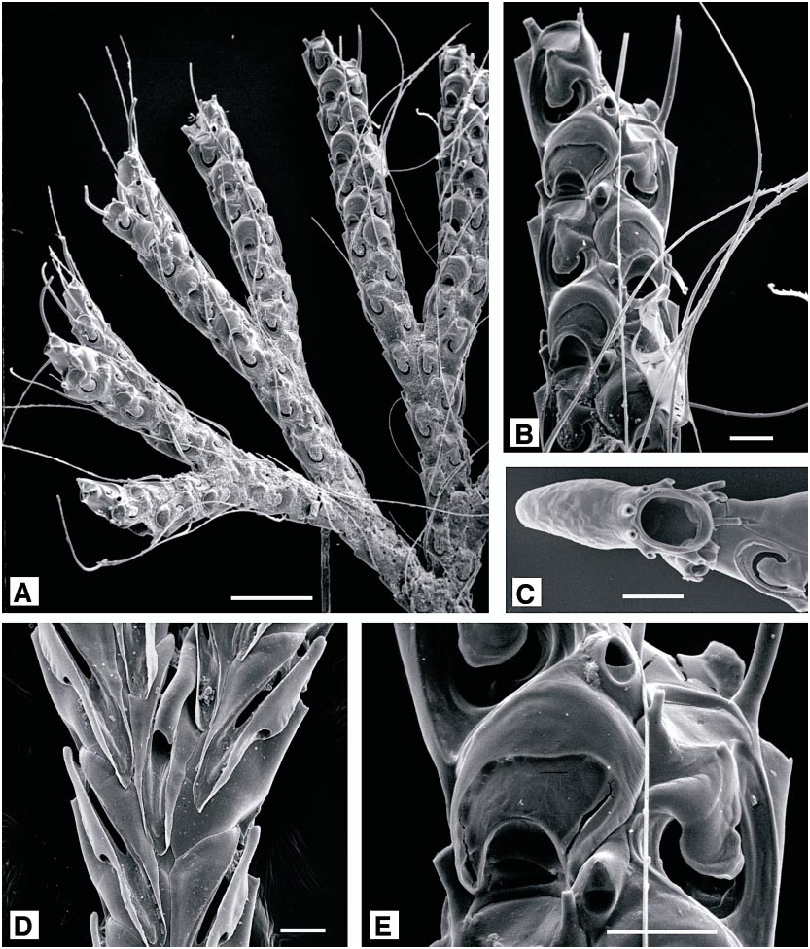
Caberea boryi ( Audouin, 1826) View in CoL Figure 12A–E View Fig
Crisia boryi Audouin, 1826: 242 . Savigny, 1809: pl. 12 figs 4.1–4.6.
Caberea boryi: Gautier, 1962: 93 View in CoL . Prenant and Bobin, 1966: 449. Hayward and Ryland, 1998: 252.
DESCRIPTION (AMNH 917–919; CMRR 2216): Colonies stiff, erect fans, to 8 mm high in present material, with cylindrical branches dividing dichotomously at regular intervals; without visible joints at the dichotomies. Autozooids in two alternating longitudinal series, or three just prior to dichotomies, defining the frontal surface of the branch, commonly 0.35 X 0.2 mm. Frontal planes of the two autozooid series angled at about 90° to each other; longitudinal axis of each autozooid slightly oblique to branch axis, inclining medially. Frontal surfaces of alternate autozooids slightly overlapped; opesia and operculum together constituting fourfifths of total frontal length, bordered proximally and laterally by broad, finely granular cryptocyst, no frontal gymnocystal calcification present. Three distal oral spines present: two on inner distal angle of autozooid and one, very much longer, on the out er distal angle. Much of opesia obscured by thick, oval scutum, attached by a thick stalk just proximal to inner distal spines; its distal edge straight, abutting with a blunt process projecting from opposite, outer edge of the opesia, and defining the proximal border of the operculum, which is terminal in position and tilted basally at a marked angle to frontal plane. A small adventitious avicularium on the inner distal angle of many autozooids, with short triangular mandible, distally directed; frequently, a much larger avicularium proximal to dichotomy, with broadly triangular rostrum frontally directed and almost perpendicular to frontal plane. Ovicell recumbent on distally succeeding autozooid, inclined medially; broader than long, with highly arched aperture bordered by broad band of uncalcified ectooecium. Abfrontal surface of colony distinctive: basal wall of each autozooid obscured by a large, proximally tapered vibraculum; seta 1.5 mm long, finely toothed along one edge, fitting into a narrow groove extending from outer distal edge of vibraculum, proximally and medially, contributing to a pronounced ridge extending length of branch. Thin kenozooidal rhizoids arising from vibracular chambers, passing proximally along median abfrontal surface of each branch and coalescing as thick bundles at base of colony.
Tentacles clear, 13; lophophores campylonemidan.
DISTRIBUTION: Caberea boryi is widespread and abundant in shallow inshore waters throughout the Mediterranean, and ranges northwards to the southwest British Isles, and southwards to South Africa. It has been
recorded from the Red Sea and the Indian Ocean, and from the West Pacific.
MEASUREMENTS (SKELETAL): DO 223 ± 34 µm, 172–310 (3, 28), OvL 122 ± 9, 110– 137 (1, 6), OvW 156 ± 14, 142–181 (1, 6), ZL 357 ± 40, 265–435 (3, 29), ZW 147 ± 11, 132–169 (1, 9). (POLYPIDE): IH 32 ± 7 µm, 20–40 (1, 9), LDMn 438 ± 27, 400– 460 (1, 6), LDMx 497 ± 37, 440–540 (1, 9), MD 20 ± 0 (1, 5), TL 443 ± 55, 360–540 (1, 10).
SUPERFAMILY MICROPOROIDEA GRAY, 1846 View in CoL FAMILY MICROPORIDAE GRAY, 1848 View in CoL GENUS CALPENSIA JULLIEN, 1888 View in CoL
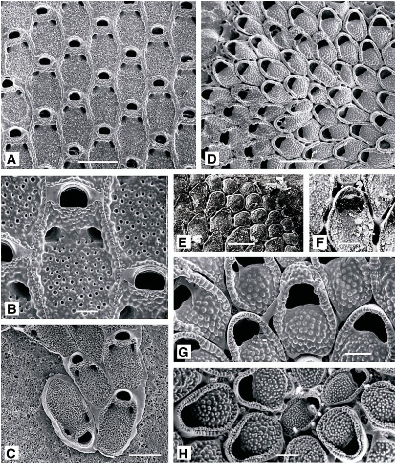
Calpensia nobilis (Esper, 1796) View in CoL
Figure 13A–C View Fig
Cellepora nobilis Esper, 1796: 145 .
Membranipora bifoveolata Heller, 1867: 95 .
Calpensia nobilis: Gautier, 1962: 59 View in CoL . Zabala,
1986: 289. Zabala and Maluquer, 1988: 90.
Hayward and Ryland, 1998: 292.
DESCRIPTION (AMNH 981, 1008; CMRR 2217): Colonies widely spreading, multiserial, flat, lightcolored grayishbrown sheets, often exceeding 10 X 10 cm; through successive phases of regeneration and overgrowth may form thick cylinders and nodules on every kind of hard substratum. Autozooids simple rectangles or hexagons with a thick, densely perforated cryptocyst, a small terminal opesia, equivalent to about 12% total autozooid length and coincident with the operculum, and paired opesiules almost as large as opesia. Outer epitheca glistens in dried material. First asexual zooid budded from distal end of ancestrula but with growth direction oriented 120° with respect to ancestrula and lying beside ancestrula, establishing early colony growth in direction opposite the polarity of the ancestrula (fig. 13C). Occasional reparative autozooidal budding. No polymorphs.
Cystids are occupied by a succession of up to four generations of polypides, and the brown body remains of previous polypides are stored in the proximal portions of zooids. Proximal portions of large colonies are senescent and commonly are bright green in color due to a thin growth of chlorophyte algae.
Tentacles clear, 19–22; lophophores bell shaped, radially symmetrical, supported on very long introverts, commonly overlapped with adjacent lophophores up to 300 µm.
REMARKS: This common and conspicuous Mediterranean species is sufficiently distinctive to be recognized with the unaided eye. Poluzzi and Coppa (1991) described growth and regeneration in C. nobilis , in relation to spatial competition. McKinney and Jaklin (1993) reported ephemeral populations of freelying colonies composed of single zooidthick sheets, up to several centimeters in diameter, off the Istrian Peninsula 30 to 50 km southsouthwest of Pula. Ristedt (1991) described the ancestrula and early astogeny, but sexual reproduction has not been described.
OCCURRENCE: This species encrusts virtually every type of nontoxic firm to hard substratum, including rock, shell debris, pottery, glass, other bryozoans, thecate ascidians, and—elsewhere in the Adriatic and Mediterranean ( Poluzzi and Coppa, 1991)—marine grasses.
DISTRIBUTION: The species is widespread throughout the Mediterranean and occurs sparsely northwards to the Gulf of St. Malo, and on the northwest African coast.
MEASUREMENTS (SKELETAL): DO 530 ± 93 µm, 366–684 (2, 20), OpL 96 ± 8, 80–107 (2, 20), OpW 150 ± 17, 119–174 (2, 20), ZL 817 ± 70, 719–935 (2, 20), ZW 383 ± 36, 322–439 (2, 20). (POLYPIDE): IH 495 ± 283 µm, 220–1200 (4, 21), LD 955 ± 128, 480–1140 (4, 40), MD 34 ± 8, 20–50 (3, 9), TL 654 ± 63, 560–820 (4, 18).
GENUS ROSSELIANA JULLIEN, 1888 View in CoL
Rosseliana rosselii ( Audouin, 1826) View in CoL Figure 14A–E View Fig
Flustra rosselii Audouin, 1826: 240 .
Membranipora rosselii: Hincks, 1880: 166 .
Rosseliana rosselii: Canu and Bassler, 1925: 17 View in CoL . Hayward and Ryland, 1998: 294.
Rosseliana View in CoL ‘‘sp. nov.’’ Gautier, 1962: 63.
DESCRIPTION (AMNH 1053–1055; CMRR 2218): Colonies multiserial encrusting, flat, beige colored. Autozooids arched oval distally and tapered proximally (proximal shape determined by shape of immediately preceding zooids), with boundaries marked by prominently raised lateral walls around arched distal end. Gymnocyst absent; cryptocyst flat to gently arched transversely, with coarsegrained or finely tuberculate surface. Opesia terminal, semielliptical with very slightly concave proximal edge, equivalent to about 40% total autozooid length. Ancestrula similar to autozooids; first three asexually budded zooids distal and distolateral; primary astogenetic zone of change only three or four generations Ovicell small, largely immersed, apparent as shallow crescentic cap at distal end of zooid.
Tentacles clear, 15; lophophores bellshaped, radially symmetrical away from colony margin between chimneys, obliquely truncate adjacent to chimneys and colony margin; introverts long.
REMARKS: Gautier (1962: 63–64) used R. rosselii for Rosseliana with large zooids (length 0.70–0.82 mm) and distinguished an unnamed new species for colonies with smaller zooids (length 0.40–0.68 mm). We encountered only colonies with the smaller zooids, essentially equivalent in size to the zooids of colonies attributed to R. rosselii around Britain ( Hayward and Ryland, 1998: 294). Prenant and Bobin (1966: 346–348) noted the two forms recognized by Gautier but indicated that they seem to cooccur ecologically and geographically and could not at the time be separated into two distinct species. Although only one of the forms has been found by us in the northern Adriatic, suggesting that the geographic ranges of the two ‘‘forms’’ or perhaps species are not fully coincident, we have preferred to retain the name R. rosselii for the northern Adriatic specimens.
DISTRIBUTION: This species is widespread in the eastern temperate Atlantic, from the Cape Verde Islands to Shetland, and throughout the Mediterranean.
MEASUREMENTS (SKELETAL): DO 452 ± 46 µm, 363–520 (2, 20), OpL 246 ± 24, 218– 308 (2, 20), OpW 227 ± 27, 185–277 (2, 20), ZL 591 ± 30, 533–668 (2, 20), ZW 375 ± 45, 307–457 (2, 20). (POLYPIDE): IH 366 ± 127 µm, 200–720 (2, 22), LDMn 560 ± 62, 400–630 (2, 13), LDMx 622 ± 72, 420– 840 (2, 24), MD 24 ± 3, 20–30 (2, 10), TLMn 476 ± 62, 340–590 (2, 23), TLMx 588 ± 113, 400–790 (2, 25).
GENUS MOLLIA LAMOUROUX, 1816 View in CoL
Mollia circumcincta ( Heller, 1867)
Figure 13D–H View Fig
Membranipora circumcincta Heller, 1867: 96 .
Mollia circumcincta: Gautier, 1962: 61 . Zabala
and Maluquer, 1988: 92.
NEOTYPE (chosen here): UIIZ 116.
DESCRIPTION (AMNH 920, 921; CMRR 2219): Colonies light orange, encrusting, multiserial, unilaminar sheets. Autozooids oval, 0.375–0. 50 mm; disjunct, each linked to its neighbors by about 12 short, tubular communication organs. Frontal body wall membranous, underlain for about twothirds of its extent by a vitreous, granular cryptocyst; opesia semielliptical, the proximal edge straight or slightly convex, and gently arched frontally, with indistinct opesiular indentations at each corner. Vertical walls of the autozooid develop a crenulate mural rim, which is especially raised distally, forming a cowl above the distal edge of the opesia. Spines absent, except in first two or three astogenetic generations. Ovicell hemispherical, partially immersed, with a crenulate vertical wall bordering a vitreous, nodular cryptocystal frontal surface. No polymorphs other than kenozooids, which may occur in clusters.
Tentacles clear, 15; lophophores bellshaped, radially symmetrical away from colony margin between chimneys, obliquely truncate adjacent to chimneys and colony margin; introverts long.
REMARKS: Specimens of Mollia circumcincta collected from the Rovinj area are conspecific with bryozoan specimen 116 in the University of Innsbruck Institute of Zo ology, labeled as ‘‘ Caleschara patellaria var. circumcincta Heller. Adria (S. Heller) 9837.’’ There is no annotation that this specimen is one of Heller’s original types, but we illustrate it here as at least an indication of long consistency in concept of the species and designate it as neotype for the species. If eventually discovered to be a specimen on which Heller originally described Membranipora circumcincta , it should be redesignated as lectotype.
DISTRIBUTION: This pretty species was first described from the Adriatic ( Heller, 1867) and has been reported from very few additional localities. It is known from the Siculo Tunisian shelf and the northwest Mediterranean ( Gautier, 1962), but does not seem to range outside of the Mediterranean.
MEASUREMENTS (SKELETAL): DO 336 ± 36 µm, 264–412 (2, 20), OpL 86 ± 14, 62–107 (2, 20), OpW 120 ± 19, 84–159 (2, 20), OvL 102 ± 10, 82–115 (2, 17), OvW 174 ± 12, 156–206 (2, 17), ZL 410 ± 35, 371–479 (2, 20), ZW 267 ± 24, 234–338 (2, 20).
SUPERFAMILY CELLARIOIDEA FLEMING, 1828 View in CoL FAMILY CELLARIIDAE FLEMING, 1828 View in CoL GENUS CELLARIA ELLIS AND SOLANDER, 1786 View in CoL
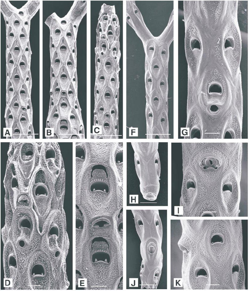
Cellaria fistulosa ( Linnaeus, 1758) View in CoL Figure 15A–E View Fig
Eschara fistulosa Linnaeus, 1758: 804 View Cited Treatment .
Cellaria fistulosa: Hayward and Ryland, 1998: 306 View in CoL .
DESCRIPTION (AMNH 922; CMRR 2220): Colonies erect, branching, consisting of rigid, cylindrical internodes formed from alternating whorls of three to six autozooids, linked by flexible, chitinous nodes at which dichotomous branching occurs. Internodes 2–9 mm long, typically about 5 mm, in present material, with diameter 0.45–0.7 mm, typically about 0.55 mm. Colonies attach by bundles of tubular rhizoids developed from the frontal cuticles of the proximal autozooids of the colony. Autozooids diamondshaped or hexagonal in outline; successive autozooids of longitudinal rows commonly but not always in contact along common transversely linear border and completely bounded laterally by the two adjacent offset rows of autozooids; less commonly succes sive zooids in longitudinal rows separated by line marking contact between the two adjacent offset rows of autozooids. Frontal membrane distinct and glistening in living material; in cleaned specimens, cryptocystal frontal shield concave between prominent ridges marking boundaries of autozooids. Calcification finely nodular; opesia wider than long, equivalent to onesixth zooid length, distal rim arched and finely beaded, proximal rim convex, with a blunt, frontally curved denticle in each proximal corner. A pair of wellmarked longitudinal cryptocystal ridges apparent on each side of the opesia, extending proximal to it, converging but not meeting. Avicularia about onethird length of autozooids, roundedquadrangular, with a narrow, semielliptical opesia; mandible narrowly crescentic, supported on an arched rostrum, directed distally or distolaterally. Ovicell aperture small and rounded, situated immediately distal to opesia, partially occluded in older parts of colonies by calcified plate extending from proximal edge, as has been not ed elsewhere for C. salicornioides ( Hayward and Ryland, 1998) .
Tentacles clear, 13; lophophores bellshaped, radially symmetrical.
DISTRIBUTION: Common in shallow, coastal waters throughout the Mediterranean, and northwards to the British Isles, western Norway and Iceland.
MEASUREMENTS (SKELETAL): DO 314 ± 23 µm, 252–345 (2, 16), OpL 71 ± 8, 60–87 (2, 20), OpW 107 ± 8, 97–126 (2, 17), ZL 564 ± 21, 510–601 (2, 20), ZW 262 ± 14, 241–283 (2, 15). (POLYPIDE): IH 67 ± 27 µm, 20–100 (1, 6), LD 534 ± 38, 490–580 (1, 7) MD 30 (1, 1), TL 437 ± 29, 400–480 (1, 6).
Cellaria salicornioides Lamouroux, 1816 View in CoL Figure 15F–K View Fig

No known copyright restrictions apply. See Agosti, D., Egloff, W., 2009. Taxonomic information exchange and copyright: the Plazi approach. BMC Research Notes 2009, 2:53 for further explanation.
|
Kingdom |
|
|
Phylum |
|
|
Class |
|
|
Order |
|
|
Family |
|
|
Genus |
Sertularia scruposa Linnaeus, 1758: 815
| HAYWARD, PETER J. & McKINNEY, FRANK K. 2002 |
Cellaria fistulosa : Hayward and Ryland, 1998: 306
| Hayward, P. J. & J. S. Ryland 1998: 306 |
Caberea boryi : Gautier, 1962: 93
| Hayward, P. J. & J. S. Ryland 1998: 252 |
| Prenant, M. & G. Bobin 1966: 449 |
| Gautier, Y. V. 1962: 93 |
Calpensia nobilis : Gautier, 1962: 59
| Gautier, Y. V. 1962: 59 |
Rosseliana
| Gautier, Y. V. 1962: 63 |
Mollia circumcincta : Gautier, 1962: 61
| Gautier, Y. V. 1962: 61 |
Rosseliana rosselii : Canu and Bassler, 1925: 17
| Hayward, P. J. & J. S. Ryland 1998: 294 |
| Canu, F. & R. S. Bassler 1925: 17 |
Membranipora rosselii :
| Hincks, T. 1880: 166 |
Membranipora bifoveolata
| Heller, C. 1867: 95 |
Membranipora circumcincta
| Heller, C. 1867: 96 |
Crisia boryi
| Audouin, J. V. 1826: 242 |
Flustra rosselii
| Audouin, J. V. 1826: 240 |