
Centroderes impurus, Sørensen & Gąsiorowski & Randsø & Sánchez & Neves, 2016
|
publication ID |
https://doi.org/10.5281/zenodo.4502533 |
|
publication LSID |
lsid:zoobank.org:pub:819AC644-37BC-43DB-8E11-984D77804AFE |
|
DOI |
https://doi.org/10.5281/zenodo.4776223 |
|
persistent identifier |
https://treatment.plazi.org/id/03D187A7-DA35-4E0A-3B2C-FF14FE72FD6B |
|
treatment provided by |
Carolina |
|
scientific name |
Centroderes impurus |
| status |
sp. nov. |
Centroderes impurus sp. nov.
( Figs. 2–5 View Fig View Fig View Fig View Fig )
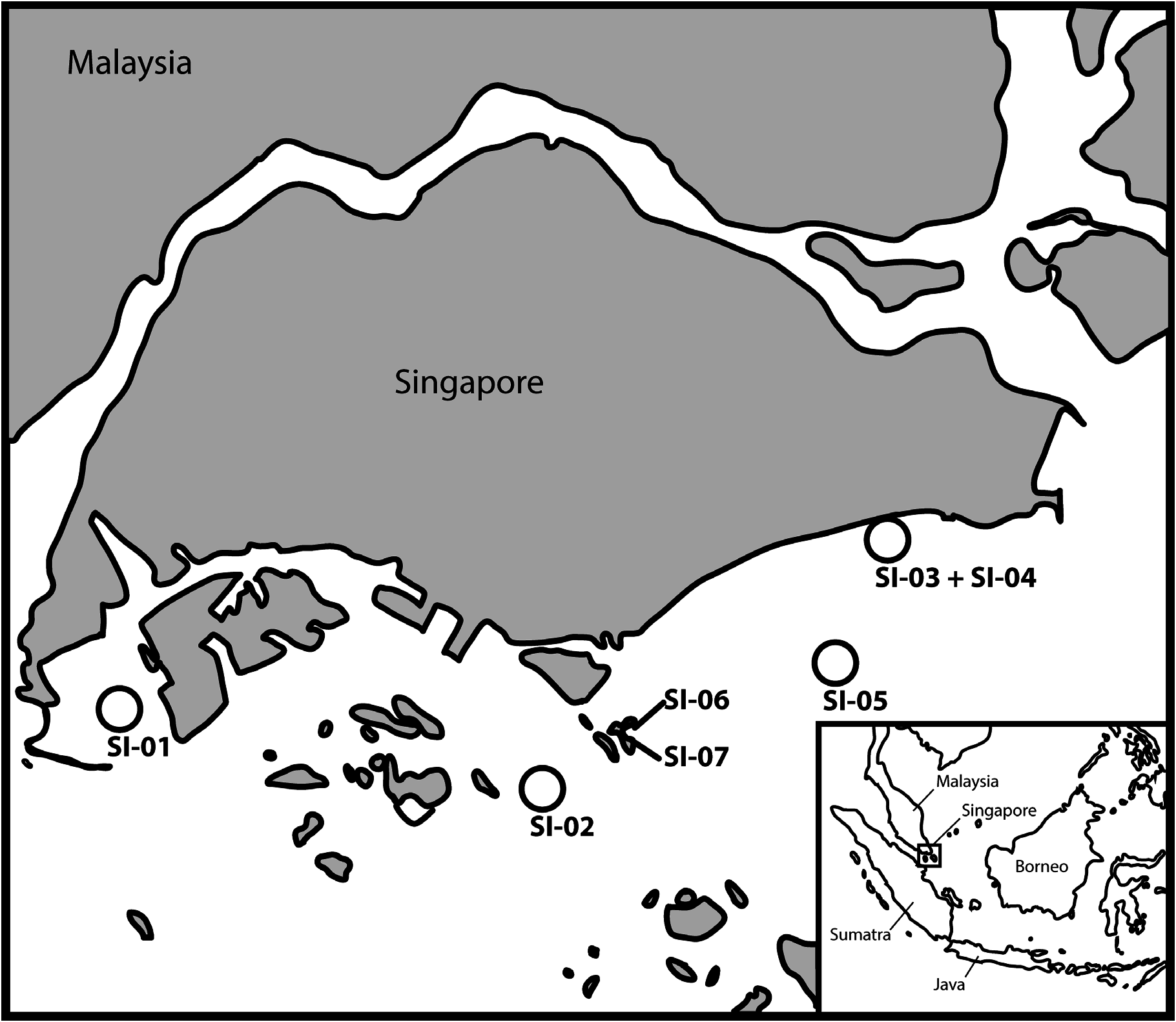
Material examined. Holotype adult stage- 2 male, collected from sand with mud on 16 May 2014, at station SI-03 ( Fig. 1 View Fig , Table 1), at 9 m depth, between Bedok Jetty and Sungei Bedok in the southeast part of Singapore Island ( 01°18.387’N, 103°57.591’E), mounted in Fluoromount G, deposited at the Lee Kong Chian Natural History Museum, under catalogue number ZRC.MIS.0001 GoogleMaps . Paratypes include two specimens of uncertain sex, probably either adult stage- 1 male or female, or eventually preadult stages (J6), collected at same locality as holotype, mounted in Fluoromount G, and deposited at the Natural History Museum of Denmark, under catalogue numbers ZMUC KIN-846 and KIN-847 . Additional, non-type material, includes one adult stage- 2 male and two putatively stage- 1 specimens of uncertain sex, collected at same locality as holotype, and mounted for SEM. The SEM specimens are very dirty, and contributed only with limited information GoogleMaps .
Diagnosis. Centroderes with middorsal acicular spines on segments 1–9 and 11; long, flexible, ventromedial acicular spines on segment 1; rigid lateroventral acicular spines on segments 8 and 9, with spine on segment 9 being conspicuously stout and robust.Male stage-2 with middorsal and midlateral crenulated spines on segment 10. Putative adult stage- 1 specimens, or alternatively preadults, with middorsal and midlateral acicular spines on segment 10, and with minute tubes present in ventrolateral positions on segment 2 and lateroventral positions on segment 5. Sensory spots present paradorsally on segments 2–6 and 8–9, subdorsally on segment 1 (in male stage-2 only) and segment 11 (two pairs), laterodorsally on segments 1 (in putative J6 or stage- 1 specimen only) and 3–10, midlaterally on segment 2, in a lateral accessory position on segments 1, 3–4 and 6–9 and ventromedially on segments 3–4 and 6–10.
Etymology. The species is named after the Latin word “ impurus ”, meaning “unclean” or “covered with dirt”, referring to the severe dirt problems that were experienced during the examination of the specimens.
Description. Adult with head, neck and eleven trunk segments ( Figs. 2 View Fig , 3A, B View Fig , 4A, B View Fig ). For measurements and dimensions, see Table 2. Distribution of cuticular structures (spines, tubes and sensory spots) is summarised in Table 3.
The head consists of a retractable mouth cone and an introvert. The mouth cone is equipped with nine outer oral styles, each consisting of two joined units, arranged as one style anterior to each introvert sector, except at the middorsal sector 6. A double fringe consisting of numerous tips is located basally to each style. Scalid arrangement could not be examined in detail.
The neck has 16 placids that dorsally and laterally alternate in size width between broader ( 15–16 µm width) and narrower ( 6–8 µm width) ones; all placids measure 15–17 µm length. The midventral placid is broader and flanked by two narrower placids on each side.
Segment 1 consists of one complete cuticular ring with middorsal acicular spine, being short and stout in male stage-2 ( Fig. 3A View Fig ), and two slender, elongated spines in ventromedial positions, which extend over two following segments ( Figs. 2 View Fig , 5B View Fig ). All spines on this and following segments are at least partly covered with minute cuticular hairs, however, frequently the proximal 1/3 of the surface is smooth. At least until segment 6 or 7, the middorsal spines appear stout in male stage-2, whereas they are more slender and acicular in the putative J6 or stage- 1 specimens (compare Fig. 3A View Fig with 3B). A midventral process is present between the two ventromedial spines ( Figs. 2 View Fig , 4B View Fig ). Sensory spots are located medially on the segment in either subdorsal (male stage-2) or laterodorsal (putative J6 or stage- 1 adults) positions, and always in lateral accessory position ( Fig. 4C View Fig ). Sensory spots in subdorsal/laterodorsal positions are circular and composed of micropapillae arranged around a central pore ( Fig. 4C View Fig ); all sensory spots on all the following segments show the same appearance. The sensory spots in the lateral accessory positions differ though, and are more oval with cuticular papillae arranged in three rows and adhering to the cuticle surface ( Fig. 4D View Fig ). Minute, densely distributed cuticular hairs are present on the posterior half of the segment.
Segment 2 and all remaining segments consist of one tergal and two sternal plates. A middorsal spine is present in all specimens. Furthermore, minute ventrolateral tubes were observed in the putative J6 or stage- 1 specimens ( Fig. 5B View Fig ). In male stage-2 no such structure was observed neither in LM nor in SEM; in the single specimen examined with SEM it was evident that the position where the tube would attach was filled with densely arranged prominent hairs. Similar hairs were spotted on other segments as well ( Fig. 4F View Fig ). Sensory spots are present in paradorsal and midlateral position. The paradorsal sensory spots are located very close to the posterior segment margin, next to the base of the middorsal spine. Secondary pectinate fringe consisting of minute cuticular hairs present on anterior part of segment; the fringe may be partially covered by the posterior margin of preceding segment. Additional hairs are densely and evenly distributed over the posterior half of the segment. Hairs in the tergosternal junction region are distinctly longer, especially anteriorly. A similar arrangement of hairs is present on segments 3–10 ( Figs. 2 View Fig , 4E View Fig ).
Segments 3 and 4 with middorsal acicular spine. Sensory spots are present in paradorsal, laterodorsal, lateral accessory and ventromedial positions ( Figs. 2 View Fig , 5A View Fig ). Cuticular hairs as on preceding segment.
Segment 5 with middorsal acicular spine. Minute lateroventral tubes are present in putative J6 or stage- 1 specimens ( Figs. 4E View Fig , 5D View Fig ). Stage- 2 specimens without such tubes, and instead with densely arranged prominent hairs in the areas where the tubes would attach ( Fig. 4F View Fig ). Sensory spots are present in paradorsal and laterodorsal positions ( Figs. 2 View Fig , 5A View Fig ). Cuticular hairs as on preceding segment.
Segment 6 with middorsal acicular spine. Sensory spots present in paradorsal, laterodorsal, lateral accessory and ventromedial positions ( Figs. 2 View Fig , 5A View Fig ). Cuticular hairs as on preceding segment.
Segment 7 with middorsal acicular spine. Sensory spots present in laterodorsal, lateral accessory and ventromedial positions. On this and the following segments, the laterodorsal sensory spots are located slightly closer to the midlateral position. Putative J6 or stage- 1 specimens show no indication of female specific glands, as observed in other Centroderes (see Neuhaus et al., 2014). Cuticular hairs as on preceding segment.
Segment 8 with prominent, long middorsal acicular spine ( Figs. 2 View Fig , 3A View Fig ). Small acicular spines present in lateroventral positions ( Figs. 4G View Fig , 5E, F View Fig ). Sensory spots present in paradorsal, laterodorsal, lateral accessory and ventromedial position. Putative J6 or stage- 1 specimens show no indication of female specific glands. Cuticular hairs as on preceding segment.
Segment 9 with prominent, long middorsal acicular spine ( Figs. 2 View Fig , 3A View Fig ). Conspicuously robust acicular spines are present in lateroventral positions; these spines can be twice as long as the corresponding spines on segment 8 ( Figs. 4G View Fig , 5E, F View Fig , see also Table 2), especially in the male stage-2. Sensory spots present in paradorsal, laterodorsal, lateral accessory and ventromedial position. Cuticular hairs as on preceding segment.
Segment 10 with middorsal and midlateral acicular spines in putative J6 or stage- 1 specimens ( Figs. 3B View Fig , 5F View Fig ) and with middorsal and midlateral crenulated spines in stage- 2 males ( Figs. 2 View Fig , 3A View Fig , 4H View Fig , 5C, E View Fig ). Sensory spots present in laterodorsal and ventromedial positions. Cuticular hairs as on preceding segment.
Segment 11 with middorsal, lateral terminal, lateral terminal accessory, and midterminal spines ( Figs. 2 View Fig , 3 View Fig , 4A, B View Fig , 5F View Fig ). The midterminal spine is considerably longer than the other spines on this segment ( Table 2). Two pairs of subdorsal sensory spots, one pair being more anterior than the other, are present. The whole cuticular surface is covered with minute cuticular hairs which turn slightly longer and denser in the terminal part of the segment.


No known copyright restrictions apply. See Agosti, D., Egloff, W., 2009. Taxonomic information exchange and copyright: the Plazi approach. BMC Research Notes 2009, 2:53 for further explanation.
|
Kingdom |
|
|
Phylum |
|
|
Class |
|
|
Order |
|
|
Family |
|
|
Genus |