
Leydigia cf. ciliata (Gauthier, 1939)
|
publication ID |
https://doi.org/ 10.5281/zenodo.277273 |
|
DOI |
https://doi.org/10.5281/zenodo.5695790 |
|
persistent identifier |
https://treatment.plazi.org/id/03D28785-3008-3F20-FF76-FCA6FA9BFBDD |
|
treatment provided by |
Plazi |
|
scientific name |
Leydigia cf. ciliata (Gauthier, 1939) |
| status |
|
Leydigia cf. ciliata (Gauthier, 1939) View in CoL
Sars, 1903, p. 14, Pl. 1: figs 4, 4a (propinqua); Gauthier, 1939, p. 168–173, fig. 9 (propinqua var. Ciliata ); Rey and Sain-Jean 1968, 105, fig. 21; Biswas 1971, 129, fig. 12F (acanthocercoides); Smirnov 1971, 454–458, figs 562, 564–56; Dumont & Van de Velde 1977, p. 90–91, figs 8–9 (propinqua ciliata ); Chiang & Du 1979, 208–209, fig. 139A–F (acanthocercoides) p. 209–210, fig. 140 (propinqua); Dumont et al. 1984, p. 167, figs 1–5; Prasad, Santa Kumari et Bose, 1986, p. 99–107, figs 1–15 (ankammaraoi); Jeje 1988, p. 113–116, figs 1–8. (macrodonta macrodonta); Tanaka 1989, p. 4–5, Pl. 11: figs 11–12; Kotov et al. 2003b, p. 180–190, figs 1–63: Kotov 2009: 68–71 (for full reference list see Kotov, 2009).
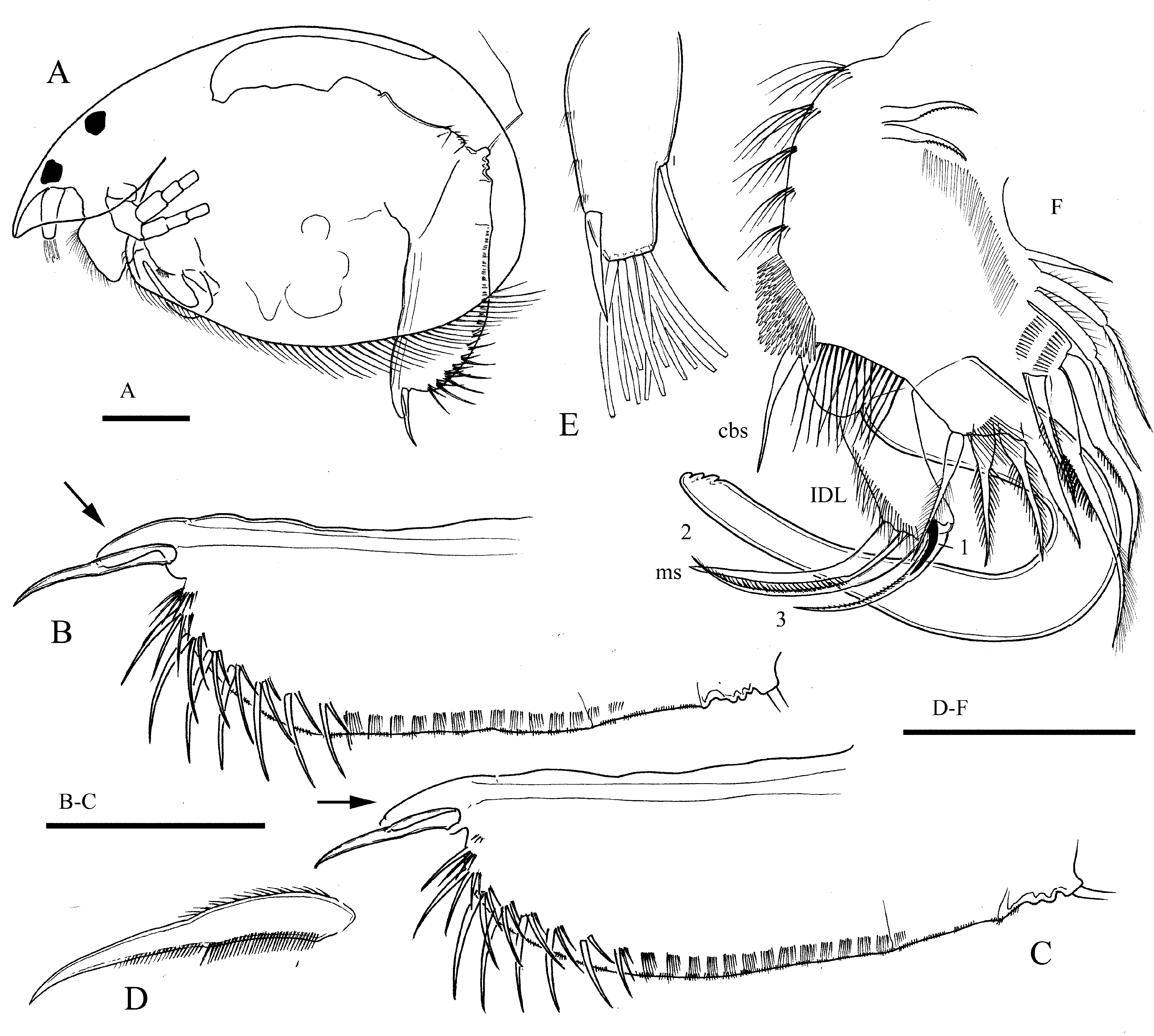
Description. Male. Length of studied specimens 0.64 and 0.65 mm. Body egg-shaped, high, widening posteriorly ( Fig. 4 View FIGURE 4 A), height/length ratio about 0.63. Maximum height in the third fourth of the body. Dorsal, posterior and ventral margins of valves evenly convex, posteroventral and posterodorsal angles broadly rounded. Head large, broad, protruding forward. Rostrum short, protruding forward and downward. Eye and ocellus large, of similar size.
Postabdomen ( Fig. 4 View FIGURE 4 B,C) long, moderately broad, with almost parallel dorsal and ventral margins in central portion, narrowing in the distal fourth. Length about 2.5 height. Ventral margin wavy, slightly concave, sperm duct openings located at the end of short process (penis) protruding between the bases of claws. Penis less than half length of the claw, without stylet. Distal part of postabdomen rounded, with no defined distal angle. Distal part of postabdomen 6–7 times longer than preanal, postanal portion 4 times longer than anal. Preanal margin very short, with several hillocks. Anal margin almost straight, postanal margin straight in basal half and convex in distal half. Both postanal and preanal angles weakly defined. Dorsal margin with numerous clusters of extremely short setules. Distal portion of postabdomen bears 7–8 lateral fascicles of very large setules, followed by about 14 much smaller fascicles. Distal fascicles consisting only of 2–4 (usually 3) setules each, length of distal setule in fascicle up to 2/3 height of postabdomen. Postabdominal claw ( Fig. 4 View FIGURE 4 D) almost straight, slender, 3 times longer than preanal margin, without basal spine, with a pecten of long spinules in basal half.
Antennule ( Fig. 4 View FIGURE 4 E) moderately broad, length/width ratio about 2.5, with 12 terminal aesthetascs of variable length, longest about 2/3 length of antennule. Male seta moderately long, thick, about 1/2 length of antennule, arising at 1/4 antennule length from the tip.
Thoracic limb I ( Fig. 4 View FIGURE 4 F) massive, broader than that of female, copulatory hook U-shaped, very large, as long as limb itself. Copulatory brush present, a cluster of about 15 very long setules located below it on the ventral face of limb. Copulatory brush seta long, slender, similar in size to setae of endite 3. IDL elongated, narrow, IDL seta 1 short, 3 times shorter than that of female, setae 2 and 3 thin, much shorter that these of female, seta 2 longer than seta 3, male seta of same size with than IDL seta 2, but thicker, curved. Morphology of endites same as in female.
Taxonomical notes. The species was recently revised ( Kotov et al. 2003; Kotov 2009), but no description of male was provided. There is only one description of L. ciliata male (Dumont & Van de Velde 1977, 90–91, Fig.9), for a population from Niger. Our specimens significantly differ from that population by the shape of postabdomen. The specimens from Niger have broader postabdomen (length about 2.5 height) with convex dorsal and ventral margins, maximum height at the middle of postabdomen, defined postanal angle, and penis longer than half length of claws. There are no significant differences between these populations in body shape and in the morphology of antenna. The first thoracic limb was not studied for the Niger population. According to Kotov (2009) there are no significant differences between parthenogenetic females of the African and Asian populations, but the differences in male morphology are quite prominent and suggest a possibility of species-complex. Investigations of L. ciliata populations should be continued, and detailed study of male and gamogenetic female from the terra typica is necessary to clarify status of the Asian populations.
Disparalona cf. hamat а (Birge, 1910)
Non Disparalona hamat а (Birge, 1910) s. str.
Description. Male. Length of two studied males were 0.50 mm. Body low oval ( Fig. 5 View FIGURE 5 A), height/length ratio about 0.5. Maximum height in the second fourth of the body. Dorsal margin of valves convex, posterior margin of valves almost straight, posteroventral and posterodorsal angles broadly rounded. Ventral margin of valves convex. Head large, narrow. Rostrum long, bend at the middle at almost right angle, its tip pointed posteriorly. Eye and ocellus large, eye 2 times larger than ocellus.
Postabdomen ( Fig. 5 View FIGURE 5 B, C) long and narrow, slightly curved and narrowing in postanal portion, length about 4 height. Ventral margin convex and wavy in postanal portion, concave at the level of postanal angle. Sperm duct openings located at the end of postabdomen, on level of incursion between the base of claws and distal angle. Distal angle small, rounded, nor prominent. Distal part of postabdomen 2.2 times longer than preanal, postanal portion 1.5 times longer than anal. Both anal and postanal margins concave. Both postanal and preanal angles defined. Dorsal margin with cluster of long setules and single large denticle at the end, following by about 15 clusters of short setules. Lateral fascicles of setules same as in female. Postabdominal claw curved, about 2/3 length of preanal margin, with thin basal spine about 0.25 length of claw.
Antennule ( Fig. 5 View FIGURE 5 D, E) large, broad, length/width ratio about 1.8, with 10 terminal aesthetascs of variable length, longest about 2/3 length of antennule, Male seta thick, very long, about 1.2–1.3 length of antennule, arising at 1/3 antennule length from the tip.
Thoracic limb I ( Fig. 5 View FIGURE 5 F,G) of moderate size, copulatory hook U-shaped, small, 2.5 times shorter than limb itself. Copulatory brush present, three clusters of numerous long setules located below it on the ventral face of limb. Copulatory brush seta long, slender, slightly smaller than setae of endite 3. IDL seta 1 absent, setae 2 and 3 similar to that of female; seta 3 massive, claw-like; seta 2 of similar length, but much thinner, setulated distally; male seta slender, curved in distal portion, about 2/3 length of seta 2. Morphology of endites same as in female.
Taxonomical notes. Disparalona hamata (syn. Pleuroxus hamata , Alonella hamulata ) was described from North America ( USA, Massachusetts). It is presumed to be cosmopolitan ( Smirnov 1996), recorded from North and South America, Europe, Africa and Southern Asia. Such wide distribution does not agree with Frey’s non-cosmopolitanism paradigm ( Frey 1982, 1987), now widely accepted in cladoceran taxonomy ( Kotov et al. 2010). This usually indicates a species-complex, as in many other Chydorinae. An African taxon, Pleuroxus chappuisi Brehm, 1934 is presumed to be a synonym of D. hamata ( Smirnov 1996) , but is possibly a separate species.
The only, partial, description of Disparalona hamat a male, from Washington, USA, was provided by Kiser (1950, p. 247, Pl.2). Unlike our specimens, male of North American D. hamata s. str. has much shorter, evenly curved rostrum and different morphology of postabdomen. It is evenly narrowing distally, not curved in postanal portion, and has no expressed distal angle and no large denticle at the end. Such differences obviously mean that our population does not belong to D. hamata s. str., but to a new, not yet described sibling-species. A revision of D. hamata complex should be conducted. A similar situation was revealed for the D. rostrata complex by Michael & Frey (1984), with D. rostrata s. str. distributed only in Palearctic, and D. leei (Chien, 1970) in North America.

No known copyright restrictions apply. See Agosti, D., Egloff, W., 2009. Taxonomic information exchange and copyright: the Plazi approach. BMC Research Notes 2009, 2:53 for further explanation.