
Regadrella okinoseana Ijima, 1896
|
publication ID |
https://doi.org/ 10.11646/zootaxa.3628.1.1 |
|
publication LSID |
lsid:zoobank.org:pub:37D2D7F2-FA0C-40E9-B6D0-9C74EBB6C7F0 |
|
DOI |
https://doi.org/10.5281/zenodo.5261624 |
|
persistent identifier |
https://treatment.plazi.org/id/03D287B2-FFA0-3632-9AD7-FC052BEFFE57 |
|
treatment provided by |
Felipe |
|
scientific name |
Regadrella okinoseana Ijima, 1896 |
| status |
|
Regadrella okinoseana Ijima, 1896 View in CoL
( Figs. 12 View FIGURE 12 & 13 View FIGURE 13 , Table 6)
Synonymy: Regadrella okinoseana Ijima, 1896: 250 ; 1901: 223; Levi & Levi 1982: 292; Reiswig 1992: 33; Stone et al., 2011: 25.
Regadrella cylindrica Ijima, 1927: 335 View in CoL .
Regadrella decora Schulze, 1900: 30 View in CoL .
Material examined. USNM# 1196553 About USNM , ROV 'Jason II' from RV 'Roger Revelle', dive J2102, 03 August 2004, S Amchitka Pass, 27.4 km W of Amatignak Island, Delarof Islands, Aleutian Islands , Alaska, 51º17.469'N, 179º32.585'W, 1386 m, partial specimen, dry & ethanol GoogleMaps .
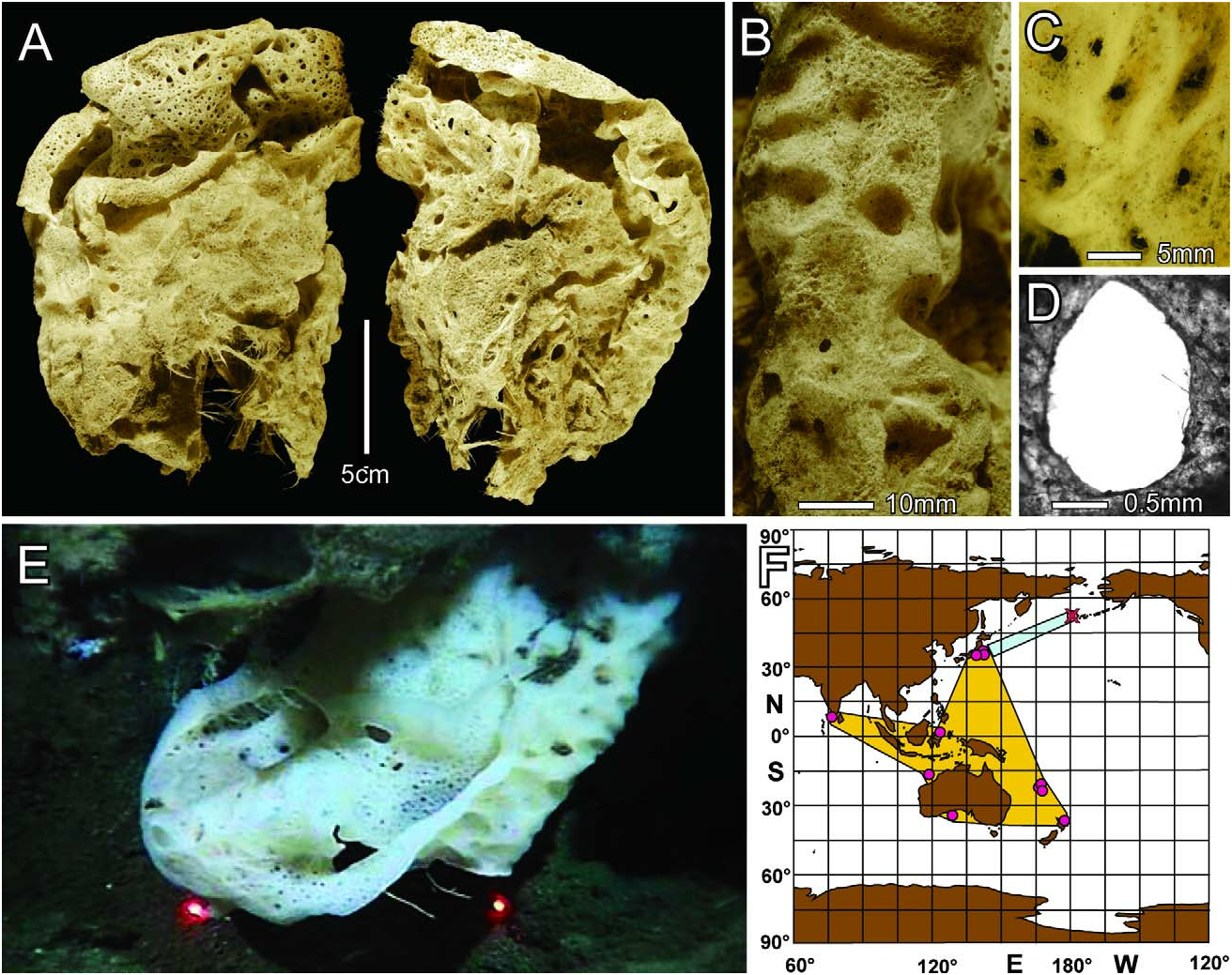
Description. The single specimen from the Aleutian Islands is a soft flattened sac with a wide but subdivided oscular aperture ( Fig. 12A View FIGURE 12 ); dimensions of the specimen lacking the attachment base (not collected) are 18.8 cm tall, 12.8 cm wide, 9.1 cm thick. The body wall is 3–4 mm thick. Prostalia are not present. The characteristic Regadrella surface pattern of pits and ridges with small parietal oscula at the center of pits ( Table 6) is developed only on the external body edges that remain free of contact with the surrounding substrate ( Figs. 12 B, C View FIGURE 12 ). The larger frontal surfaces that contact the substratum lack this pattern but still have parietal oscula scattered irregularly across their surface. Parietal oscula are ovoid with mean major diameter of 2.6 mm ( Fig. 12D View FIGURE 12 ). The main oscular edge is curled outward ( Fig. 12A View FIGURE 12 ) but a cuff and sieve plate are absent. Within the single oscular margin, the distal atrial cavity is subdivided into four separate passages by tissue bridges joining the atrial wall surfaces. One of these bridges extends longitudinally at least half the length of the body, thereby flattening and subdividing the distal atrial cavity into two main passages. In the lower half of the saccate body, the two longitudinal atrial passages join to form a single open atrial cavity. Before collection, the specimen was attached under the edge of a large mudstone mound with the osculum exposed ( Fig. 12E View FIGURE 12 ). Color in life was white; dried and ethanol-preserved samples are light beige. The new location is quite remote from the known distribution of this species ( Fig. 12F View FIGURE 12 ).
The main supporting skeleton is a network of loose interwoven unfused diactins resulting in a very soft texture in the upper body. In the lower body quarter, internal diactins are haphazardly fused at contact points, resulting in a stiff but still flexible texture in a soft surface in this area. Main dermal hexactins and atrial pentactins remain unfused throughout the body surfaces.
Megascleres (spicule dimensions are given in Table 6): Parenchymal megascleres are thick principal diactins and thin intermediate and comital diactins. Thick diactins are slightly curved, smooth, with slight medial swelling and rounded inflated tips ( Fig. 13L View FIGURE 13 ). Thin diactins are similar ( Fig. 13K View FIGURE 13 ) but have acutely pointed tips. Dermalia are mainly hexactins with a very few subhexactins. The primary hexactins are large sword-shaped forms ( Fig. 13A View FIGURE 13 ) with straight, robust tapering rays that end in slightly roughened sharp tips. The distal ray is shorter and the proximal ray is much longer than the tangential rays. Smaller hexactins with all rays nearly equal in length ( Figs. 13 B, C View FIGURE 13 ) are less abundant. Atrialia are mainly pentactins ( Fig. 13D View FIGURE 13 ), with a few subhexactins ( Fig. 13E View FIGURE 13 ), hexactins ( Fig. 13F View FIGURE 13 ), irregular tetractins, stauractins with two reduced knobs ( Fig. 13G View FIGURE 13 ), triactins ( Fig. 13H View FIGURE 13 ) and diactins ( Fig. 13I View FIGURE 13 ). All atralia have smooth rays that end in slightly rough rounded tips. Parietal oscular membranes have most types of atrialia, but also small stubby hexactins and pentactins and very small distinctive diactins with four central knobs ( Fig. 13J View FIGURE 13 ) found nowhere else.
Microscleres consist mainly of floricomes, oxystaurasters, and graphiocomes, but oxyhexasters, oxystauractins, and oxydiasters occur in small numbers. Floricomes ( Fig. 13M View FIGURE 13 ) have short smooth primary rays ending in small discs; 7–15 s-shaped terminals originate from each primary disc in a single marginal whorl and together form a perianth. Internal (within perianth) surfaces of the terminals are smooth but the outer surfaces bear a continuous band of small spines extending up under the 2–4 large claws on the outer margin of the terminal swelling. Oxystauractins ( Fig. 13N View FIGURE 13 ) are sturdy microscleres with only four nearly smooth, crucial, primary rays, each of which supports 2–5 longer terminal rays ornamented with reclined spines ( Fig. 13P View FIGURE 13 ). Oxyhexasters ( Fig. 13O View FIGURE 13 ) are similar to the staurasters, but bear the full set of six primary rays, each of which carry 3–6 straight or slightly curved rough terminal rays. Graphiocome centers ( Fig. 13Q View FIGURE 13 ) have short, strong, nearly smooth primary rays, each of which ends in a small furry disc entirely covered by the ca 120 short bases of the invariably broken off terminal rays; articulation facets are only 0.2 µm in diameter. The loose terminal rays ( Fig. 13R View FIGURE 13 ) are slightly sinuous (perhaps spiralled?) with an articulation base bearing a facet and two teeth; the raphidial ray is completely but sparsely ornamented with small spines, basally proclined and distally reclined. These spicules must be truly spectacular when they are intact.
Remarks. Although the body form of the Aleutian Islands specimen differs in many respects from a typical Regadrella okinoseana , its spiculation agrees entirely with that species. The site of collection of the new specimen is ca 3700 km distant from the nearest reported occurrence of R. okinoseana in Japan, and thus represents a very striking range extension ( Fig. 12F View FIGURE 12 ).
Review of all video footage collected with the ROV 'Jason II' indicates that it is an uncommon species occurring singly on mudstone, bedrock and possibly hexactinellid skeletons at depths between 1071 and 1395 m.

| RV |
Collection of Leptospira Strains |
No known copyright restrictions apply. See Agosti, D., Egloff, W., 2009. Taxonomic information exchange and copyright: the Plazi approach. BMC Research Notes 2009, 2:53 for further explanation.
|
Kingdom |
|
|
Phylum |
|
|
Class |
|
|
Order |
|
|
Family |
|
|
Genus |
Regadrella okinoseana Ijima, 1896
| Reiswig, Henry M. & Stone, Robert P. 2013 |
Regadrella cylindrica
| Ijima, I. 1927: 335 |
Regadrella decora
| Schulze, F. E. 1900: 30 |