
Montandoniola pictipennis ( Esaki, 1931 )
|
publication ID |
https://doi.org/ 10.5281/zenodo.196531 |
|
DOI |
https://doi.org/10.5281/zenodo.3510434 |
|
persistent identifier |
https://treatment.plazi.org/id/03D387CE-FF90-5C70-FF7E-FCF99335C163 |
|
treatment provided by |
Plazi |
|
scientific name |
Montandoniola pictipennis ( Esaki, 1931 ) |
| status |
|
Montandoniola pictipennis ( Esaki, 1931)
( Figs. 5, 6 View FIGURES 1 – 13 , 15 View FIGURES 14 – 16 , 17–20 View FIGURES 17 – 22 )
Ectemnus pictipennis Esaki, 1931: 264 (sp. n.); Esaki, 1932: 1673 (illustration, diagnosis); Esaki, 1950: 256 (illustration, diagnosis).
Teisocoris pictipennis : Hiura, 1959: 2 (n. comb., illustration, note).
Montandoniola pictipennis: Carayon, 1961: 543 (note); Carayon & Ramade, 1962: 208 (note); Herring, 1966: 93 [syn. with M. moraguesi (Puton) ]. Restored by Pluot-Sigwalt et al., 2009: 33 View Cited Treatment (illustration, diagnosis).
Montandoniola moraguesi (Puton, 1896) : Miyamoto & Yasunaga, 1989: 167 (list); Yasunaga, 1993: 169 (note); Yasunaga, 2001: 287 (note) (misidentifications).
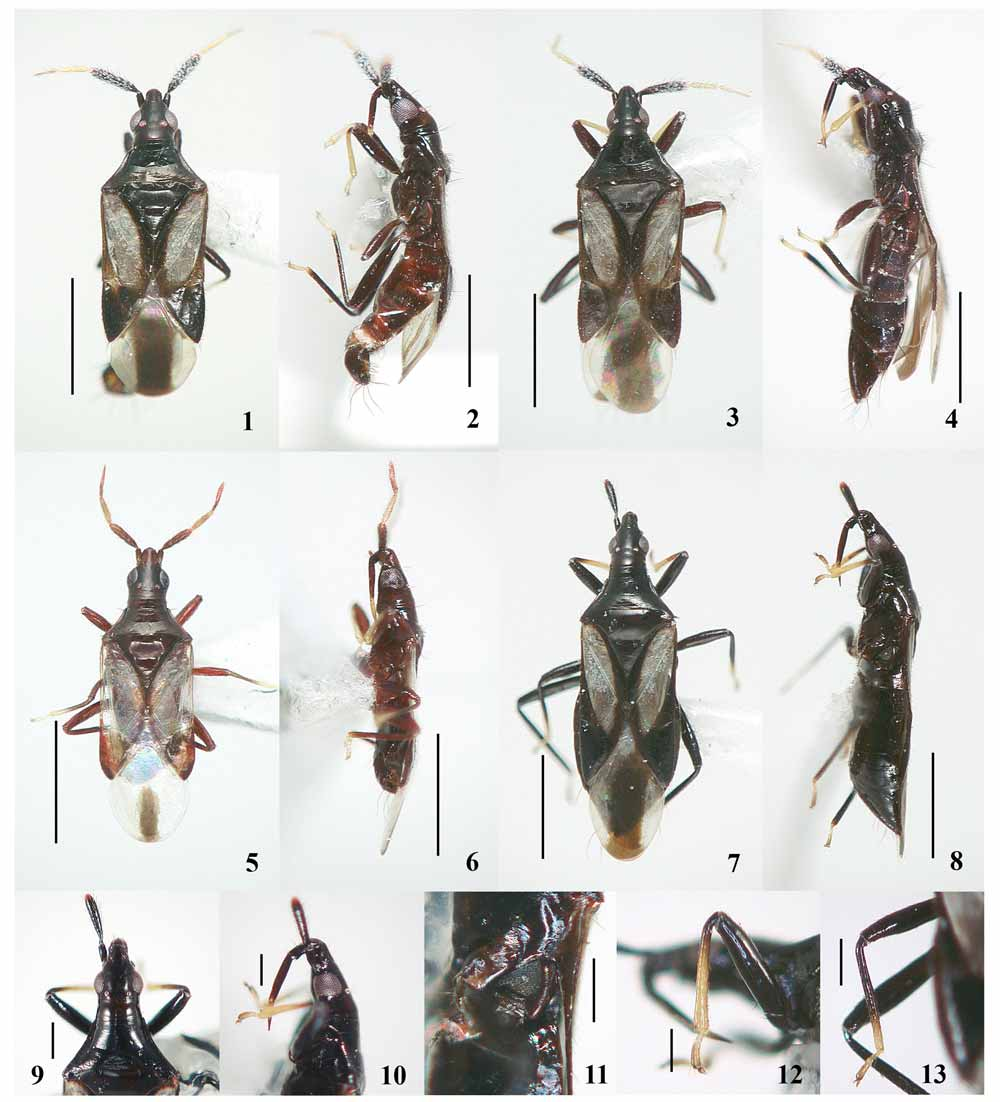
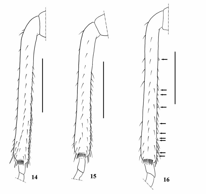
Diagnosis. Head ( Fig. 5 View FIGURES 1 – 13 ) dark brown to black; length excluding neck as long as width across eyes. Antennal segments I and II dark brown, segment II lighter at apex, segments III and IV pale yellow, segment IV reddish brown tinge ( Fig. 5 View FIGURES 1 – 13 ); segment II ( Fig. 5 View FIGURES 1 – 13 ) narrower than that of M. thripodes , 0.81–0.84 times as long as head width across eyes; segment IV weakly flattened. Labium ( Fig. 6 View FIGURES 1 – 13 ) dark, extreme apex of segment III pale yellow, segment IV pale yellow except apex dark, extending a little beyond the anterior margin of prosternum. Pronotum ( Fig. 5 View FIGURES 1 – 13 ) dark brown to black; anterior margin about as wide as mesal length; lateral margin nearly straight; lateral carina well developed anteriorly; posterior margin slightly concave, 2.33–2.56 times as wide as anterior margin. Scutellum ( Fig. 5 View FIGURES 1 – 13 ) overall black. Hemelytra mostly transparent or whitish; inner margin of clavus and endocorium narrowly dark brown to black and cuneus widely dark brown to black; male embolium mostly transparent ( Fig. 5 View FIGURES 1 – 13 ); membrane centrally with distinct blackish stripe except for basal transparent area, male with narrow stripe on apical half ( Fig. 5 View FIGURES 1 – 13 ). Legs ( Fig. 6 View FIGURES 1 – 13 ) dark brown; fore tibiae pale yellow, except for extreme base dark; mid tibiae pale yellow at apical half; tarsi pale yellow with dark apex; male fore tibiae armed with a ventral row of 18 minute, fuscous teeth, female without minute teeth ( Fig. 15 View FIGURES 14 – 16 ). Ostiolar peritreme angular posteriorly. Abdomen ( Fig. 6 View FIGURES 1 – 13 ) black, tinged with reddish brown.0
Female genitalia: Copulatory tube fused on left side near posterior margin of sternum VII in dorsal view, close to base of ovipositor, long, curved toward midline.
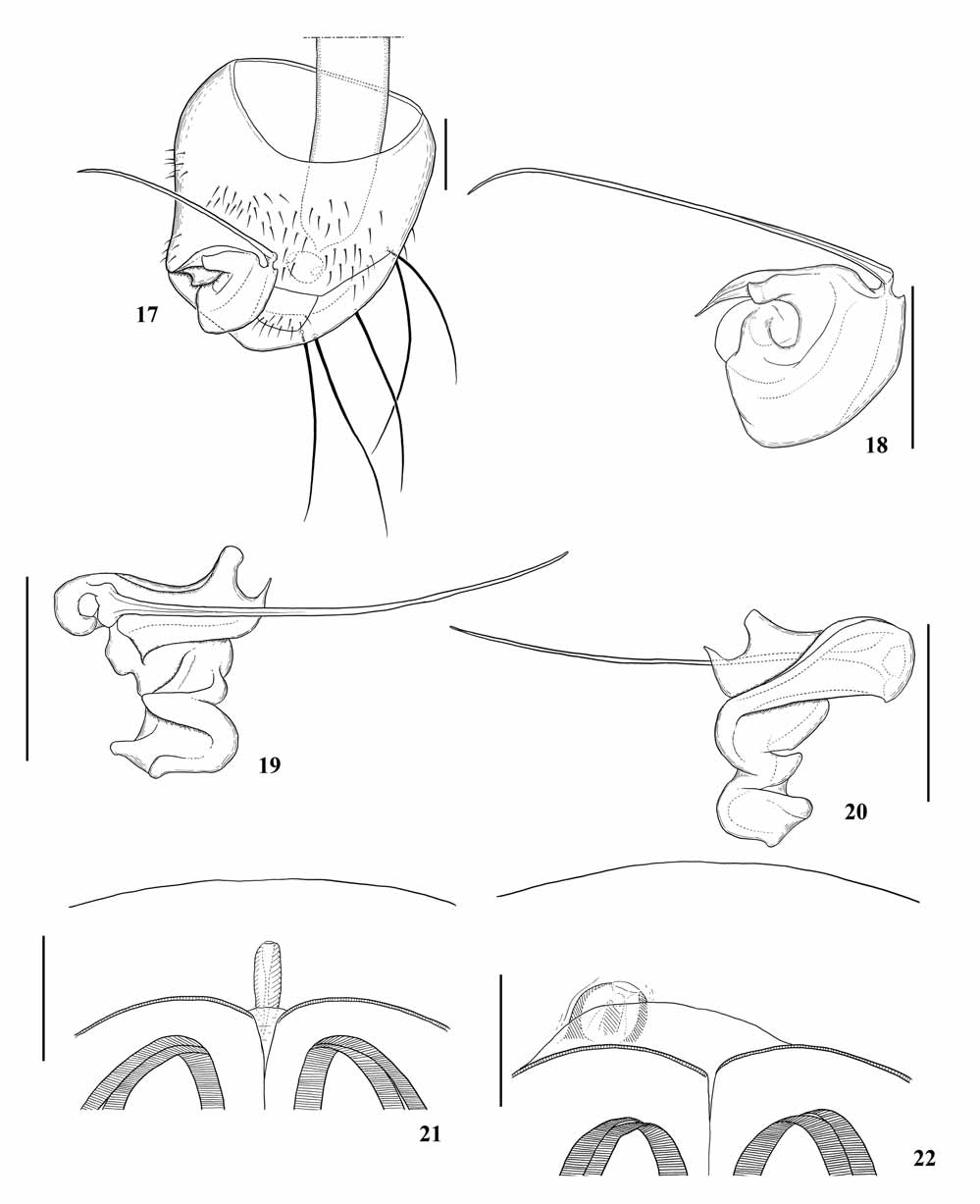
Description of male genitalia. Ventral surface of pygophore ( Fig. 17 View FIGURES 17 – 22 ) with densely distributed, simple setae and five long, stout setae posteriorly (the longest setae are about as long as the total length of pygophore); flagellum ( Figs. 17, 18 View FIGURES 17 – 22 ) straight, long, thin, apically curved, about twice as long as maximum width of cone, extending well beyond left edge of pygophore; cone ( Figs. 18–20 View FIGURES 17 – 22 ) very thin, acute apicad in dorsal view, apically well elevated and bifurcate in lateral view, with a weak indentation at base.
Measurements [% (n = 2)/ Ψ (n = 8)]. Body length 2.40–2.45/ 2.68–2.85; head length (excl. neck) 0.33–0.34/ 0.34–0.36; head width across eyes 0.33–0.34/ 0.34–0.36; vertex width 0.16–0.17/ 0.19–0.20; width between ocelli 0.12/ 0.13–0.14; length of antennal segments I-IV 0.10–0.11/ 0.13, 0.28–0.29/ 0.28–0.29, 0.23–0.25/ 0.23–0.24, and 0.25–0.26/ 0.24–0.25; length of labial segments II-IV 0.10/ 0.09–0.11, 0.28–0.29/ 0.31, and 0.23/ 0.23–0.24; anterior pronotal width 0.27–0.31/ 0.33–0.34; mesal pronotal length 0.28–0.30/ 0.33–0.34; basal pronotal width 0.69–0.71/ 0.75–0.81; length of embolial margin 0.63/ 0.69–0.73; length of cuneal margin 0.41–0.42/ 0.48–0.50; maximum width across hemelytra 0.72–0.74/ 0.78–0.81.
Material examined. JAPAN: Honshu: [Tochigi Pref.] 1Ψ, Shimominagawa, Ôhira-machi, 31.x.2003, S. Maehara, 1% ( Figs. 5, 6 View FIGURES 1 – 13 , 17–20 View FIGURES 17 – 22 ), 21.iv.2004, 1Ψ ( Fig. 15 View FIGURES 14 – 16 ), 19.ix.2005, 1Ψ, 25.x.2006, 1Ψ, 20.iv.2007, 1Ψ, 2.vii.2007 ( TKPM); 1%, Hasama, Ashikaga-shi, 19.ix.2009, S. Maehara ( TKPM). [Gunma Pref.] 1Ψ, Shimonino, Fujioka-shi, 3.i.1998, S. Arai ( TKPM). [Saitama Pref.] 1Ψ, Kanasawa-jinjya, Kamikawa-machi, 25.xi.2000, S. Arai ( TKPM). [Osaka Pref.] 1Ψ, Mt. Mikusayama, Nose-chô, 24.x.2000, K. Yamada ( TKPM).
Type material examined. Holotype Ψ, Japan, Kyushu, Wakasugiyama (Chikuzen), 29–30.iii.1930, Esaki, Hori, Fujino, Cho, Hashimoto, Yasumatsu ( ELKU).
Distribution. Japan: Honshu ( Hiura 1959; present study), Kyushu ( Esaki 1931). Montandoniola pictipennis is now confirmed from Honshu (Tochigi, Gunma, Saitama, Osaka) and Kyushu (Fukuoka).
Remarks. The male of M. pictipennis is reported here for the first time. The paramere of this species is similar to that of M. bellatula Yamada, 2007 from Bali, Indonesia, in having the straight and apically curved flagellum. However, it differs from the latter by having the cone apically well elevated and bifurcate in lateral view, with a weak indentation at the base (in M. bellatula , apically not so elevated, not bifurcate, and without an indentation at base) and the flagellum being about twice as long as maximum width of cone (cf. about 1.2 times as long as maximum width of cone).
Biology. This species was collected from grasses, dead trees and beneath fallen leaves ( Esaki 1932, 1950; Hiura 1959, 1979; Yasunaga 1993). Yamada collected a female from the leaves of Quercus serrata (Fagaceae) in northern area of Osaka. According to Mr. S. Maehara (pers. comm.), a few specimens were obtained from leaves of Quercus serrata , Q. acutissima and Prunus spp. ( Rosaceae ) at Tochigi, together with some small tubuliferan thrips.
No known copyright restrictions apply. See Agosti, D., Egloff, W., 2009. Taxonomic information exchange and copyright: the Plazi approach. BMC Research Notes 2009, 2:53 for further explanation.
|
Kingdom |
|
|
Phylum |
|
|
Class |
|
|
Order |
|
|
Family |
|
|
Genus |
Montandoniola pictipennis ( Esaki, 1931 )
| Yamada, Kazutaka, Yasunaga, Tomohide & Miyamoto, Syôiti 2010 |
Montandoniola moraguesi
| Yasunaga 2001: 287 |
| Yasunaga 1993: 169 |
| Miyamoto 1989: 167 |
Montandoniola pictipennis:
| Pluot-Sigwalt 2009: 33 |
| Herring 1966: 93 |
| Carayon 1962: 208 |
| Carayon 1961: 543 |
pictipennis
| Hiura 1959: 2 |
Ectemnus pictipennis
| Esaki 1950: 256 |
| Esaki 1932: 1673 |
| Esaki 1931: 264 |