
Montandoniola kerzhneri, Yamada, Kazutaka, Yasunaga, Tomohide & Miyamoto, Syôiti, 2010
|
publication ID |
https://doi.org/ 10.5281/zenodo.196531 |
|
DOI |
https://doi.org/10.5281/zenodo.6206634 |
|
persistent identifier |
https://treatment.plazi.org/id/03D387CE-FF93-5C71-FF7E-FB6C93F0C286 |
|
treatment provided by |
Plazi |
|
scientific name |
Montandoniola kerzhneri |
| status |
sp. nov. |
Montandoniola kerzhneri sp. nov.
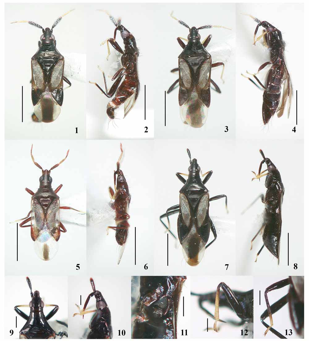
( Figs. 7–13 View FIGURES 1 – 13 , 16 View FIGURES 14 – 16 , 22 View FIGURES 17 – 22 )
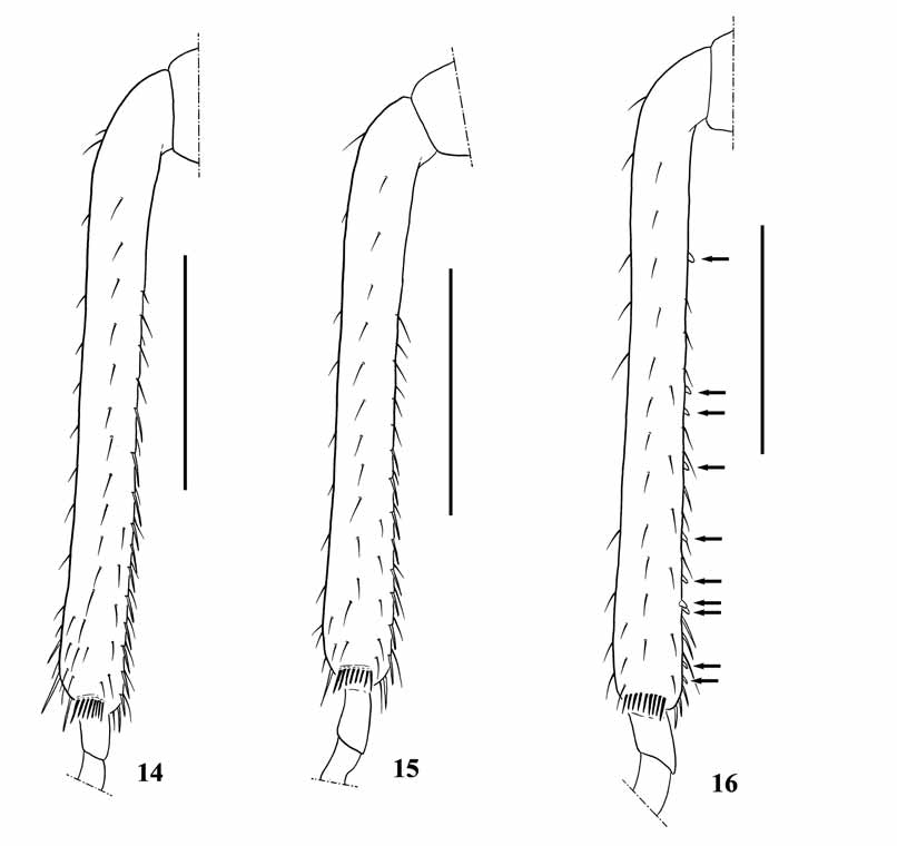
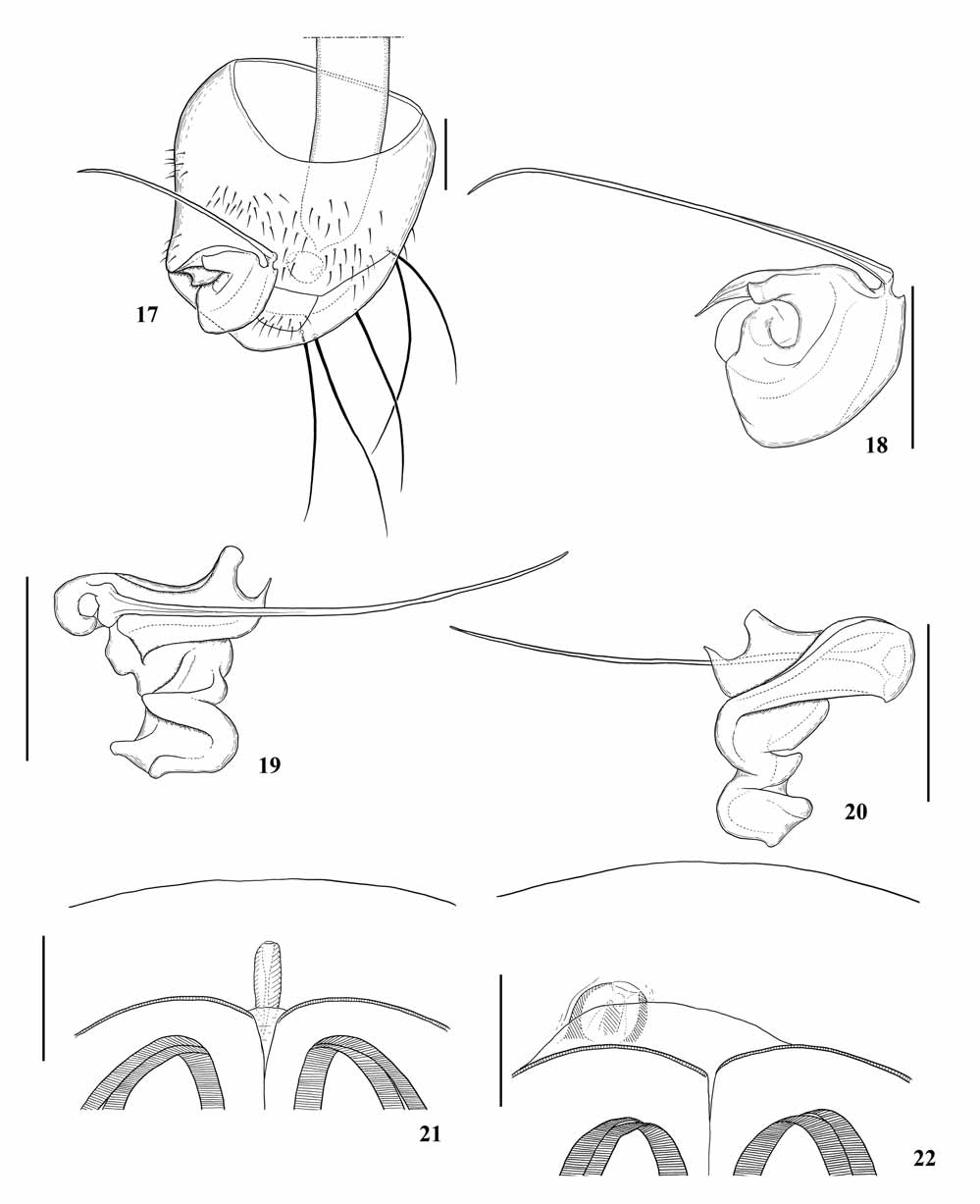
Diagnosis. Recognized by the following characters: Body larger (3.45 mm) ( Figs. 7, 8 View FIGURES 1 – 13 ). Antennal segments I and II ( Fig. 9 View FIGURES 1 – 13 ) dark brown; segment II about as long as head width across eyes. Labium ( Fig. 10 View FIGURES 1 – 13 ) dark brown, except for basal half of segment IV pale yellow. Ostiolar peritreme ( Fig. 11 View FIGURES 1 – 13 ) strongly angular posteriorly, slightly curved interiorly, not touching anterior margin of metapleura. Legs ( Figs. 7, 8 View FIGURES 1 – 13 ) dark brown; fore tibiae ( Fig. 12 View FIGURES 1 – 13 ) pale yellow except for darkened base; mid tibiae ( Fig. 13 View FIGURES 1 – 13 ) pale yellow at apical 1/4; hind tibiae uniformly dark brown; fore tibiae of female ( Figs. 12 View FIGURES 1 – 13 , 16 View FIGURES 14 – 16 ) armed with 10 small, fuscous teeth on ventral side. Copulatory tube ( Fig. 22 View FIGURES 17 – 22 ) fused on left side of intersegmental membrane between sterna VII and VIII in dorsal view, adjacent to base of ovipositor, ovoid, shortened, consisting of tiny, membranous tube surrounded by well sclerotized wall.
Description. Female (holotype). Coloration. Head and pronotum ( Figs. 7–10 View FIGURES 1 – 13 ) uniformly black; eyes reddish black; margin of ocellus red. Antennal segments I and II ( Figs. 7–9 View FIGURES 1 – 13 ) dark brown; segment II somewhat reddish at tip; segments III and IV missing. Labium ( Figs. 8, 10 View FIGURES 1 – 13 ) dark brown, except for basal half of segment IV pale yellow. Scutellum ( Fig. 7 View FIGURES 1 – 13 ) overall blackish. Clavus ( Fig. 7 View FIGURES 1 – 13 ) black along inner margin and claval commisure; endocorium ( Fig. 7 View FIGURES 1 – 13 ) narrowly black along corium-membrane boundary; embolium and cuneus ( Fig. 7 View FIGURES 1 – 13 ) wholly black; membrane ( Fig. 7 View FIGURES 1 – 13 ) semi-transparent, centrally with distinct, broad, blackish stripe, except for semi-transparent base; remaining area of hemelytra whitish. Venter of thorax ( Fig. 8 View FIGURES 1 – 13 ) black. Outer margin of ostiolar peritreme tinged with reddish brown. Legs ( Figs. 7, 8 View FIGURES 1 – 13 ) dark brown; fore tibiae ( Fig. 12 View FIGURES 1 – 13 ) pale yellow, except for darkened base; mid tibiae ( Fig. 13 View FIGURES 1 – 13 ) pale yellow at apical 1/4; hind tibiae uniformly dark brown; all tarsi pale yellow tinged with fuscous apex. Abdomen ( Fig. 8 View FIGURES 1 – 13 ) black, tinged with reddish brown.
Structure. Body ( Figs. 7, 8 View FIGURES 1 – 13 ) elongate, shiny, sparsely covered with short, silky setae. Head ( Figs. 7–10 View FIGURES 1 – 13 ) cylindrical, impunctate, as long as width across eyes, sparsely covered with long, erect setae intermixed with short, suberect setae; long, erect seta on each side of tylus, anteromesad of each eye, and between eye and ocellus; anteocular region ( Fig. 9 View FIGURES 1 – 13 ) about as long as length of eye in dorsal view; vertex ( Fig. 9 View FIGURES 1 – 13 ) about 2.3 times as wide as eye in dorsal view; postocular region distinctly long; eye ( Figs. 9, 10 View FIGURES 1 – 13 ) oblong, not exceeding level of ventral surface and dorsal surface of head in lateral view. Antennal segment I ( Fig. 9 View FIGURES 1 – 13 ) just reaching apex of head, sparsely covered with short setae; segment II ( Fig. 9 View FIGURES 1 – 13 ) thickened, about as long as head width across eyes, densely covered with suberect setae which are shorter than width of the segment; segments III and IV missing. Labium ( Fig. 10 View FIGURES 1 – 13 ) robust, reaching near fore coxae, sparsely covered with very short, reclining setae; segments II with a pair of long, erect setae near middle; segment III about 2.6 times as long as segment II; segment IV about 0.64 times as long as segment III.
Pronotum ( Figs. 7, 9 View FIGURES 1 – 13 ) with long, stout, erect setae on anterolateral and posterolateral corners and a pair of similar setae behind the collar; surface smooth, with scattered short, suberect setae and fine, erect setae along lateral margin; anterior margin nearly straight, slightly shorter than mesal length; lateral margin shallowly concave inwardly; lateral carina well expanded anteriorly, gradually more obscure posteriad; posterior margin concave, about 2.5 times as wide as anterior margin; collar about 0.25 times as long as mesal pronotal length, with scattered short setae, weakly rugose; callus demarcated by a shallow transverse impression; posteromedial region of pronotum widely depressed. Scutellum ( Fig. 7 View FIGURES 1 – 13 ) smooth, slightly wider than long, anteriorly swollen and gradually more depressed posteriad. Hemelytra ( Fig. 7 View FIGURES 1 – 13 ) subparallel-sided, slightly narrowed toward posteriorly, impunctate, sparsely covered with short, suberect setae; endocorium 1.75 times as wide at maximum as embolium; cuneal margin 0.66 times as long as embolial margin; membrane with two weak veins, inner vein arising from base of membrane, outer vein a little remote from outer margin. Ostiolar peritreme ( Fig. 11 View FIGURES 1 – 13 ) broad, elbowed at middle, strongly anglar posteriorly, gradually narrowed anteriad, slightly curved interiorly, not touching anterior margin of metapleura; outer margin of ostiolar peritreme strongly raised above level of surrounding evaporative area. Legs densely covered with short, reclining setae; all tibiae lacking distinct long spines; fore tibiae ( Figs. 12 View FIGURES 1 – 13 , 16 View FIGURES 14 – 16 ) armed with 10 small, fuscous teeth on ventral side; hind tibiae ( Fig. 13 View FIGURES 1 – 13 ) weakly flattened; mid- and hind coxae widely separated. Abdomen ( Fig. 8 View FIGURES 1 – 13 ) ventrally covered with short, suberect setae at posterior portion of respective segments.
Female genitalia ( Fig. 22 View FIGURES 17 – 22 ): Genital segments (segments VII to IX) laterally covered with long, stout setae; copulatory tube ( Fig. 22 View FIGURES 17 – 22 ) fused on left side of intersegmental membrane between sterna VII and VIII in dorsal view, adjacent to base of ovipositor, ovoid, shortened, consisting of tiny, membranous tube surrounded by well sclerotized wall.
Male. Unknown.
Measurements (holotype, Ψ). Body length 3.45; head length (excl. neck) 0.38; head width across eyes 0.39; vertex width 0.21; width between ocelli 0.15; length of antennal segments I-II 0.15, 0.40, (III and IV missing); length of labial segments II-IV 0.13, 0.34, and 0.22; anterior pronotal width 0.36; mesal pronotal length 0.40; basal pronotal width 0.89; length of embolial margin 0.91; length of cuneal margin 0.60; maximum width across hemelytra 0.91.
Holotype. Ψ (TKPM-IN-13226; Figs. 7–13 View FIGURES 1 – 13 , 16 View FIGURES 14 – 16 , 22 View FIGURES 17 – 22 ), labeled “[ JAPAN] / Shirahama-rindô, / Iriomote Is., / The Ryukyus, / 1. III. 2002 / T. Ishikawa leg.” [white square] ( TKPM). Right antenna and left antennal segments III and IV are missing.
Distribution. Japan (the Ryukyus: Iriomote Is.).
Etymology. The specific name is dedicated to the late Dr. I. M. Kerzhner, one of the world’s leading heteropterists, who regretfully passed away in May, 2008.
Remarks. Montandoniola kerzhneri sp. nov. is unique among the other congeners in having the female fore tibiae furnished with the 10 small, fuscous ventral teeth; and with shortened ovoid copulatory tube, consisting of tiny, membranous tube surrounded by well sclerotized wall. In addition to these characters, this new species undoubtedly differs from Japanese congeners M. thripodes and M. pictipennis by the coloration of the labium and legs.
Biology. Unknown.
No known copyright restrictions apply. See Agosti, D., Egloff, W., 2009. Taxonomic information exchange and copyright: the Plazi approach. BMC Research Notes 2009, 2:53 for further explanation.
|
Kingdom |
|
|
Phylum |
|
|
Class |
|
|
Order |
|
|
Family |
|
|
Genus |