
Gieysztoria bimaculata, Lu, Yan-Hong, Wu, Cheng-Chen, Xia, Xiao-Jie & Wang, An-Tai, 2013
|
publication ID |
https://doi.org/ 10.11646/zootaxa.3745.5.5 |
|
publication LSID |
lsid:zoobank.org:pub:D86E6EEE-5B0B-4E7A-9F90-63DA2A76E9F1 |
|
DOI |
https://doi.org/10.5281/zenodo.5629391 |
|
persistent identifier |
https://treatment.plazi.org/id/03D887EA-3728-FFA8-E5F4-FB19E1682DCF |
|
treatment provided by |
Plazi |
|
scientific name |
Gieysztoria bimaculata |
| status |
sp. nov. |
Gieysztoria bimaculata View in CoL n. sp. Wang,Lu & Wu
( Figs.1–2 View FIGURE 1 View FIGURE 2 )
Localities. 1) Constructed wetland system (pH=6.78, 27.5?) in artificial lake in Shenzhen University campus, Shenzhen, Guangdong province, China (22° 31' 44"N, 113° 55' 52"E).
2) Landscape ponds in Shenzhen Garden Expo Park, Shenzhen, Guangdong province, China (22° 32' 25"N, 113° 59' 47"E).
3) Fairy lake in Seven Star Crags, Zhaoqing, Guangdong province, China (23° 4' 33"N, 112° 28' 43"E).
Material. Holotype (PLA–G0005): permanent slides of specimen strained by H.E. Method. Paratype (PLA– G0006–10): PLA–G0006–8: permanent slides of specimens strained by H.E. Method. PLA–G0009–10: permanent slides of stylet in polyvinyl-lactophenol.
Etymology. The species has two clavate pigmentations dorsally between pharynx and intestine.
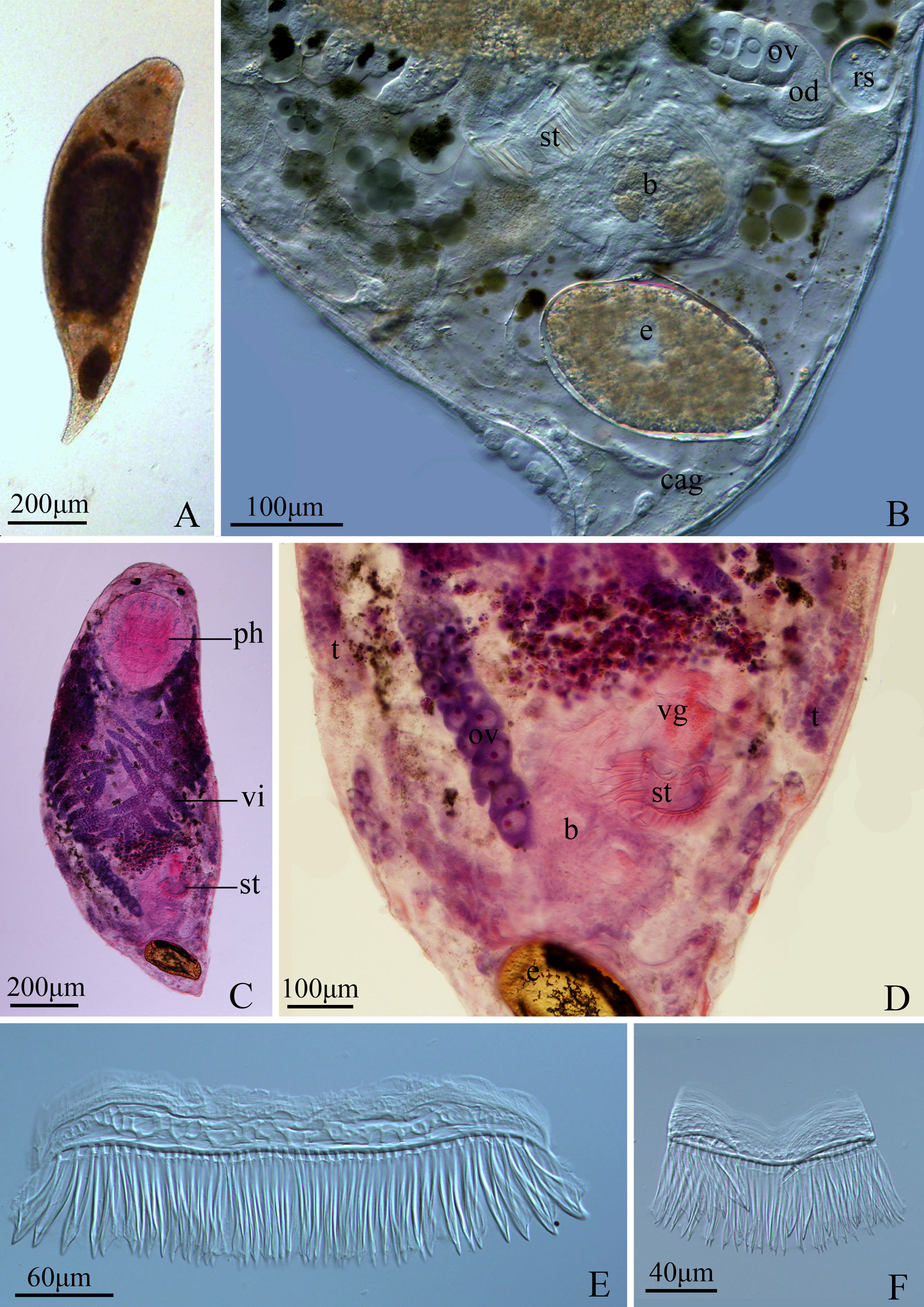
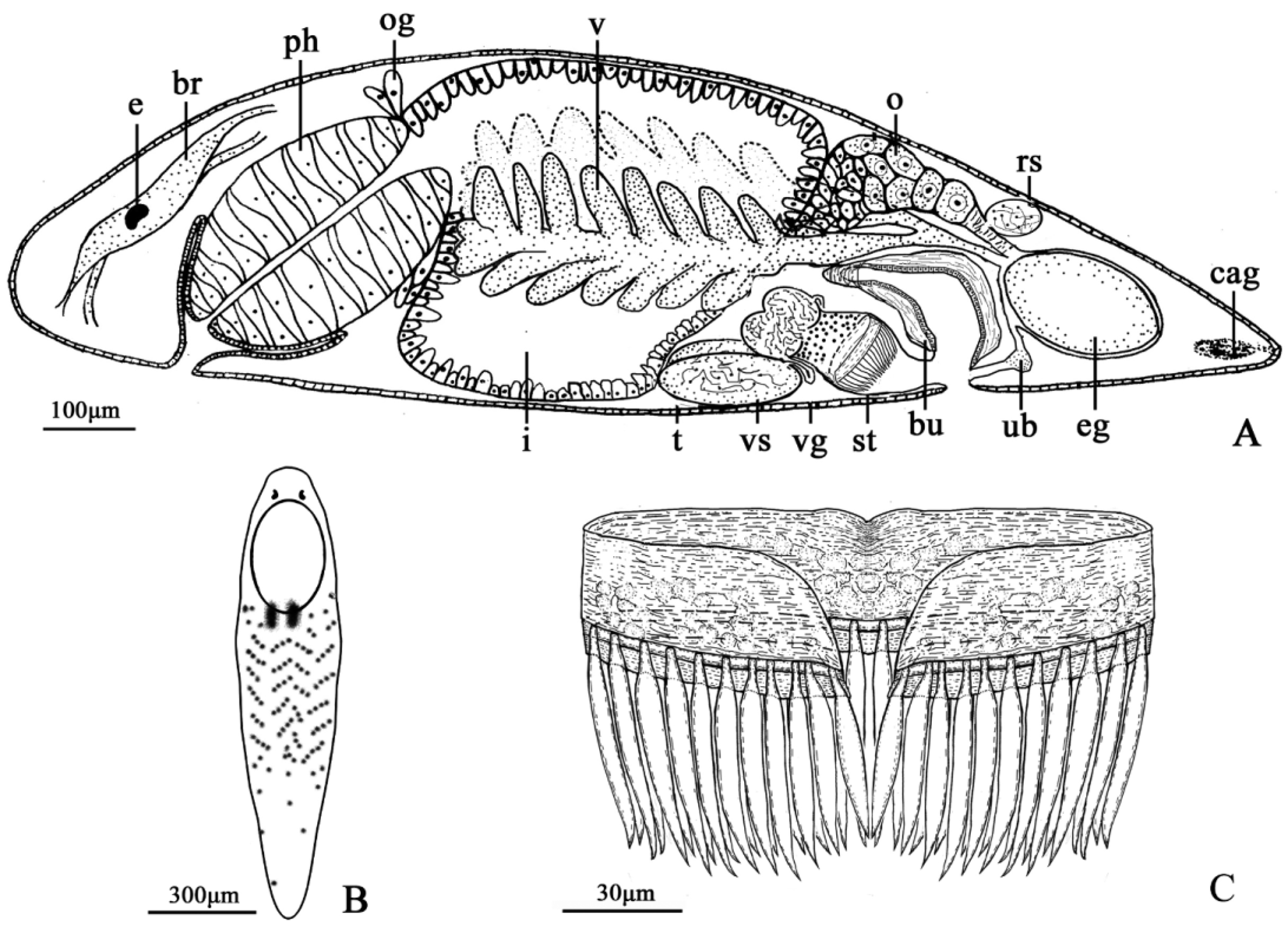
Description. Living adults are 1315–1367 µm long and approximately 290µm wide. Animals, conspicuously, have two clavate pigmentations dorsally between pharynx (ph) and intestine (i) ( Fig.1 View FIGURE 1 A; Fig. 2 View FIGURE 2 B). With 6µm long cilia densely covering the whole body, animals have a blunt anterior end and a tapering posterior end. Sensory cilia, 17µm average length, are also distributed over the body but especially rich on the dorsal side. Pair of eyes (ey) are in kidney-shaped and present on the dorsal anterior end; each eye spot consists of a collection of pigment spheres ( Fig. 2 View FIGURE 2 B).
Oral pore lies antero-ventrally. At 1/5 of the body length, barrel-shaped pharynx (ph) is 323µm long and 243µm wide, with oesophageal glands (og) at the posterior end of the pharynx (ph) ( Figs. 1 View FIGURE 1 A & C).
Female reproductive system comprising a thick rod-like ovary (ov), which is 245µm long and 43.5µm wide, lies at the left side of the body and dorsal to the posterior end of the intestine (i) ( Fig. 1 View FIGURE 1 B). Immature oocytes arranged in piles at the anterior portion of the ovary (ov) and enlarge gradually to the distal part ( Figs. 1 View FIGURE 1 B–D; Fig. View FIGURE 2
2A). Ovary (ov) ends into a short oviduct tightly associated to a separate seminal receptacle (rs) and later connects to the uterus by a variable female duct (fd). Seminal receptacle (rs), diameter 96µm, is filled with allo-sperms ( Fig. 2 View FIGURE 2 A). Uterus with thick walls, connects to the genital atrium. Irregular-oval-shaped eggs (e) are approximately 182µm in length and 81µm in width ( Figs.1 View FIGURE 1 A & B). Vitellaria (vi), about 525µm long, start from both side at the trailing edge of the pharynx (ph), end dorsally at the end of intestine (i), and enter into a common female duct (fd). The vitellaria (vi) have numerous finger-like branches; each branch is 115µm in average length ( Fig. 1 View FIGURE 1 C; Fig. 2 View FIGURE 2 A). Gonopore lies ventro-caudally.
Male reproductive system consists of two oval testes (t) ( Figs. 1 View FIGURE 1 C–D; Fig. 2 View FIGURE 2 A), 118µm long and 83µm wide, located under both ventro-caudal side of the intestine (i). Vasa deferentia (3µm wide) start from the middle of each testis (t) and project into a double-bulb shape seminal vesicula (vs) subapically (72µm diameter of each) ( Fig. 2 View FIGURE 2 A). The seminal vesicle (vs) connects with the distal columnar prostate vesicle (pv) (84µm in height and diameter 92µm), which is tightly associated to the sclerotic stylet (st) ( Figs. 1 View FIGURE 1 E–F; Fig. 2 View FIGURE 2 C).
The sclerotic stylet (st) ( Figs. 1 View FIGURE 1 E–F; Fig. 2 View FIGURE 2 C) comprises two distinct portions: a belt-like girdle and numerous distal spines in similar shape and size. The maximum length of the stylet (st) is 95µm with a maximum girdle height of 44.9µm. The girdle, with a maximum circumference ca. 250µm, curves into a crescent shape. Countless hollow concaves, considered as muscular attachment points, are present in every single separated stylet (st) ( Figs. 1 View FIGURE 1 E–F; Fig. 2 View FIGURE 2 C). Distally, dagger-shaped spines arise from the proximal girdle. The number of spines ranges from 40 to 46. The first two spines on both side of the girdle are 46±2.9µm (n=17) in average and the rest are 55±3.4µm (n=140) ( Fig. 2 View FIGURE 2 C).Spines are 5-6µm embedded in the proximal girdle, arising as hollow and straight. Spines (ca.1µm thick wall) widen to 10µm around the mid-point and then form a point at the end.
Remarks. The stylet morphology firmly suggests that G. bimaculata n. sp. is a typical “Aequales” species of Gieysztoria . Based mainly on stylet morphology and configuration, G. bimaculata n. sp. can be compared to the following six species: Gieysztoria expedita (Hofsten 1907) , G. bicoronaria (Fulinski & Szynal 1933) and G. expeditoides (Luther 1955) from Europe; G. bergi (Beklemischev 1927) in Asia; G. superba (Hartenstein & Dwine 2000) and G. queenslandica (Hochberg & Cannon 2001) in Australia. G. superba is the species with the most similar stylet to G. bimaculata n. sp..This two species differ in size, pigmentation pattern and the structure of the male copulatory organ (see Table 1 View TABLE 1 ). G. superba is a small species which is 550µm long and 200µm wide with posterior red pigment bands and scattered brown pigment spots covering the head and trunk. G. bimaculata n. sp. is a larger one which is 1350µm long and 290µm wide with scattered brown spots covering from posterior end of the pharynx to the tapering end of the body. Furthermore, conspicuously, G. bimaculata n. sp., present two dark brown clavate pigmentation both ventrally and dorsally between pharynx and intestine. However, the main taxonomic character is the structure of the sclerotic stylet(Luther 1955). The girdle of G. superba is 20µm of height and the one of G. bimaculata n. sp. is much higher as 35µm in average, additionally, 45µm at maximum. The circumference of the girdle of G. superba is ca.100µm and the one of the new species is ca.250µm. The number of distal spines of G. superba is ca.40 comparing to 44~46 spines of the new species. The shape and length of the spines of both species are different. The spines of G. superba are straight and fine which are equal length and approx. 43µm long. Nevertheless, spines of G. bimaculata n. sp. are dagger-shaped with fine tips slightly bent. It is worth mentioning that the first spine (46±2.9µm) on both side girdle are ca.10µm shorter than the spines (55±3.4µm) in the middle. As for G. queenslandica , the Australian species has a stylet with 11µm girdle and 27–32 distal spines. G. queenslandica with spines which are ca. 7µm less than the spines of the new species, shows additional differences in girdle height as well as the number and length of spines. G. expedita and G. expeditoides have a small stylet less than 40µm in length and 20–24 spines. G. b e rgi has a 107µm stylet and 24 distal spines. G. bicoronaria has a stylet with 31 fine spines. Within the “Aequales” group, G. bimaculata n. sp. has the maximum number of spines (44–46) and girdle circumference (250µm).
No known copyright restrictions apply. See Agosti, D., Egloff, W., 2009. Taxonomic information exchange and copyright: the Plazi approach. BMC Research Notes 2009, 2:53 for further explanation.
|
Kingdom |
|
|
Phylum |
|
|
Class |
|
|
Order |
|
|
Family |
|
|
Genus |