
Pattersoniomyces tillandsiae (F. Patt.) Piątek, M. Lutz, M.F. Landell & C.A. Rosa 2017
|
publication ID |
https://doi.org/ 10.1007/s13127-017-0340-8 |
|
DOI |
https://doi.org/10.5281/zenodo.4562818 |
|
persistent identifier |
https://treatment.plazi.org/id/03D93021-FFDE-0E7C-2494-2E65FD3DFA03 |
|
treatment provided by |
Donat |
|
scientific name |
Pattersoniomyces tillandsiae (F. Patt.) Piątek, M. Lutz, M.F. Landell & C.A. Rosa |
| status |
comb. nov. |
Pattersoniomyces tillandsiae (F. Patt.) Piątek, M. Lutz, M.F. Landell & C.A. Rosa View in CoL , comb. nov.
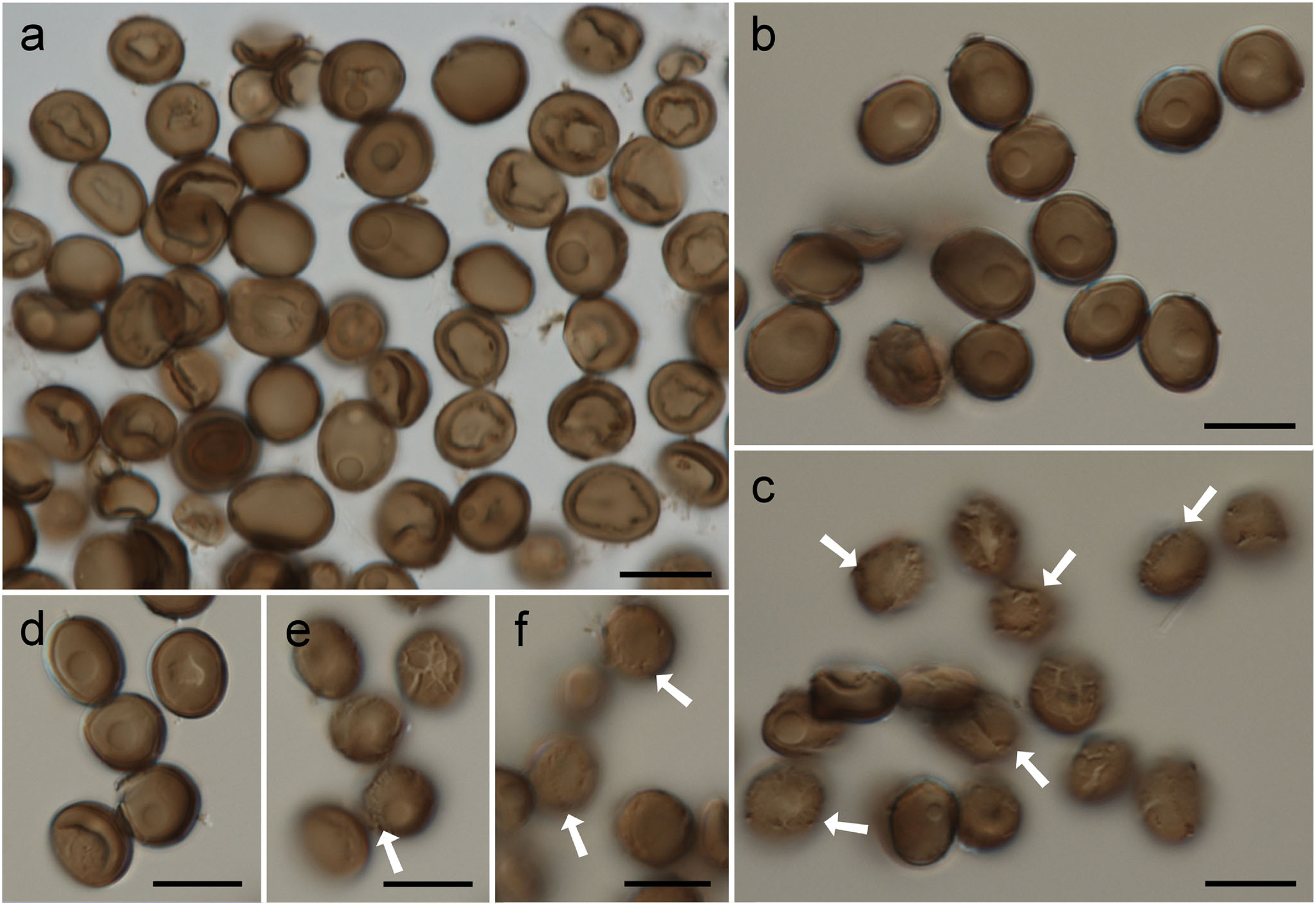
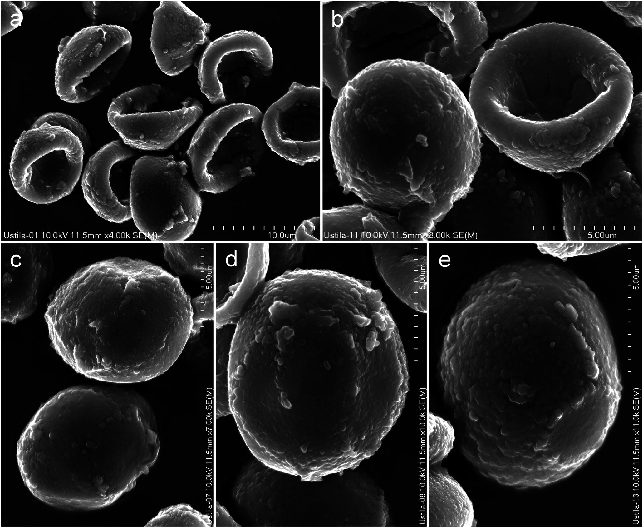
Figs. 2 View Fig , 3 View Fig , 4 View Fig , 5 View Fig , and 6
MycoBank no. MB821984
Basionym: Ustilago tillandsiae F. Patt., J. Mycol. 8: 135 (1902)
Description of the teleomorph: Sori in all flowers of the inflorescences of Tillandsia spp. suggesting systemic infection, 1 – 2 cm long, producing naked, dusty spore masses around the rudimentary developed innermost floral organs —staminal filaments and ovary remain partly intact in the sorus center, anthers not developed, peridium lacking, sori protected only by closed perianths and bracts. Spores usually single, sometimes glued together forming small groups, variable in color, shape, and size (also between collections, see Table 1 View Table 1 ), pale brown to olive-brown, globose or subglobose, more or less collapsed or hemispherically cupped, (5.0 –)6.0 – 11.5(−16.0) × 5.0 – 10.5 μm, usually with one or exceptionally two spherical bodies (lipid granules) in the cytoplasm; wall even 0.4 – 0.8(−1.0) μm thick, somewhat darker than the rest of spore, often somewhat lighter on one side, surface variably ornamented, smooth or with brittle epispore breaking up into thin polygonal areas, cracked or covered with fine ridges as seen by LM, almost smooth, granulose or covered with fine, circular, or irregular ridges as seen by SEM.
Specimens examined: Guatemala ( intercepted at Miami- 010189-Florida ), on Tillandsia leiboldiana , 23 Jan. 1975, leg. E.B. Lee ( BPI 168186 ); Honduras ( intercepted at Miami-014910-Florida ), on Tillandsia leiboldiana , 17 Nov. 1976, leg. F. Matthews ( BPI 168184 ); Mexico, Valleė de Cordova, on Tillandsia leiboldiana , 23 Jan. 1865, leg. Bourgeau 64 ( BPI 168185 – paratype); Mexico, Vera Cruz ( intercepted at Brownsville-007786-Texas ), on Tillandsia flabellata , 21 Nov. 1977, leg. J.M. Van Valkenburgh ( BPI 168187 ).
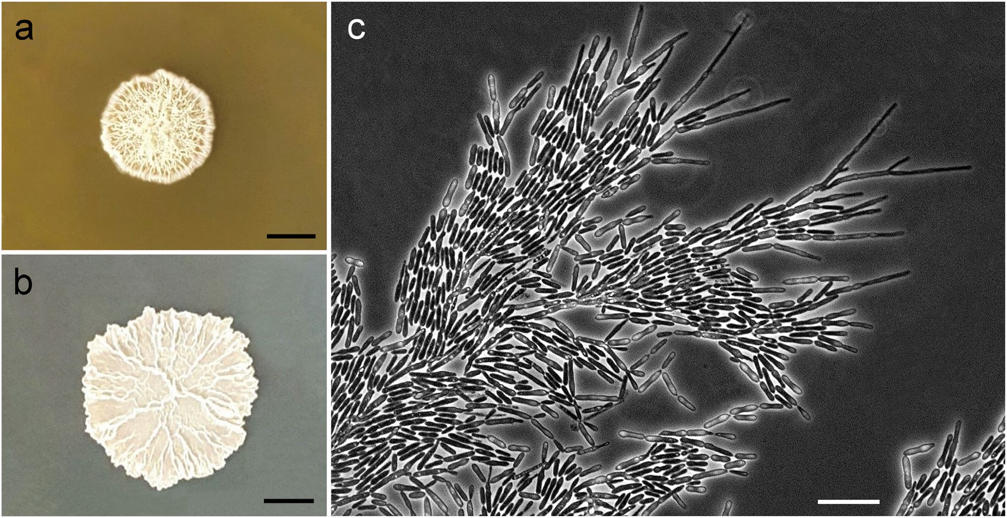
Description of the anamorph: Growth on YM agar after 3 days at 25 °C: colonies smooth to wrinkled, butyrous, glistening, cream-colored to light salmon with an entire margin, cells ellipsoidal to fusoid and variable in size, 3 – 9 × 1.5 – 3 μm, budding is polar on a short denticle. Growth in Dalmau plate culture on cornmeal agar after 3 weeks: pseudomycelium and true mycelium formed. Sexual reproduction not observed. Ballistoconidia not produced. Fermentation absent. Assimilation of carbon sources: D- glucose, inulin, sucrose, raffinose, melibiose, galactose, lactose, trehalose (slow), maltose, melezitose, cellobiose (slow), salicin (slow), L-sorbose, D- xylose, L- arabinose, D- ribose (slow), glycerol, erythritol, ribitol (slow), D- mannitol, D- glucitol, myoinositol (latent), succinic acid, citric acid, D- gluconate, D-glucosamine (latent), N -acetyl-D- glucosamine and xylitol (variable) positive. No growth was detected in L- rhamnose, D- arabinose, ethanol, methanol, galactitol, DL- lactate and hexadecane. Assimilation of nitrogen compounds: positive for nitrate, lysine and cadaverine (latent). Growth in aminoacid-free medium negative. Growth at 37 °C negative. Growth on YM agar with 10% sodium chloride negative. Growth in 50% glucose negative. Acid production negative. Starch-like compounds not produced. In 1 0 0 μg cycloheximide mL −1 growth is negative. Diazonium Blue B reaction positive.
Yeast strains were isolated from the phylloplane of Canistrum improcerum and water tanks of Vriesea minarum . The strains have been deposited in the Collection of Microorganisms and Cells of the Federal University of Minas Gerais (Coleção de Micro-organismos e Células da Universidade Federal de Minas Gerais, UFMG), Belo Horizonte, Minas Gerais, Brazil, as strains UFMG-CM-Y 1455, UFMG-CM-Y 1466, UFMG-CM-Y6109 (= BSS144), and UFMG-BRO-110B, and are permanently preserved in a metabolically inactive state.
Host plants/source and distribution: Tillandsia flabellata Baker , Tillandsia leiboldiana Schltdl. , Tillandsia L. sp. (teleomorph), Canistrum improcerum Leme & J.A. Siqueira , Vriesea minarum L.B. Sm. (anamorph) ( Bromeliaceae ) — the distribution of Pattersoniomyces tillandsiae is neotropical. The sequenced teleomorphic material is from Honduras (new country report), the remaining morphologically analyzed smut specimens are from Guatemala (new country report) and Mexico, and this smut has been additionally reported from Costa Rica and Mexico ( Clinton 1902; Zundel 1953; Durán 1987; Piepenbring 1996, 2003; Vánky 2012). Yeast strains of this species are from Brazil (new country report).

No known copyright restrictions apply. See Agosti, D., Egloff, W., 2009. Taxonomic information exchange and copyright: the Plazi approach. BMC Research Notes 2009, 2:53 for further explanation.
|
Kingdom |
|
|
Phylum |
|
|
Class |
|
|
Order |
|
|
Family |
|
|
Genus |