
Sagmatocythere rectum (Brady) (Loxoconchidae)
|
publication ID |
https://doi.org/10.11646/zootaxa.5244.4.1 |
|
publication LSID |
lsid:zoobank.org:pub:82968DC8-49F4-4E1E-9DFA-EE71531EE121 |
|
DOI |
https://doi.org/10.5281/zenodo.7665202 |
|
persistent identifier |
https://treatment.plazi.org/id/03DA682B-2E17-FFB6-A398-FF56FB273BD2 |
|
treatment provided by |
Plazi |
|
scientific name |
Sagmatocythere rectum (Brady) (Loxoconchidae) |
| status |
|
? Sagmatocythere rectum (Brady) (Loxoconchidae) comb. nov.—A case of homeomorphy
? Sagmatocythere rectum ( Brady, 1868) is a Recent (presumed living) rare species, whose original description, based on the examination of only one carapace, was brief and without illustration.The analysed specimen was from Lerwick Bay, Shetland, dredged from a depth of c. 21–25 m, and the species was first attributed to the genus Cytheropteron , subfamily Cytheropterinae . Later, the species was found in other localities (west of Ireland, Bay of Biscay), and figured with some drawings in Brady & Robertson (1869, pl. XX, figs 6–8) and in Brady & Norman (1889, pl. XIX, figs 13, 14). Brady & Norman (1889) considered the species as belonging to the genus Bythocythere Sars, 1866 , family Bythocytheridae . Athersuch & Horne (1983), studying specimens of Nannocythere pavo ( Malcomson, 1886) and of Cytheropteron rectum Brady, 1868 , two species considered by Brady & Norman (1889) as only one, with N. pavo being the juveniles of C. rectum , moved both species, completely distinct, to the family Loxoconchidae .
Our attention was initially drawn to this species by its resemblance to members of the genus Microceratina , however, closer examination and comparison with Microceratina species especially from the Mediterranean demonstrated that we are dealing with an example of homeomorphy that merits clarification.
The illustrations in Brady & Norman (1889), together with SEM images of one specimen from St Magnus Bay, Shetland (Norman Collection 1911.11.8 M3820), allow us to identify some Portuguese material (loose valves) collected in modern sediments of the Eastern Algarve continental shelf, off Tavira, and of the Mira estuary, SW Portugal, as belonging to the same species. The Portuguese specimens have a gongylodont hinge, NPC of two types and StPC (description below), and are considered as Loxoconchidae , in accordance with Athersuch & Horne (1983). We tentatively place the rectum species in the genus Sagmatocythere , described by Athersuch (1976), due to its dimensions> 0.35 mm (L = 0.43–0.45 mm), the sub-quadrangular to sub-rectangular outline, the pitted/reticulate ornamentation, the presence of few and simple marginal pore canals and of the gongylodont hinge with the terminal elements “comma-shaped” and lobate. However, the genus was described using morphological features of the valves and soft parts, the latter not available in the Portuguese material with only loose valves found. The Loxoconchidae genus Nannocythere Schäfer, 1953 possesses some characteristics in common with rectum species but has a smaller size (< 0.35 mm), conspicuous pore conuli and an anterior marginal flange. A more complete description of the carapace’s morphology, important for its taxonomy and comparison with Microceratina , is presented below.
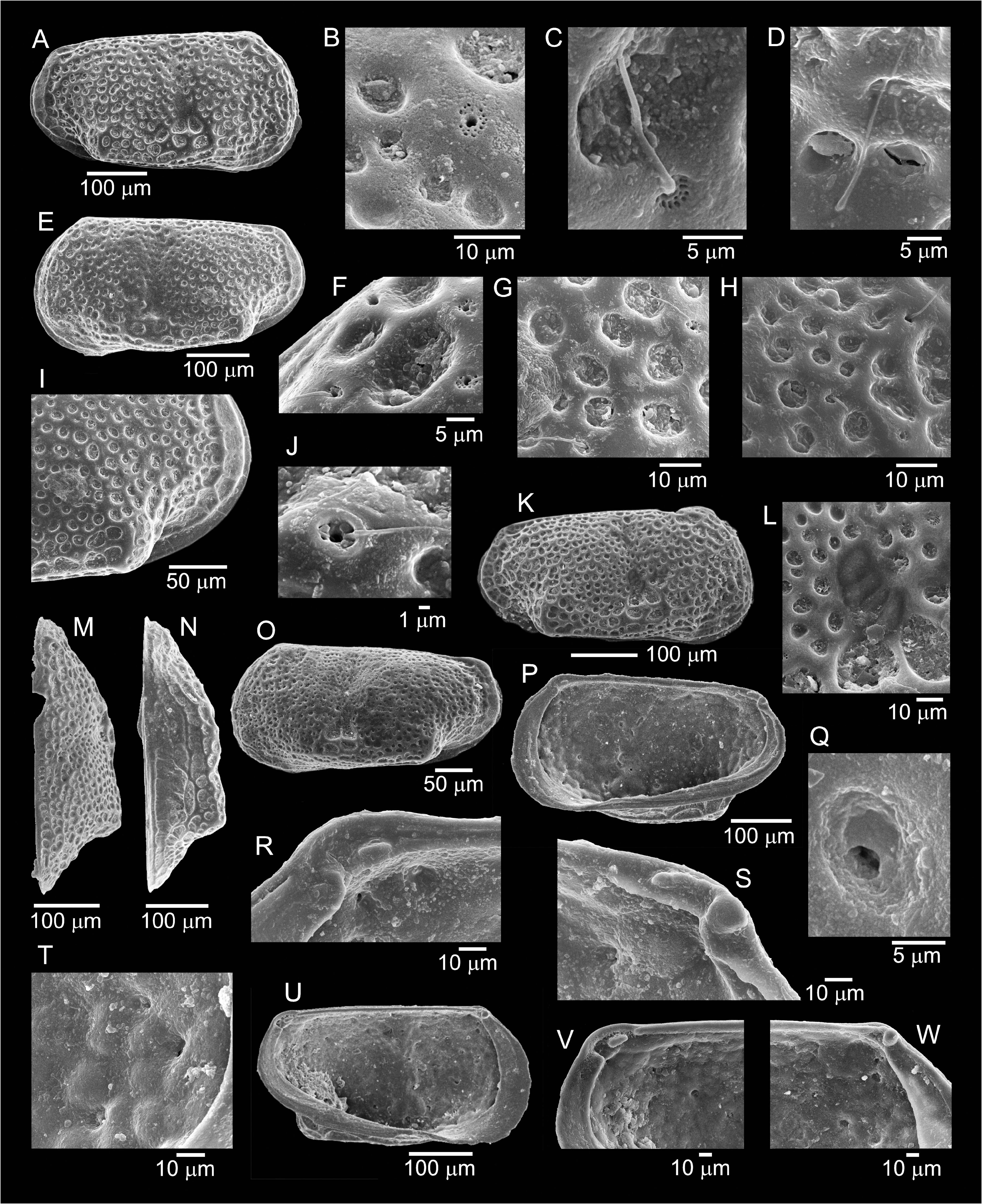
The carapace is small, sub-rectangular in lateral outline. L < 0.5 mm with greatest length at mid-height, anterior margin equicurvate. Greatest height located at the anterior third of L; H/L% is 50–51%. Greatest width at approximately three-quarters of length from anterior, slightly larger than one-quarter of length, corresponding to the maximum development of the ventral inflation. The anterior margin displays a lamellar AMZ, representing 2.5% of L ( Fig. 15E View FIGURE 15 ), and which continues along the ventral and posterior margins of the valve ( Figs 15E, I View FIGURE 15 ).
Lateral surface with a dorso-median sulcus ( Figs 15A, E, K, O View FIGURE 15 ), ending in an enlarged subcentral depression where the adductor muscle scars can be seen ( Figs 15K, L View FIGURE 15 ). Mid and postero-ventral zones inflated, forming a longitudinal expansion almost parallel to the dorsal margin, ending in a round alar protuberance, emphasised by a thickening of the reticulation net ( Fig. 15I View FIGURE 15 ). Eye tubercle not obvious. In dorsal view the carapace is inflated, with posterior end strongly prominent and dorsal-median sulcus visible ( Fig. 15M View FIGURE 15 ); in ventral view it has a sagittate outline, with a weak compression in the alar processes and the posterior margin extending beyond them ( Fig. 15N View FIGURE 15 ). Dorsal margin straight, with obtuse cardinal angles. Ventral margin weakly sinuous, tapering towards the posterior. Anterior margin rounded, infracurvate, with extremity below mid-height. Posterior margin flattened, rounded to slightly pointed, infracurvate. The valve surface is covered by large cells, generally of round shape. The diameter of the cells illustrated in Figs 15B, H, I View FIGURE 15 : M = 6.5 µm (r = 4.6–9.25, N = 19). There is no reticular polygonal pattern of muri covering the surface of the valve. Simple NPC without a rim (A’- type) with sensilla are visible on the tectum of the valve ( Fig. 15D View FIGURE 15 ); the diameter for the sensillum illustrated is 1.25 µm at its basal part. There are other simple NPC (A”- type) opening either on a flat conulus ( Figs 15F, H View FIGURE 15 ) or surrounded by a rim ( Fig. 15J View FIGURE 15 ); their diameters vary between 1–1.5 µm. StPC represented by a central pore with sensillum and a perforated surrounding area (C-type pore) are distributed on the tectum of the valve ( Figs 15B, C, F View FIGURE 15 ). The StPC of? S. rectum are never located in the cells. Such StPC have a diameter of about 3.2–3.5 µm with the central pore having a diameter of 1–1.5 µm. The sensillum emerging from inside the pore is slender, e.g., those from Fig. 15F View FIGURE 15 have a diameter of about 0.5 µm. The area around the central pore is perforated with 10–15 small pores opening within tubuli which penetrate the valve. There is one, sometimes two rows of such pores within the area surrounding the central pore ( Figs 15B, C, F View FIGURE 15 ). These minute pores have a size of: M = 0.3 µm (r = 0.18–0.43, N = 11) .
Internally the selvage of the RV extends up to the OM of the valve ( Fig. 15P View FIGURE 15 ). The diameter of pores apparently emerging from a StPC ( Fig. 15Q View FIGURE 15 ) is 3.33 µm; two tubules with a diameter of about 0.6–0.8 µm are visible inside the larger cavity. Other pores on the internal side of the valve are visible in Figs 15T, V, W View FIGURE 15 ; their diameter is approximately 5 µm. Hinge gongylodont ( Figs 15P, R, S, U–W View FIGURE 15 ) with smooth median element and “comma-shaped” and lobate terminal elements (clearer in the posterior part, Figs 15S, V View FIGURE 15 ). Marginal zone rather wide anteriorly, relatively narrow posteriorly, both with vestibules, the posterior being very narrow; MPC straight and simple, about seven in the posterior zone, number anteriorly could not be determined; four slightly elongated adductor muscle scars, arranged in a curved vertical row, seen externally in the subcentral depression ( Figs 15K, L View FIGURE 15 ).
| NPC |
National Pusa Collection |
No known copyright restrictions apply. See Agosti, D., Egloff, W., 2009. Taxonomic information exchange and copyright: the Plazi approach. BMC Research Notes 2009, 2:53 for further explanation.
|
Kingdom |
|
|
Phylum |
|
|
Class |
|
|
Order |
|
|
Family |
|
|
Genus |