
Munida manqingae, Liu & Lin & Huang, 2013
|
publication ID |
https://doi.org/10.11646/zootaxa.3734.3.7 |
|
publication LSID |
lsid:zoobank.org:pub:3383F82F-5907-45B9-861C-86B5CE12103C |
|
DOI |
https://doi.org/10.5281/zenodo.5271692 |
|
persistent identifier |
https://treatment.plazi.org/id/03DA87AD-FFF6-6E6B-86B9-5516991AD0BC |
|
treatment provided by |
Felipe |
|
scientific name |
Munida manqingae |
| status |
sp. nov. |
Munida manqingae View in CoL sp. nov.
Material examined. HOLOTYPE: ♀ (CL 6.9 mm) ovigerous, Southwest Indian Ridge, hydrothermal vent field, 36.1010°S 53.2552°W, 2218 m, TVG17, 16 February 2009.
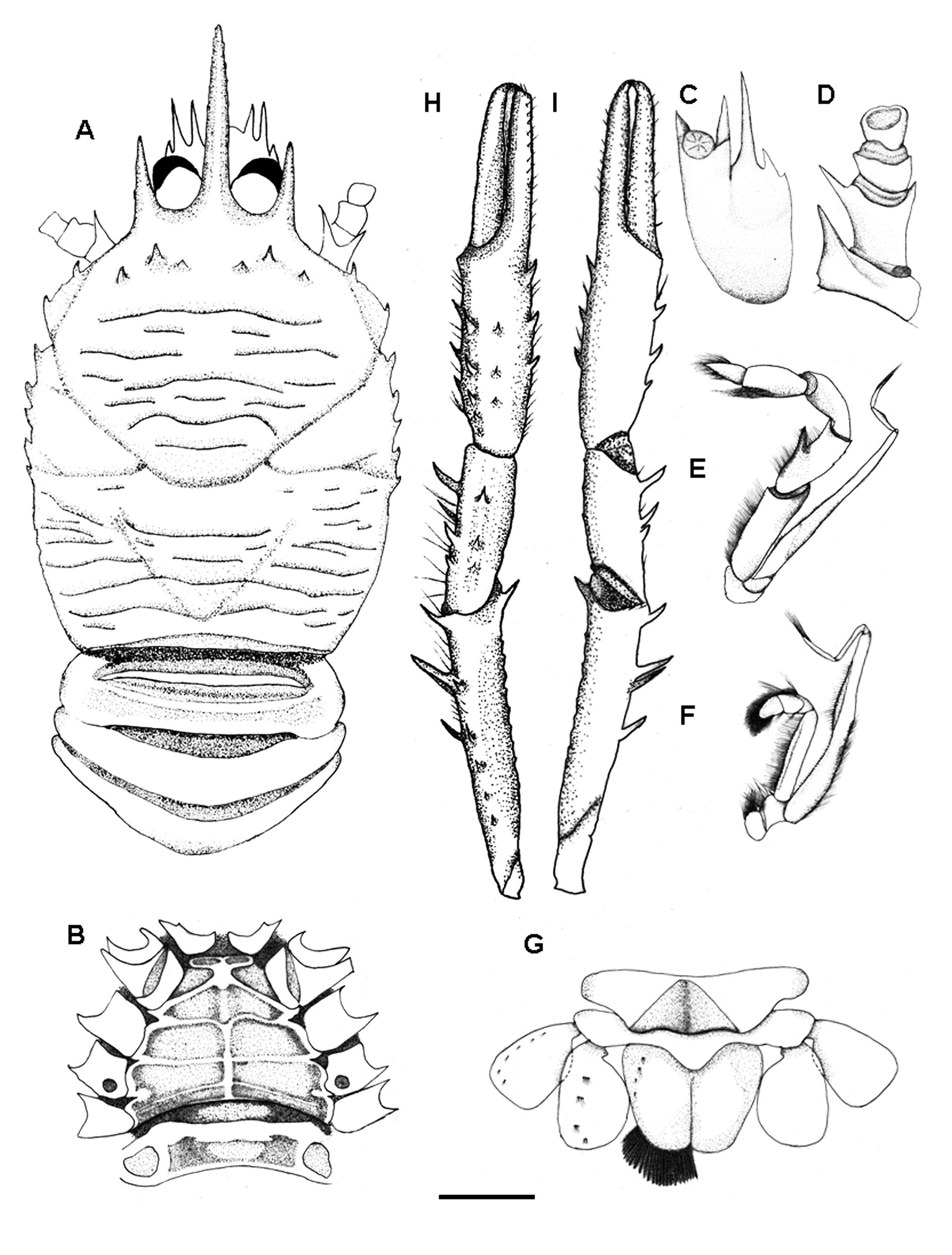
Description. Carapace (excluding rostrum) 1.2 times longer than broad, with gastric region anteriorly elevated and hepatic region depressed. Few secondary striae between main transverse ridges. Ridges sparsely provided with short non-iridescent setae, some ridges uninterrupted as illustrated. Branchial margins with 5 small spines. Gastric region with 3 pairs of epigastric spines, second spine largest; other dorsal regions without spines. Anterior branchial regions nearly smooth. Frontal margins feebly oblique. Lateral margins slightly convex. Anterolateral spine relatively short. Hepatic margins strongly convex, each with spine smaller than anterolateral spine. Rostrum spiniform, about 0.4 of remaining carapace length, straight and horizontal. Supraocular spines barely reaching midlength of rostrum, distinctly overreaching ocular peduncles, parallel with rostrum, directed upwards ( Fig. 1A View FIGURE 1 ). Surfaces of thoracic sternites smooth, without setae. Anterior margin of fourth sternite clearly narrower than third sternite. Third thoracic sternite about 6.5 times wider than long ( Fig. 1B View FIGURE 1 ).
Abdominal somites unarmed. Second abdominal somite slightly narrower than third somite, with 2 elevated transverse ridges on tergum; third to fifth somites without ridges ( Fig. 1A View FIGURE 1 ). Sixth abdominal with deep triangular depression. Telson 1.3 times as broad as long; median suture clearly delimited ( Fig. 1G View FIGURE 1 ).
Eyes small; corneas not dilated, width narrower than basal width of ocular peduncle and distance between sinus formed by supraocular and rostral spines, 0.15 of carapace length.
Basal segment of antennule (distal spines excluded) about 0.15 length of carapace, nearly reaching half of rostrum, distinctly overreaching distal margins of corneas, with 2 distal spines, mesial spine clearly shorter than lateral; 3 spines on lateral margin, proximal spine very short, median spine located at mid-length of proximal and distal spines, distal spine 4 times length of median spine and overreaching distolateral spine ( Fig. 1C View FIGURE 1 ).
First segment of antennal peduncle with moderately long distomesial spine nearly reaching distal margin of second segment; second segment with 2 distal spines, mesial spine nearly 2 times longer than lateral spine and reaching end of third segment; third and fourth segments unarmed ( Fig. 1D View FIGURE 1 ).
Third maxilliped with setae mostly on flexor margin of the articles. Ischium about 2 times length of merus measured along extensor margin, with mesioflexor margin denticulate. Merus with 2 well-developed spines on flexor margin, distal spine slightly smaller than proximal spine arising at about midlength; extensor margin minutely dentiticulate but without distinct spines ( Fig. 1E View FIGURE 1 ). Second maxilliped with exopod, distinctly longer than endopod; articles of endopod and exopod covered with simple and plumose setae ( Fig. 1F View FIGURE 1 ).
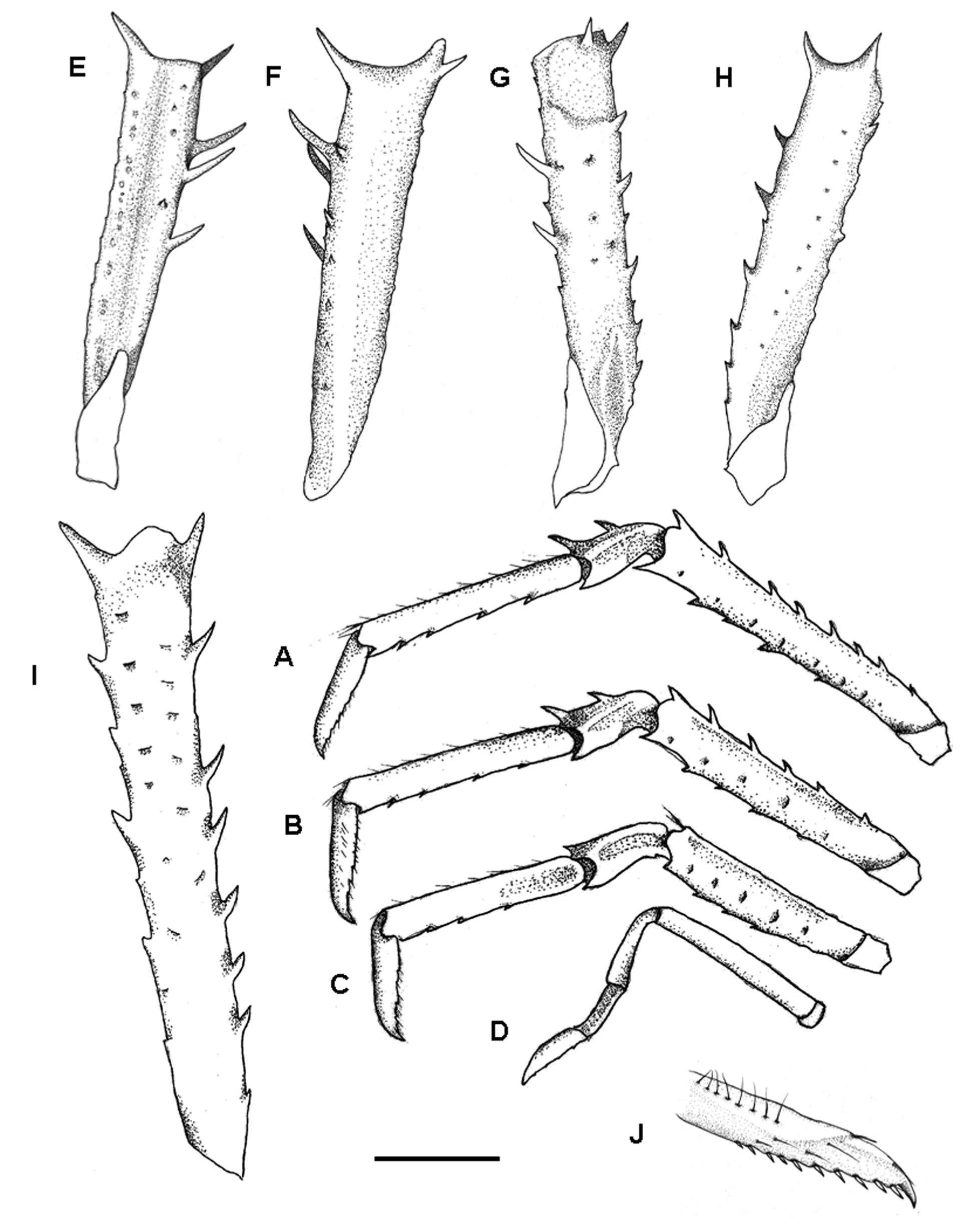
Chelipeds (P1) subequal in length, slightly squamous, about 2.6 times as long as carapace( Fig. 1H, I View FIGURE 1 ). Merus with 6 small spines on dorsal surface laterally and 2 spines mesially (second spine stronger than spine at distomesial angle); lateral face with spinulose tubercles ventrally and with 1 strong spine on ventrodistal angle; ventromesial margin with 2 (right) or 3 (left) strong spines; ventral surface smooth; mesial face with short setae ( Fig. 2E–H View FIGURE 2 ). Carpus twice as long as wide; dorsal surface with 3 small spines along midline; dorsolmesial margin with 2 spines (distal spine stronger); ventromesial margin with 1 prominent spine; mesial face with short setae and 1 spin located at about mid-length; ventral face smooth and with 1 spin at ventral angle. Palm about 3 times longer than wide; dorsal surface with row of 3 very small spines along midline; lateral face with 3 spines and short setae; dorsolmesial margin with 5 spines increasing in size distally; ventromesial margin with 3 spines; ventral surface smooth without setae. Dactylus as long as palm; fingers unarmed, except terminal spines, distally curving, ending in sharp claw; lateral margin with short setae ( Fig. 2J View FIGURE 2 ).
Second to fourth pereopods (P2–4) moderately long and slender, decreasing in length posteriorly. P2 about 2 times length of carapace; merus about 0.9 times as long as carapace, dorsal margin with row of 7 spines and 1 spinule increasing in size distally, ventral margin with 5 spines and 3 spinule, spine at ventrodistal angle largest, ventral surface smooth, with few long setae ( Fig. 2I View FIGURE 2 ); carpus with 2 strong distal extensor spines and 1 prominent distal flexor spine, lateral surface nearly smooth; propodus flexor margin with row of 5 moveable spines, with extensor margin unarmed, ventral surface with scattered tiny granules and with 1 spine on ventrodistal angle; dactylus about 0.5 times as long as propodus, slightly curved distally, with 10 movable spines on flexor margin ( Fig. 2A, I View FIGURE 2 ). P3 about 1.9 times length of carapace; merus about about 0.9 length of that of second pereopod, with 6 spines on dorsal margin increasing in size distally and 4 spines on ventral margin; carpus with 1 strong extensor spines in addition to 1 extensor distal spine and 1 small flexor distal spin; propodus with 5 moveable spines on flexor margin; dactylus with 8 moveable spines on flexor margin ( Fig. 2B View FIGURE 2 ). P4 about 1.7 times length of carapace; merus about 0.7 length of that of second pereopod, with 1 prominent extensor distal spine and 3 spinules on ventral margin; carpus with extensor distal and flexor distal spines much smaller than those of preceding pereopods; propodus with 5 moveable spines on flexor margin; dactylus with 8 moveable spinules on flexor margin ( Fig. 2C View FIGURE 2 ). P5 as long as carapace, carpus, slightly longer than propodus, all segments unarmed, dense setae present on entire dactylus ( Fig. 2D View FIGURE 2 ).
Twenty-four eggs preserved under abdomen, rotund or oval, measuring 0.8–1.0 mm in diameter.
Coloration. Anterior part of carapace (including hepatic, gastric, cardiac and anterior branchial region), ocular peduncles, antennal peduncles and antennule peduncles deep pink, posterior part of carapace (including posterior branchial region and intestinal region), abdomen pink. P1–5 and telson light pink.
Etymology. The species is dedicated to the newborn niece of the first author.
Remarks. Munida manqingae sp. nov. belongs to the group of deep-sea species in having five spines on the branchial margins of the carapace, smooth thoracic sternites, and small eyes. This informal group includes M. clevai Macpherson 2009 , M. endeavourae Ahyong & Poore, 2004 , M. parvioculata Baba, 1982 , and M. tiresias Macpherson, 1994 . The new species appears closest to M. tiresias in the anterolateral and hepatic marginal spine being small and not well developed and the absence of spines on the anterior transverse ridge of the second abdominal somite. Munida clevai , M. endeavourae , and M. parvioculata have at least two distinct spines on the second abdominel somite. However, M. manqingae sp. nov. is distinguished from M. tiresias by the following features: 1) the transverse ridges on the posterior parts of the branchial and cardiac regions of the carapace are uninterrupted; 2) three spines are present on the lateral margin of the basal antennular segment (only two lateral spines in M. tiresias ); 3) the distal spine on the lateral margin of the basal antennular segment overreaches the end of the distolateral spine (it does not reach the tip of the distolateral spine in in M. tiresias ); 4) the distomesial spine of the first antennal segment nearly reaches the end of the second segment (this spine falls far short of the distal margin of the second segment in M. tiresias ).
The new species is also similar to M. magniantennulata Baba & Türkay, 1992 , but there are some distinctions between the two species. The frontal margin of the carapace of M. manqingae is more strongly oblique than in M. magniantennulata and the anterolateral spine of the carapace is much shorter in the new species. The supraocular spines clearly overreach the distal margins of the cornea in the new species, but they do not reach those margins in M. magniantennulata . The antennular basal segment of the new species reaches only to half the length of the rostrum, instead of clearly overreaching it in M. magniantennulata . The distomesial spine of the first antennal segment nearly reaches the distal margin of the second segment in the new species, whereas it falls far short of that margin in M. magniantennulata .
No known copyright restrictions apply. See Agosti, D., Egloff, W., 2009. Taxonomic information exchange and copyright: the Plazi approach. BMC Research Notes 2009, 2:53 for further explanation.
|
Kingdom |
|
|
Phylum |
|
|
Class |
|
|
Order |
|
|
Family |
|
|
Genus |