
Discocyrtus cerayanus ( Roewer, 1929 ) Carvalho & Kury, 2022
|
publication ID |
https://doi.org/ 10.5252/zoosystema2022v44a9 |
|
publication LSID |
urn:lsid:zoobank.org:pub:5CE1923C-C671-4B5E-BDE0-9E489D160029 |
|
DOI |
https://doi.org/10.5281/zenodo.6522275 |
|
persistent identifier |
https://treatment.plazi.org/id/03DB8798-FFE3-FF83-FF29-F9E66561FA28 |
|
treatment provided by |
Felipe |
|
scientific name |
Discocyrtus cerayanus ( Roewer, 1929 ) |
| status |
comb. nov. |
Discocyrtus cerayanus ( Roewer, 1929) View in CoL n. comb.
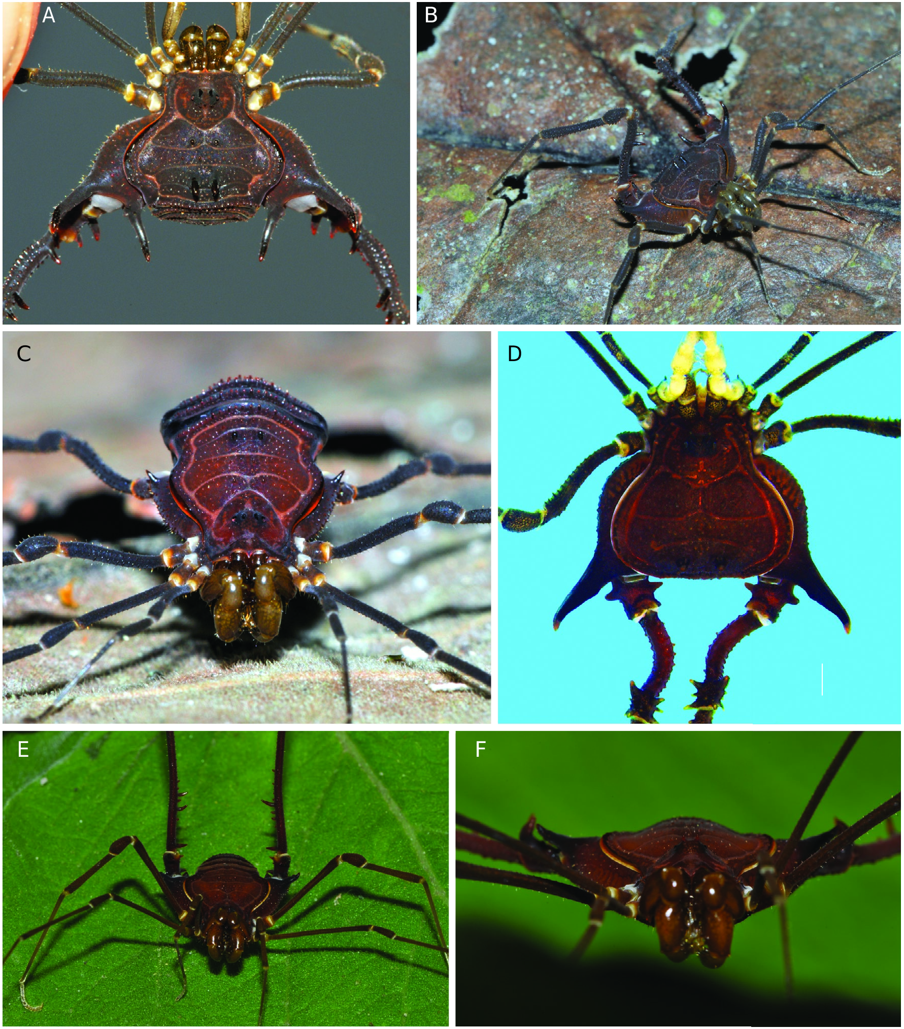
( Figs 1B View FIG ; 4B, D, F View FIG ; 6 View FIG ; 7 View FIG ; 8 View FIG )
Paradiscocyrtus cerayanus Roewer, 1929: 247 View in CoL , fig. 29; 1931: 104. — Mello-Leitão 1932: 206. — Soares & Soares 1954: 286. — Acosta 1996: 221. — Kury 2003: 185.
TYPE DATA. — Brazil • ♂ holotype (examined), “Ceraya” [= Minas Gerais, Serra do Caraça], wrongly identified as “Ceará” by Roewer (1931), see discussion in the geographical remarks section below; SMF RII 996/53 .
RECORDS. — Without further literature records.
MATERIAL EXAMINED. — Brazil • 1 ♂, 1 ♀, 2 juv; Minais Gerais, Caeté, Projeto Apolo, AP-09; 05-09.VII.2011; A. Giupponi, D. Pedroso, C. Sampaio leg.; MNRJ 7244 View Materials † ( Fig. 6 View FIG ) • 5 ♂, 10 ♀; same data; MNRJ 7245 View Materials †.
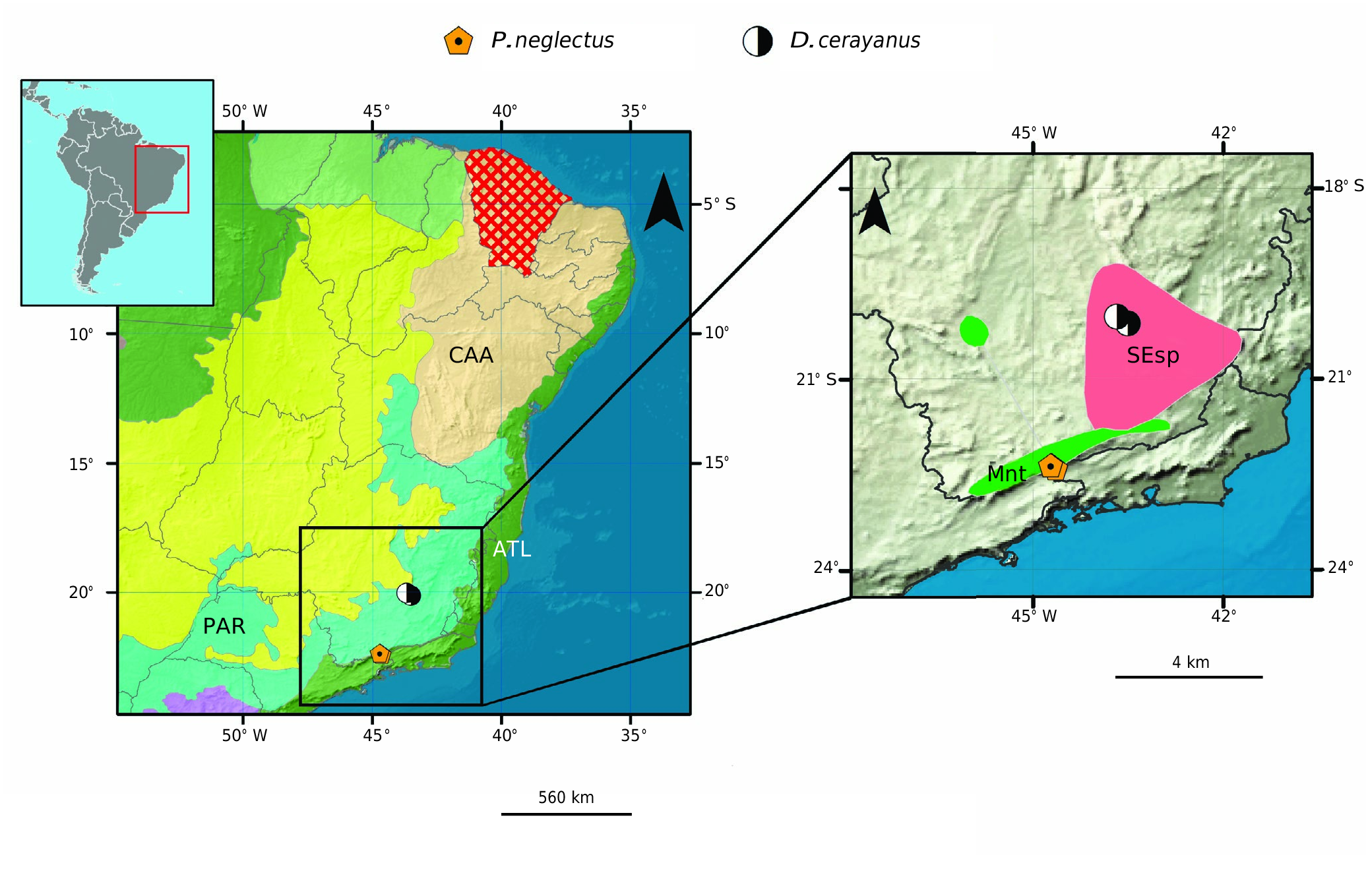
DISTRIBUTION. — Brazil: Minas Gerais: Caeté, new record; Serra do Caraça ( Fig. 5 View FIG ).
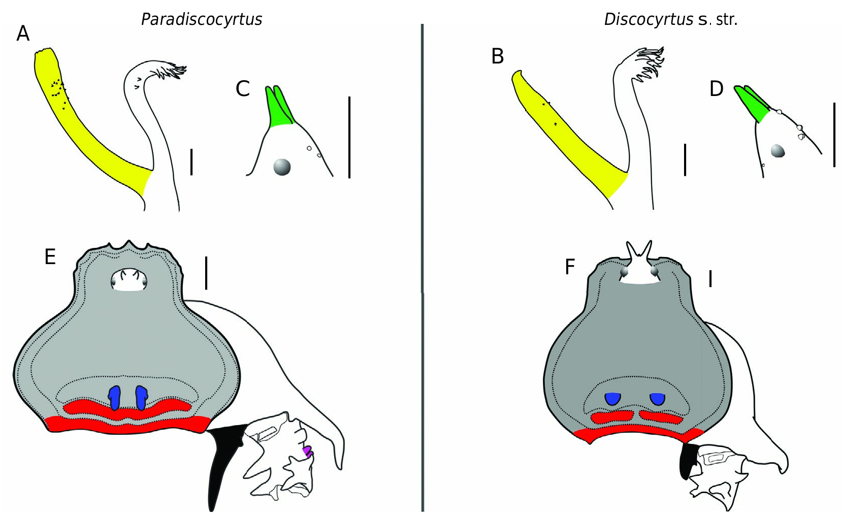
DIAGNOSIS. — Area IV divided into left and right halves by a median groove ( Fig. 4F View FIG ; 8A View FIG ) (as in D. crenulatus and D. testudineus ; entire in D. flavigranulatus ). Stigmatic area inverted T-shape ( Figs 6B View FIG , 8D View FIG ) (inverted Y-shape in D. crenulatus , D. flavigranulatus and D. testudineus ). Tr IV with a protuberance oriented to dorsal, which resembles a hook ( Figs 6A, B View FIG ; 8A, E View FIG ) (armature absent or reduced in D. crenulatus , D. flavigranulatus and D. testudineus ). Pa IV with proximal retroventral apophysis ( Fig. 7G, J View FIG ) (as in D. flavigranulatus ; absent in D. crenulatus and D. testudineus ). Ti IV dorsal portion with outstanding tubercles ( Fig. 7E, F, H, I View FIG ) (as in D. crenulatus , ordinary tubercles in D. flavigranulatus and D. testudineus ). DS outline of female lambda-shaped (gamma-pyriform-shaped in D. crenulatus , D. flavigranulatus and D. testudineus ). Glans ventral process almost the same height as the stylus ( Fig. 8A, B View FIG ) (as in D. flavigranulatus ; with half of the height in D. crenulatus and D. testudineus ).
REDESCRIPTION
Male specimens
SMF RII 996/53 for the external body illustrations; DS, measurements: CW 3.6, CL 2.1, AW 6.1, AL 3.6; legs I-IV measurements in the Table 4 View TABLE ; right / left tarsal (distitarsal) counts: 6(3) / 5(3) - 9(3) / 10(3) - 7 / 7 - 7 / 7. MNRJ 7244† for color references and genitalic illustrations.
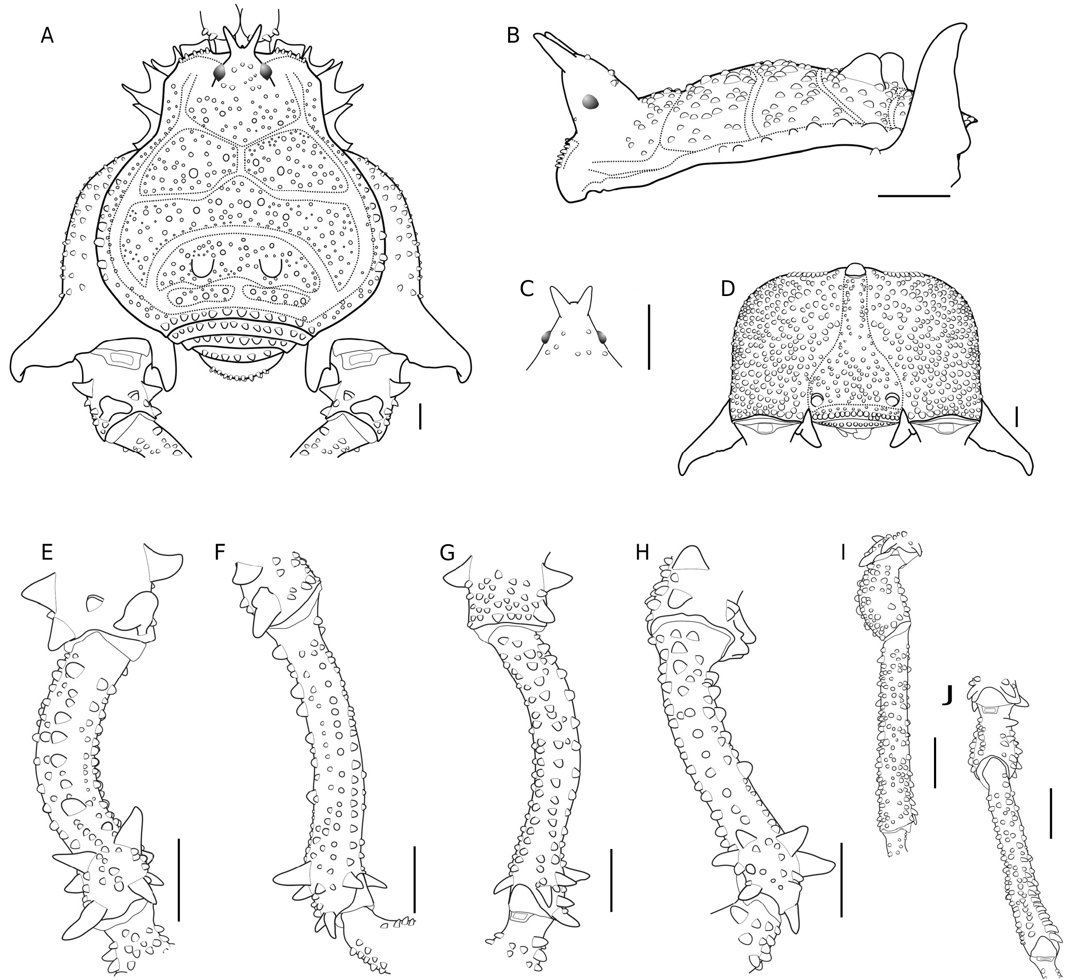
Dorsum. DS gamma-pyriform, as long as wide, with lateral margins of the AS convex, widest at areas II-III and thickest at area III, with concave posterior margin ( Figs 6A, C, D View FIG ; 8A, B View FIG ). DS anterior margin with two sets of five acuminated tubercles, divided by a small apophysis in the center and a pair of shallow cheliceral sockets ( Fig. 7A View FIG ). Carapace with many tubercles on the posterior portion, with a transversal row of four prominent tubercles ( Fig. 7A View FIG ). Ocularium elliptical (in dorsal view), high (c. 4.5× the eye diameter), inclined frontwards, placed in the anterior portion of the carapace ( Figs 6A, C View FIG ; 8A, B View FIG ). Ocularium armed with a pair of divergent spines (c. 3× the eye diameter) fused at baseline ( Figs 6D View FIG ; 8 View FIG A-C). Mesotergum is divided into four clearly defined areas ( Fig. 7A View FIG ). Areas I and IV are divided into left and right halves by a median groove ( Fig. 7A View FIG ). AS lateral borders with two rows of tubercles: one external, composed by six or seven prominent tubercles at areas II-III ( Fig. 7A, B View FIG ); other internal with ordinary tubercles from the anterior portion of carapace backward ( Fig. 7A View FIG ). All areas tuberculate ( Fig. 7A, B View FIG ). Area I with two pairs of dome-shaped paramedian tubercles, twice the size of the ordinary tubercles ( Fig. 7A, B View FIG ); area II with three pairs of prominent tubercles. Area III with an outstanding pair of domed-shaped tubercles, each one surrounded by prominent tubercles (two external and three medial, with c. twice the ordinary tubercles). Area IV with two transversal sets of three prominent tubercles (c. twice the ordinary) interspersed by ordinary ones ( Fig. 7A View FIG ). DS posterior border and free tergites with a transversal row of larger tubercles ( Fig. 7A View FIG ). Anal operculum covered by rows of ordinary tubercles ( Fig. 7A View FIG ).
Venter. Cx I-III parallel to each other ( Fig. 6B View FIG ), each with ventral transverse rows of 8-13 setiferous tubercles (Cx I rows with higher and sharper tubercles than the others). Cx II with a retroventral distal row of seven acuminate tubercles. Cx III with a retroventral distal row of nine acuminate tubercles ( Fig. 7D View FIG ). Cx IV much larger than the others, directed obliquely ( Figs 6B, C View FIG ; 8D View FIG ). Stigmatic area Y-inverted-shaped, clearly sunken concerning Cx IV distal part ( Figs 6B View FIG ; 8D View FIG ). Cx IV covered by prominent tubercles ( Figs 6B, C View FIG ; 8D View FIG ). Cx IV posterior margin and stigmatic area with a transversal row of the prominent tubercles ( Figs 6B, C View FIG ; 8D View FIG ). Intercoxal bridges well-marked ( Fig. 6B View FIG ). Stigmata visible ( Figs 6B View FIG ; 8D View FIG ). Free sternites with a transverse row of ordinary tubercles ( Fig. 6B, C View FIG ).
Chelicera. Basichelicerite elongate, bulla well-marked, with marginal setiferous tubercles – two ectal, two posterior ( Fig. 7A View FIG ), one mesal; hand not swollen.
Pedipalpus. Tr with two geminate ventral setiferous tubercles. Fe with a ventral basal and a mesal apical setiferous tubercle. Pa unarmed ( Fig. 6A View FIG ).Ti with two rows of setiferous tubercles: four (IiIi) ventro-mesal; (IiIi) ventro-ectal ( Fig. 6A View FIG ). Ta with two rows of setiferous tubercles: three (IIi) ventro-mesal and four (IIIi) ventro-ectal.
Legs. Tr I-III each with several ventral tubercles. Fe I and III sub-straight; Fe II straight ( Fig. 6A, B View FIG ). Fe and Ti I-III with all faces containing rows of small tubercles; Fe III and Ti III with proventral and retroventral two rows of small acuminate tubercles. Fe II-III with an apical retrodorsal spur. Pa I-III covered dorsally by tubercles. Cx IV reaching the posterior margin of DS ( Figs 6A View FIG ; 8A View FIG ). Cx IV tuberculate between prodorsal and ventral faces ( Figs 6 View FIG A-C; 8 View FIG A, D). Cx IV with a thick prolateral distal conical apophysis, swollen at the basis, with apical portion slightly curved backwards ( Figs 6 View FIG A-C; 8 View FIG A, B, D). Cx IV with a retrolateral spiniform apophysis, fused with a small secondary branch ( Figs 6A, B View FIG ; 8A, D View FIG ). Tr IV square-shaped ( Figs 6A, B View FIG ; 8A View FIG ). Tr IV proximal with conical prolateral and retrolateral apophyses, both curved to the dorsal portion ( Figs 6A, B View FIG ; 8A View FIG , E-H).Tr IV dorsal central with a pair of highlighted tubercles, longitudinally arranged ( Figs 6A View FIG ; 8A, E, F, H View FIG ). Tr IV distal retrolateral with a conical apophysis, without acuminated apex ( Fig. 7E, F, H View FIG ). Tr IV prodorsal distal with a protuberance oriented to dorsal, resembling a hook ( Fig. 7A, E, F View FIG ). Tr IV ventral face tuberculate ( Fig. 7 View FIG F- H). Fe IV C-shaped (using the right femur as a reference, in dorsal view), arched on the central portion towards dorsal ( Figs 6C View FIG ; 8 View FIG E-H). Fe IV proximal-medial portion with 1) a dorsal row of five prominent tubercles ( Fig. 7E, F, H View FIG ) and 2) prodorsal, prolateral, proventral, retroventral, retrolateral and retrodorsal rows of ordinary tubercles ( Fig. 7E, F, H View FIG ). Fe IV distal portion with: 1) two prodorsal spines ( Fig. 7E, H View FIG ); 2) a proventral and retroventral apical spur each ( Fig. 7E, F View FIG ); 3) three retrolateral spines ( Fig. 7G, H View FIG ); and 4) one retrodorsal spine ( Fig. 7E, H View FIG ). Pa IV dorsally tuberculate ( Fig. 7I View FIG ). Pa IV retroventral and retrodorsal with conical apophyses ( Fig. 7G, H, J View FIG ). Pa IV proventral and retroventral with rows of six and three spines, respectively ( Fig. 7I, J View FIG ). Ti IV with all faces tuberculate ( Fig. 7I, J View FIG ); retroventral central-distal with a row of 10 spines ( Fig. 7J View FIG ). Mt IV prodorsal, proventral, retroventral and retrodorsal with rows of setiferous tubercles. Mt IV proventral and retroventral faces with a spur.
Color (in alcohol) ( Fig. 6 View FIG A-D). Ocularium, DS background and its borders Strong Orange Yellow (68). Pp Vivid Yellow (82). Chelicerae, Tr-Pa I-III, Ti-Ta II Vivid Orange Yellow (66). Ti- Ta I and III Moderate Orange Yellow (71). Ocularium pair of spines Brownish Orange (54). Area III pair of domed-shaped tubercles Dark Red (16). Articular membranes Moderate Orange Yellow (71). Cx IV medial and Tr IV Deep Orange Yellow (69). Cx IV proximal and distal and Tr IV apophyses Strong Brown (55). Fe-Ti medial IV Strong Orange Yellow (68). Ti medial- Mt IV Vivid Yellow (82).Ta II-IV Light Greenish Yellow (101).
Male genitalia. VP slightly divided into two regions: distal part forming a rectangle with latero-apical flaps, proximal part elliptical ( Fig. 8A, C View FIG ). VP ventral surface entirely covered with microsetae of type 1 ( Fig. 8B, C View FIG ). All macrosetae inserted on lateral of VP. MS A1-A3 cylindrical, thick, and acuminate, forming a triangle (with A2 more ventral than the other two) on the basal third of VP ( Fig. 8B, C View FIG ). MS B1 small, inserted ventrally, proximal to A2 ( Fig. 8B, C View FIG ). MS C1-C3 similar to the MS A, inserted on the ventrolateral border, forming a longitudinal row on the distal third of VP ( Fig. 8 View FIG A-C). MS D1 small, inserted on VP ventrolateral border, closer to C3 than to A1 ( Fig. 8 View FIG A-C). MS E1-E2 small, located on the laterodistal flange of VP – E1 between the height of MS C1-C2, E2 between the height of MS C2-C3 ( Fig. 8B View FIG ). Glans sac arising from the middle bulge on the podium, not extended as a dorsal process ( Fig. 8A, B View FIG ). Stylus and its ventral process axis fused basally (forming a short pedestal) at a 45° angle ( Fig. 8A, B View FIG ). Stylus cylindrical, almost straight, without clearly defined head and armed with a few small subdistal setae ( Fig. 8A, B View FIG ). Ventral process with almost the same stylus’s length, with a ventro-apical flabellum ( Fig. 8A, B View FIG ). Flabellum curved proximally, scallop-shaped, measuring about 40% length of the ventral process stem ( Fig. 8A, B View FIG ).
Intraspecific variation. The material studied does not present minor morph males. It was also not found intraspecific variation among the major morph males and females.
Female (MNRJ 7244 †)
DS lambda type. Area III with a pair of paramedian spines, with an elevated basis. Free tergites with a transversal row of ordinary tubercles. Cx IV narrower than in males, with the prodorsal apophysis reduced to a single spine and without the retroventral apophysis. Tr IV with a proximal retrolateral apophysis.Fe IV thinner compared to male, moderately curved in the proximal portion.
GEOGRAPHICAL REMARKS
The male holotype of Paradiscocyrtus cerayanus (SMF RII 996/53) was originally reported as from the locality “ Brasilien (Ceraya)” by Roewer (1929: 248). As noted by Roewer (1931: 104), Mello-Leitão explained to him in a personal correspondence that Ceraya was a misspelling of the locality name and extrapolated this as being the state of “ Ceará ” in northeastern Brazil. Roewer added that all his records contained the misspelling and accepted without question Mello-Leitão’s interpretation. Subsequent authors did not pursue this question and later cataloguers – Soares & Soares (1954: 286) and Kury (2003: 185) – simply repeated “ Ceará ”. However, comparing that locality with the geographical data gathered and studied here, one cannot make sense of such an interpretation because the northeastern Brazilian fauna is quite different from the one in the Southeast (e.g. Kury 2003). Diacritics are often a source of confusion in toponyms, as exemplified in the misinterpretation by Henriksen (1932: 420) of the names “ Boyacá ” and “Choachí” (Cundinamarca, Colombia) respectively as “Boydla” and “Choact” ( Medrano et al. 2020). With the benefit of cumulative decades of study of Brazilian Gonyleptidae , for us it is easier to understand that a cedilla under a C could easily have been misspelled by European collectors as a “Y”. This toponym “Ceraya” does not appear in further harvestmen literature, however, in the same paper, Roewer (1929: 255) described another Gonyleptidae as from “Caraya”, which gives us the remaining piece of the puzzle.
This way, by replacing only a single letter, Caraya (or its further distortion Ceraya) can be easily retrieved as “Caraça”, a small mountain range in Minas Gerais state, southeastern Brazil. Further material of D. cerayanus n. comb. studied by us corroborates this new interpretation, because the specimens hail from Caeté, Minas Gerais, which is extremely close to the Serra do Caraça (about 20 km in a straight line). Therefore, we herein rectify the incorrect interpretation of Mello-Leitão/ Roewer (1931) of “Ceraya” as “ Ceará ” to [Serra do] Caraça, state of Minas Gerais, Brazil, which should be the correct type-locality of the species.


| SMF |
Forschungsinstitut und Natur-Museum Senckenberg |
No known copyright restrictions apply. See Agosti, D., Egloff, W., 2009. Taxonomic information exchange and copyright: the Plazi approach. BMC Research Notes 2009, 2:53 for further explanation.
|
Kingdom |
|
|
Phylum |
|
|
Class |
|
|
Order |
|
|
Family |
|
|
Genus |
Discocyrtus cerayanus ( Roewer, 1929 )
| Carvalho, Rafael N. & Kury, Adriano B. 2022 |
Paradiscocyrtus cerayanus
| KURY A. B. 2003: 185 |
| ACOSTA L. E. 1996: 221 |
| SOARES B. A. M. & SOARES H. E. M. 1954: 286 |
| MELLO- LEITAO C. F. 1932: 206 |
| ROEWER C. F. 1931: 104 |
| ROEWER C. F. 1929: 247 |