
Plaumanniola Costa Lima
|
publication ID |
https://doi.org/10.11646/zootaxa.3670.3.2 |
|
publication LSID |
lsid:zoobank.org:pub:3F2904A2-A999-484A-850B-4E2E13A9DBD7 |
|
DOI |
https://doi.org/10.5281/zenodo.6148786 |
|
persistent identifier |
https://treatment.plazi.org/id/03DC5A34-FF8F-5037-DBEF-42C514ABFB4D |
|
treatment provided by |
Plazi |
|
scientific name |
Plaumanniola Costa Lima |
| status |
|
Plaumanniola Costa Lima View in CoL
Plaumanniola Costa Lima, 1962: 415 View in CoL . Type species: Plaumanniola sanctaecatharinae Costa Lima, 1962 View in CoL (monotypy).
Revised diagnosis. Head strongly broadened and flat, with lateral margins divergent caudad, with bristles on tempora and along posterior margin of vertex; occipital constriction narrower than 1/3 of HW; 'neck' region only slightly broader than occipital constriction and narrower than 1/3 of HW; mouthparts not visible in dorsal view; submentum not demarcated laterally from postcardinal parts of hypostomae; hypostomal ridges long and divergent caudad; labrum with deeply emarginate anterior margin; maxillary palps flattened dorso-ventrally, with enlarged palpomere III and subconical, pointed palpomere IV; antennae massive and clubbed; pronotum broad and flattened, without pits or grooves and with sharp lateral edges; prosternum without prosternal intercoxal carina or process; procoxal sockets closed; hypomera with incomplete hypomeral ridges; pronotosternal sutures entire; mesoventrite with keel-like, high and narrow mesoventral intercoxal process and indistinctly delimited lateral asetose impressions (= procoxal rests); each elytron with two barely discernible rudiments of asetose basal foveae; metaventral intercoxal process long and narrow, with deep and narrow posterior emargination, narrowly separating transverse metacoxae; aedeagus with complex internal armature and free (i.e., not fused with median lobe) parameres bearing apical and subapical setae.
Redescription. Body of male ( Figs. 1, 3 View FIGURES 1 – 3 ) stout and broad, with flat head ( Figs. 6–7 View FIGURES 6 – 7 ), more convex pronotum ( Fig. 7 View FIGURES 6 – 7 ) and strongly convex elytra, with deep constrictions between head, pronotum and elytra, brown and densely setose, BL 1.90–2.58 mm.
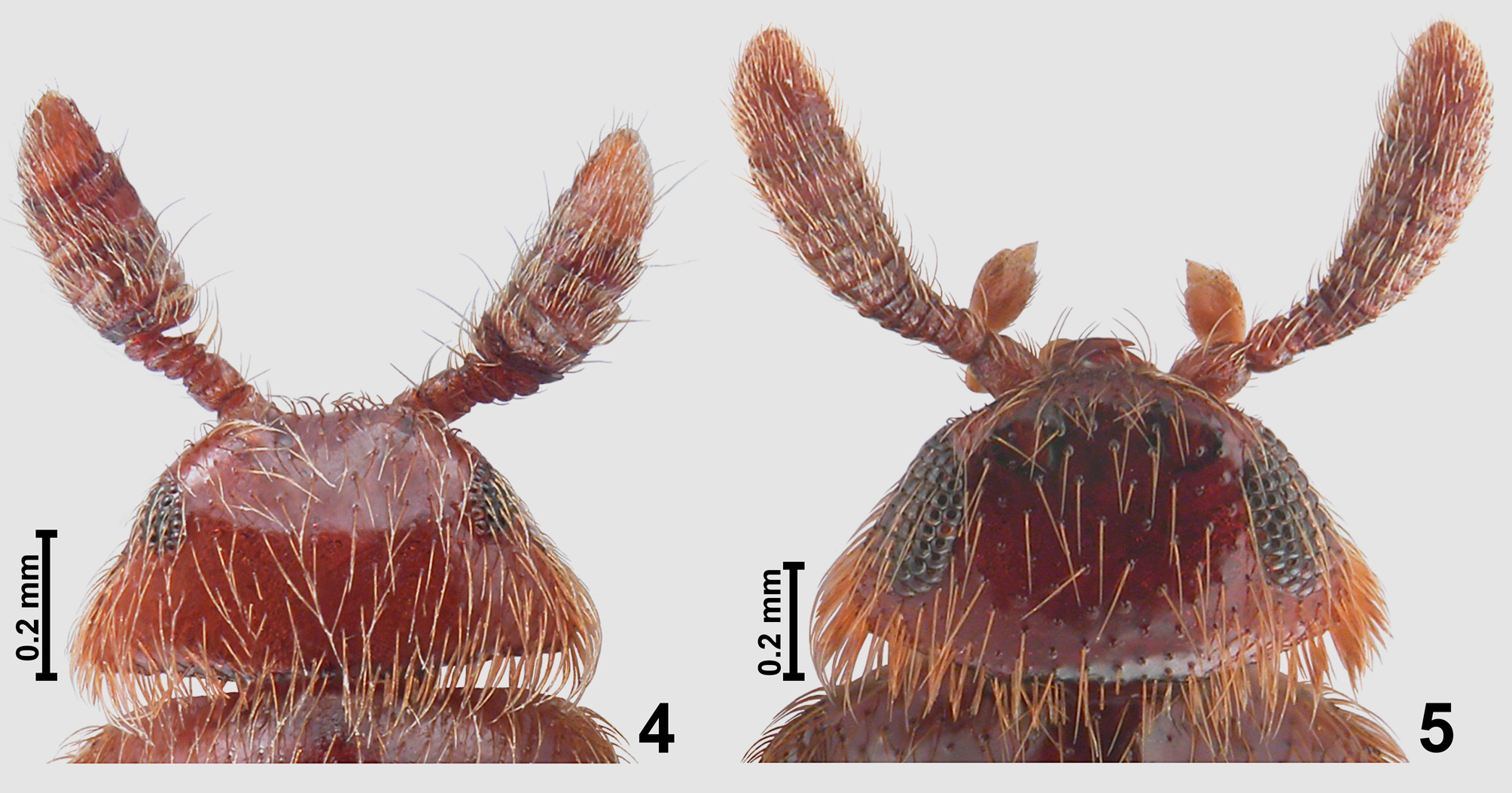
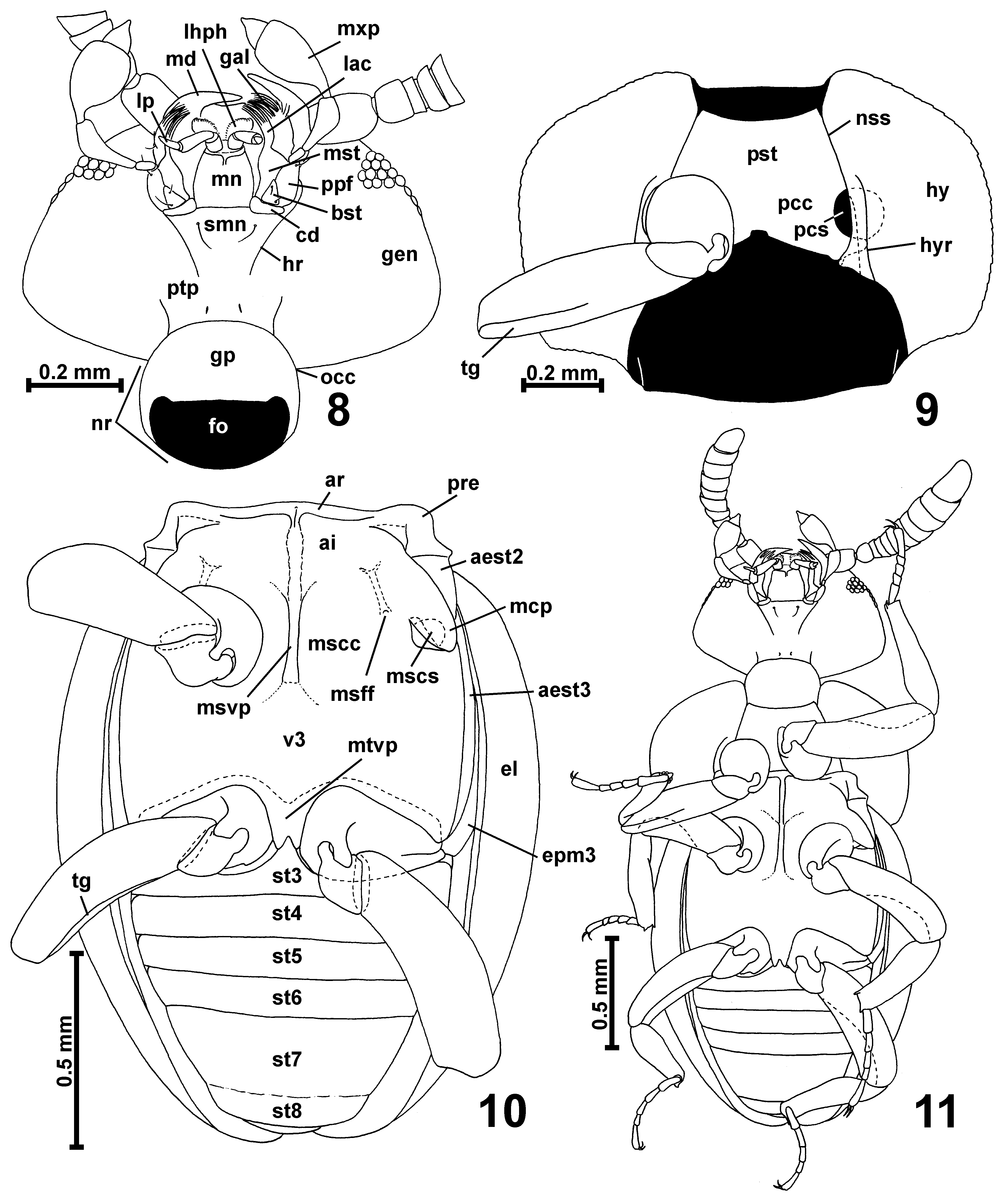
Head ( Figs. 4–7 View FIGURES 4 – 5 View FIGURES 6 – 7 , 8 View FIGURES 8 – 11 ) strongly flattened and broadened, in dorsal view approximately subtrapezoid or subtriangular, broadest near posterior margin of vertex; occipital constriction ( Fig. 8 View FIGURES 8 – 11 ; occ) in the narrowest place narrower than 1/3 of HW; 'neck' region ( Fig. 8 View FIGURES 8 – 11 ; nr) only slightly broader than occipital constriction and narrower than 1/3 of HW; tempora ( Figs. 4–5 View FIGURES 4 – 5 ) long and divergent caudad, with bristles; vertex ( Figs. 4–5 View FIGURES 4 – 5 ) strongly transverse, moderately convex, not projecting dorso-caudad, with arcuate or nearly straight posterior margin dorsally sharply demarcated from 'neck' region; frontoclypeal region ( Figs. 4–5 View FIGURES 4 – 5 ) only partly visible in dorsal view, its anterior part rapidly deflexed; fronto-clypeal groove absent; antennal insertions moderately broadly separated, located on antero-ventral margin of head; compound eyes ( Figs. 4–7 View FIGURES 4 – 5 View FIGURES 6 – 7 ) located in anterior or median part of head, small to large, multifaceted, composed of large and coarse ommatidia, with broadly emarginate posterior margin.
Mouthparts located on ventral side of head. Labrum ( Fig. 6 View FIGURES 6 – 7 ) transverse with deeply emarginate anterior margin. Mandibles ( Figs. 6 View FIGURES 6 – 7 , 8 View FIGURES 8 – 11 ; md) directed antero-ventrad, symmetrical, each with broad basal part, without noticeable prostheca, and with slender and curved distal part, without mesal tooth. Each maxilla ( Fig. 8 View FIGURES 8 – 11 ) with subtriangular basistipes ( Fig. 8 View FIGURES 8 – 11 ; bst), elongate galea ( Fig. 8 View FIGURES 8 – 11 ; gal) and lacinia ( Fig. 8 View FIGURES 8 – 11 ; lac) and moderately long, flattened dorso-ventrally maxillary palp ( Fig. 8 View FIGURES 8 – 11 ; mxp) composed of tiny and parallel-sided palpomere I about twice as long as broad, strongly elongate, pedunculate and curved palpomere II broadest in distal third, broad and stout palpomere III broadest in distal third and about twice as long as broad, and small, subconical and pointed palpomere IV with rapidly narrowing small apical part.
Labium ( Fig. 8 View FIGURES 8 – 11 ) with large and transverse submentum ( Fig. 8 View FIGURES 8 – 11 ; smn) not demarcated from gular plate ( Fig. 8 View FIGURES 8 – 11 ; gp) and laterally fused with postcardinal parts of hypostomae, subtrapezoidal mentum ( Fig. 8 View FIGURES 8 – 11 ; mn); and short prementum bearing narrowly separated at bases long 3-segmented labial palps ( Fig. 8 View FIGURES 8 – 11 ; lp). Hypostomal ridges ( Fig. 8 View FIGURES 8 – 11 ; hr) long and divergent caudad but not reaching posterior tentorial pits ( Fig. 8 View FIGURES 8 – 11 ; ptp), which are well-visible in ventral view.
Gular plate ( Fig. 8 View FIGURES 8 – 11 ; gp) large and convex; gular sutures indiscernible.
Antennae ( Figs. 4–6 View FIGURES 4 – 5 View FIGURES 6 – 7 , 11 View FIGURES 8 – 11 ) relatively short and massive, flattened dorso-ventrally, either with sharply demarcated club composed of enlarged antennomeres VII–XI or gradually thickened from antennomere IV to X, antennomere XI narrower than X; antennomeres covered with long, dense and suberect vestiture and sparse long and strongly erect setae.
Prothorax strongly flattened and broad. Pronotum ( Figs. 1–2 View FIGURES 1 – 3 ) in dorsal view oval with a large part of base abruptly expanded caudad and forming sharply separated lobe about as broad as anterior part of mesothorax; anterior pronotal corners indistinct or absent; sides rounded; posterior corners rounded; basal margin nearly straight; lateral margins forming sharp and complete edges; base of pronotum without foveae, impressions or grooves; pronotum without bristles.
Prosternum ( Fig. 9 View FIGURES 8 – 11 ; pst) slightly broader than long, with long but not demarcated precoxal (basisternal) part; procoxal cavities ( Fig. 9 View FIGURES 8 – 11 ; pcc) not separated by intercoxal carina or process; procoxal sockets ( Fig. 9 View FIGURES 8 – 11 ; pcs) closed by lateral lobes of postcoxal part of sternum. Hypomera ( Fig. 9 View FIGURES 8 – 11 ; hy) broad and elongate; hypomeral ridge ( Fig. 9 View FIGURES 8 – 11 ; hyr) visible only in posterior half of hypomeron; pronotosternal sutures ( Fig. 9 View FIGURES 8 – 11 ; nss) entire.
Mesoscutellum very small, not visible in intact specimens, subtriangular, transverse, mesoscuto-scutellar suture present.
Mesoventrite ( Fig. 10 View FIGURES 8 – 11 ) with narrow anterior ridge ( Fig. 10 View FIGURES 8 – 11 ; ar) connected in middle with anterior part of long, narrow and strongly expanding ventrad (i.e., keel-shaped) mesoventral intercoxal process ( Fig. 10 View FIGURES 8 – 11 ; msvp); mesanepisternum with long prepectus ( Fig. 10 View FIGURES 8 – 11 ; pre) and posterior part ( Fig. 10 View FIGURES 8 – 11 ; aest2) largely visible in ventral view; mesepimeron not visible in ventral view; sides of mesothorax without foveae; mesoventrite with indistinctly delimited subtriangular lateral asetose impressions ( Fig. 10 View FIGURES 8 – 11 ; ai); mesocoxal projections ( Fig. 10 View FIGURES 8 – 11 ; mcp) without posterior lobes and without bristles, with mesocoxal sockets ( Fig. 10 View FIGURES 8 – 11 ; mscs) located on their mesal surface (not exposed in ventral view); anterior and posterior margins of mesocoxal cavities ( Fig. 10 View FIGURES 8 – 11 ; mscc) without carinae; mesofurcal fovea ( Fig. 10 View FIGURES 8 – 11 ; msff) small and located in sub-anterior region of mesocoxal cavity.
Metaventrite ( Fig. 10 View FIGURES 8 – 11 ; v3) strongly transverse, anteriorly fused with mesoventrite, posteriorly deeply bisinuate and with narrow but long median subtrapezoidal metaventral intercoxal process ( Fig. 10 View FIGURES 8 – 11 ; mtvp) bearing subtriangular median emargination. Metanepisterna nearly completely, and metepimera partly visible in ventral view, narrow.
Metafurca in transparent mount not visible, possibly directed dorso-anterad or short and obscured by thick integument of posterior metaventral margin.
Elytra ( Figs. 1–2 View FIGURES 1 – 3 ) oval, each with two barely discernible asetose rudiments of basal foveae, without basal impressions; humeral calli distinct; subhumeral lines absent; elytral apices non-modified, separately rounded.
Legs ( Figs. 1–2 View FIGURES 1 – 3 , 9–11 View FIGURES 8 – 11 ) moderately long, robust; procoxae ( Fig. 9 View FIGURES 8 – 11 ) subglobose, mesocoxae ( Fig. 10 View FIGURES 8 – 11 ) oval and slightly elongate, metacoxae ( Fig. 10 View FIGURES 8 – 11 ) transverse, stout; all trochanters short and non-modified; all femora strongly flattened dorso-ventrally, each with tibial groove on posterior surface, in which posterior margin of tibia fits; tibiae flattened dorso-ventrally, with carinate posterior margin; tarsi moderately long, slender.
Abdominal sternites ( Figs. 10–11 View FIGURES 8 – 11 ) unmodified, sternites III–VI subequal in length, VII much longer, suture between VII and VIII barely marked.
Aedeagus ( Figs. 12–15 View FIGURES 12 – 15 ) stout, with narrowing and subtriangular apical part of median lobe; internal armature complex and darkly sclerotized; parameres free (i.e., not fused with median lobe) and slender, bearing apical and subapical setae.
Spermatheca not studied (due to lack of available female specimens), but Costa Lima (1962) illustrated two female specimens photographed in transparent mounts with darkly sclerotized spermatheca clearly visible inside metathorax. It is hemispherical with distinct and short duct of accessory gland ( Costa Lima 1962; Figs. 2–3 View FIGURES 1 – 3 ).
Distribution and composition. Two species are known from Brazil.


No known copyright restrictions apply. See Agosti, D., Egloff, W., 2009. Taxonomic information exchange and copyright: the Plazi approach. BMC Research Notes 2009, 2:53 for further explanation.
|
Kingdom |
|
|
Phylum |
|
|
Class |
|
|
Order |
|
|
Family |
|
|
SubFamily |
Scydmaeninae |
Plaumanniola Costa Lima
| Jałoszyński, Paweł 2013 |
Plaumanniola
| Costa 1962: 415 |