
Sundathelphusa danae, Husana, Daniel Edison M., Yamamuro, Masumi & Ng, Peter K. L., 2014
|
publication ID |
https://doi.org/10.11646/zootaxa.3815.4.6 |
|
publication LSID |
lsid:zoobank.org:pub:13A3E4D1-A4D4-4C6B-AB43-5268594EA221 |
|
DOI |
https://doi.org/10.5281/zenodo.6132780 |
|
persistent identifier |
https://treatment.plazi.org/id/03DC8797-FF83-FFB4-FF18-8694230F2D47 |
|
treatment provided by |
Plazi |
|
scientific name |
Sundathelphusa danae |
| status |
sp. nov. |
Sundathelphusa danae View in CoL sp. nov.
( Figs. 1–3 View FIGURE 1 View FIGURE 2 View FIGURE 3 , 7A, B View FIGURE 7. A, B )
Material examined. Holotype: male (25.9 × 19.8 mm) ( NMCR 39076), Bantakay Cave, Atimonan, Quezon Province, Luzon, Philippines, 14˚ 01.703N 121˚ 47.197E, coll. D.E.M. Husana, 13 January 2010. Paratypes: 1 male (21.7 × 17.9 mm) ( ZRC 2013.0274), 1 female (20.4 × 16.8 mm) ( NMCR 39093), same data as holotype.
Comparative material. Sundathelphusa holthuisi Ng, 2010 , holotype male (24.3 × 19.4 mm) ( ZRC 1989.2169), Bantakay Cave, Atimonan, Quezon Province, Luzon, Philippines, ca. 14˚00N 121˚52E Quezon Province, Luzon, Philippines, coll. D.S. Balete, 22 May 1987; 1 female (23.1 × 18.6 mm) ( ZRC 1989.2168), station C-054, National Botanic Gardens, Real, Quezon Province, Luzon, Philippines, coll. D.S. Balete, 9 September 1987; 1 male (18.5 × 15.9) (NSMT-Cr 22316) ( NMCR 39093), Bantakay Cave, Atimonan, Quezon Province, Luzon, Philippines, 14˚ 01.703N 121˚ 47.197E, coll. D.E.M. Husana, 13 January 2010; 1 male (15.6 × 13.7 mm) ( ZRC 2013.0275), Nilulubugan Cave, Atimonan, Quezon Province, Luzon, Philippines, 14˚ 01.000N, 121˚ 47.775E, coll. D.E.M. Husana, December 2009.
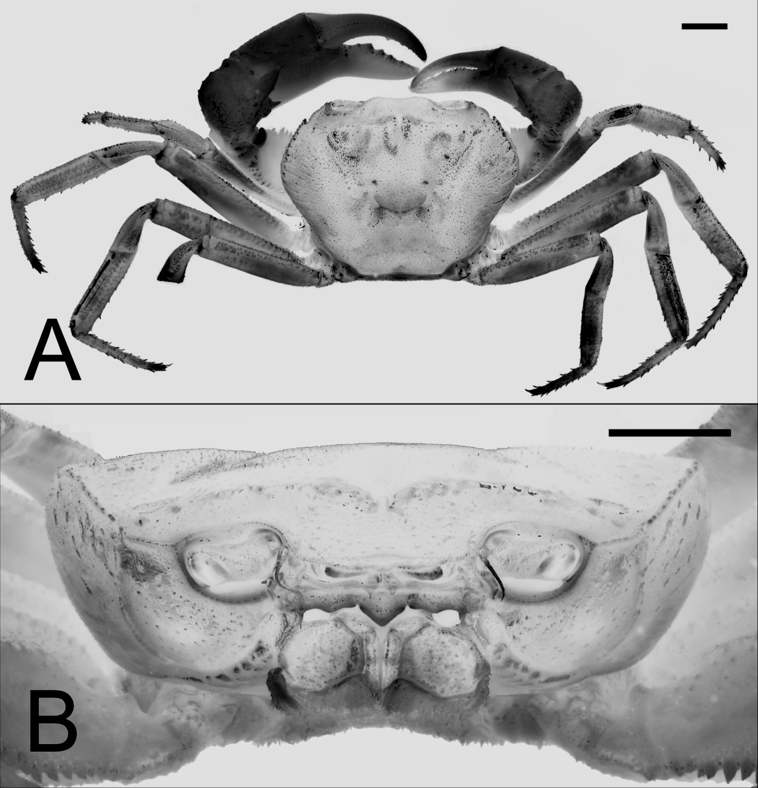
Description. Carapace quadrate to trapezoidal ( Fig. 1 View FIGURE 1 A), widest at anterior quarter, broader than long, dorsoventrally compressed, dorsal surface slightly convex longitudinally, covered with short setae, regions distinct; gastric regions covered with distinct oblique striae; cervical groove prominent; H-shaped gastric groove deep. Epigastric cristae distinct, edges sharp, separated by distinct median furrow; postorbital cristae sharp; epigastric, postorbital cristae confluent; epibranchial teeth, postorbital cristae not confluent, separated by gaps ( Fig. 1 View FIGURE 1 B). Frontal margin ( Figs. 1 View FIGURE 1 B, 2C) protruded, sinuous, deflexed. External orbital tooth triangular, produced anteriorly; outer margin concave, granular, longer than inner margin; epibranchial tooth distinct, triangular, well separated from external orbital tooth by deep U-shaped notch, tapering anteriorly; anterolateral margin straight to gently convex, upswept, serrated, clearly demarcated from posterolateral margin; posterolateral margin almost straight to gently concave, with oblique striae converging gradually towards posterior margin of carapace. Frontal median triangle distinct ( Figs. 1 View FIGURE 1 B, 2C), complete, with sharp, protruded dorsal margin, confluent with lateral margins; orbit well demarcated; supraorbital margin smooth; infraorbital margin lined with distinct granules; outer edge reaching, fused with anterolateral margin; suborbital, subbranchial regions covered with scattered oblique striae of nearly uniform size; pterygostomial region with oblique ridges on anterior dorsolateral part. Posterior margin of epistome ( Figs. 1 View FIGURE 1 B, 2D) with 3 lobes; median lobe triangular; lateral lobes sinuous, wider.
Eyes well developed ( Fig. 1 View FIGURE 1 B), well pigmented, occupying almost entire orbit. Third maxilliped ischium ( Fig. 3 View FIGURE 3 A) rectangular, much longer than broad, with distinct oblique submedian sulcus closer to mesial margin, surfaces covered with scattered short setae; merus quadrate, antero-external angle convex, upswept; tip of exopod not reaching midpoint of outer margin of merus, with short flagellum barely reaching edge of upswept mesial margin of merus.
Chelipeds not noticeably elongated, subequal, stronger in males; margins of merus serrated, dorsal margin with small subdistal tooth; carpus armed with strong distal sharply pointed inner tooth, dorso-ventrally flattened; palm with smooth outer surface; fingers robust, cutting edges armed with numerous medium-sized to large sharp teeth.
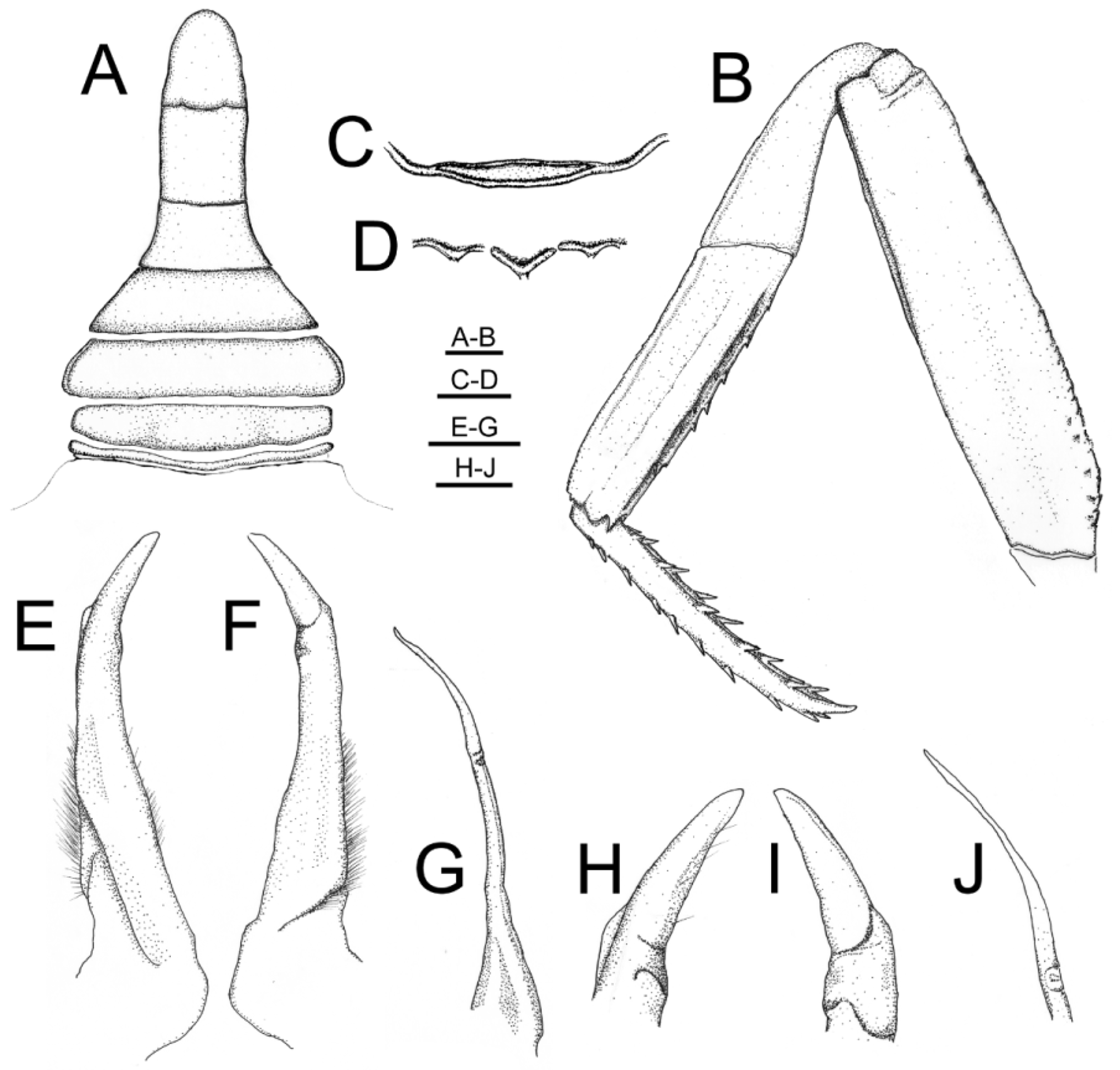
Ambulatory legs long ( Figs. 1 View FIGURE 1 A, 2B), slender, second leg longest, anterior margins of meri distinctly serrated, without subdistal tooth or spine, posterior margins smooth on all legs; second, third meri slightly shorter than carapace length; carpi short, with longitudinal submedian ridge on dorsal surface except for fourth leg, widened distally; propodi, dactyli subequal in length.
Male abdominal cavity reaching imaginary level demarcated by proximal quarter of coxae of chelipeds. Male abdomen ( Figs. 2 View FIGURE 2 A, 3B) narrow, T-shaped; somite 1 short, somite 6 longer than broad; proximal, distal margins of somites 3–5 sinuous; lateral margins of somite 2 convex; lateral margins of somite 3 convex; lateral margins of somite 4 straight, narrows gradually to distal end; somites 3–5 narrow gradually; lateral margins of somite 5 concave, proximally wide distally narrow; lateral margins of somite 6 longer than broad, straight; telson subtriangular, longer than broad, lateral margin slightly concave proximally, rounded distally. G1 slender ( Figs. 2 View FIGURE 2 E, F, H, I), subterminal segment gradually tapering towards terminal segment, proximal two-thirds straight, distal onethird slightly bent outward, segments separated by distinct suture; terminal segment straight, tapering, cylindrical, obliquely bent outwards from suture. G2 ( Figs. 2 View FIGURE 2 G, J) longer than G1, flagellum long, about half the length of basal segment.
Female. Female chelipeds subequal, neither inflated; abdomen rounded, covering entire thoracic sternum, all somites and telson freely articulating; telson broadly triangular with convex lateral margins ( Fig. 7A View FIGURE 7. A, B ). Vulvae large, round, without operculum; on median part of somite 6 ( Fig. 7B View FIGURE 7. A, B ).
Coloration. Dorsal surface of carapace is purple to maroon in life, cheliped is yellowish to reddish-orange.
Etymology. Named after a daughter of the first author, Dana Vi Husana, who accompanied him in some of his fieldwork.
Remarks. Sundathelphusa danae sp. nov. was collected from Bantakay Cave, Atimonan, Quezon Province, Luzon, the type locality of S. holthuisi Ng, 2010 . Although Sundathelphusa danae sp. nov. is sympatric with S. holthuisi (and the two species were collected together, see comparative material), and both possess a relatively slender G1 with a simple cylindrical terminal segment, they differ markedly in several other aspects. Sundathelphusa danae sp. nov. can easily be distinguished from S. holthuisi as follows: 1) the anterolateral margins are prominently more convex ( Fig. 1 View FIGURE 1 A) (margins gently convex in S. holthuisi, Ng 2010 : fig. 2B); 2) the branchial regions are proportionately more inflated ( Fig. 1 View FIGURE 1 B) (only gently swollen in S. holthuisi, Ng 2010 : fig. 2C); 3) the median triangular lobe on the posterior margin of the epistome is sharper and relatively broader ( Fig. 1 View FIGURE 1 B) (wider and the tip rounded in S. holthuisi, Ng 2010 : fig. 3B); 4) the frontal median triangle is complete ( Figs. 1 View FIGURE 1 B, 2C) (incomplete in S. holthuisi, Ng 2010: 3 B); 5) the ambulatory legs are relatively more slender and elongated, especially with regard to the merus and propodus ( Figs. 1 View FIGURE 1 A, 2B) (merus and propodus relatively shorter and stouter in S. holthuisi, Ng 2010 : fig. 2B); and 6) the G1 terminal segment is more strongly bent (Fig. E, F, H, I) (gently curved in S. holthuisi, Ng 2010 : fig. 4J–M).
Sundathelphusa danae View in CoL sp. nov. can immediately be separated from S. celer Ng, 1991 View in CoL , and S. longipes ( Balss, 1937) View in CoL (both from Luzon) by its more strongly convex anterolateral margins (cf Ng 2010: figs. 1A, 2A), proportionately longer ambulatory legs (cf. Ng 2010: figs. 1E, 2A) and more slender G1 which has the terminal segment slender and cylindrical (relatively stouter and shorter in S. celer View in CoL and S. longipes, Ng 2010 View in CoL : figs. 1H–K, 4E–H).
The spelling of the name of the type locality for S. holthuisi View in CoL – “Bautakay Cave, Station Catalina, Atomonau View in CoL ” (see also Ng 1991: 15 as Archipelothelphusa longipes ) is incorrect. It should be “Bantakay, Santa Catalina, Atimonan View in CoL ”.


| ZRC |
Zoological Reference Collection, National University of Singapore |
No known copyright restrictions apply. See Agosti, D., Egloff, W., 2009. Taxonomic information exchange and copyright: the Plazi approach. BMC Research Notes 2009, 2:53 for further explanation.
|
Kingdom |
|
|
Phylum |
|
|
Class |
|
|
Order |
|
|
InfraOrder |
Brachyura |
|
Family |
|
|
Genus |
Sundathelphusa danae
| Husana, Daniel Edison M., Yamamuro, Masumi & Ng, Peter K. L. 2014 |
S. longipes
| Ng 2010 |
S. celer
| Ng 1991 |
S. longipes (
| Balss 1937 |