
Jedediella horneri, Kontschán & Starý, 2012
|
publication ID |
https://doi.org/ 10.11646/zootaxa.3210.1.2 |
|
DOI |
https://doi.org/10.5281/zenodo.6373764 |
|
persistent identifier |
https://treatment.plazi.org/id/03DD521F-9D18-0A35-FF53-FA2C4973AC86 |
|
treatment provided by |
Plazi |
|
scientific name |
Jedediella horneri |
| status |
sp. nov. |
Jedediella horneri sp. nov.
( Figs 1–14 View FIGURES 1 – 5 View FIGURES 6 – 12 View FIGURES 13 – 14 )
Material examined. Holotype. Female ( HNHM, in alcohol), USA, California, Del Norte County , Jedediah Smith State Park , 1 April 1992, 41o50’ N, 124 o W, coniferous forest, under Tsuga heterophylla (Raf.) Sarg., sample of litter and soil, K. Horner coll. Paratypes. Four males and one female; same data as holotype. One male paratype in ISB, one male in MHNG and two males in HNHM in alcohol, one female paratype on slide in HNHM.
Description. Female. Length of idiosoma 880–890 µm, width 660–670 µm (n=2). Shape oval, idiosoma dorsally domed and strongly sclerotised.
Dorsal idiosoma ( Fig. 1 View FIGURES 1 – 5 ). Dorsal and marginal shields completely separated, all dorsal setae short (ca. 28–35 µm), smooth and needle-like. Caudal area of dorsal shield bearing a marginally strongly sclerotised, large depression. Setae on margins of depression as long as dorsal setae, but more robust. Caudal margin of dorsal shield undulate, other parts smooth. Dorsal shield without ornamentation. Marginal shield bearing smooth and needle-like setae (ca. 20–22 µm), surface strongly sclerotised near marginal setae, inner margin undulate.
Ventral idiosoma ( Fig. 2 View FIGURES 1 – 5 ). Surface of sternal shield mostly smooth, some shallow irregular pits near anterior margin of genital shield. Five pairs of smooth and needle-like sternal setae present, St1 (ca. 7 µm) placed near anterior margin of sternal shield, St2 (ca. 7 µm) at level of central area of coxae II, St3 (ca. 12 µm) at level of central area of coxae III, St4 (ca. 28 µm) at level of posterior margin of coxae III, St5 (ca. 17 µm) at level of posterior margin of coxae IV. Ventral shield without sculptural pattern and bearing smooth and needle-like setae (ca. 28–34 µm), adanal setae similar in shape and length to ventral setae, postanal seta absent ( Fig. 2 View FIGURES 1 – 5 ). Pedofossae deep, their surface smooth, without separate furrows for tarsi IV. Metapodal lines well developed, adjacent setae short and needle-like (ca. 14–17 µm). Genital shield linguiform, anteriorly rounded, its surface covered by oval pits ( Fig. 3 View FIGURES 1 – 5 .). Stigmata situated between coxae II and III, peritremes L-shaped ( Fig. 4 View FIGURES 1 – 5 ). Tritosternum with vase-shaped base, laciniae divided into two smooth lateral branches and one pilose central branch ( Fig. 5 View FIGURES 1 – 5 ).
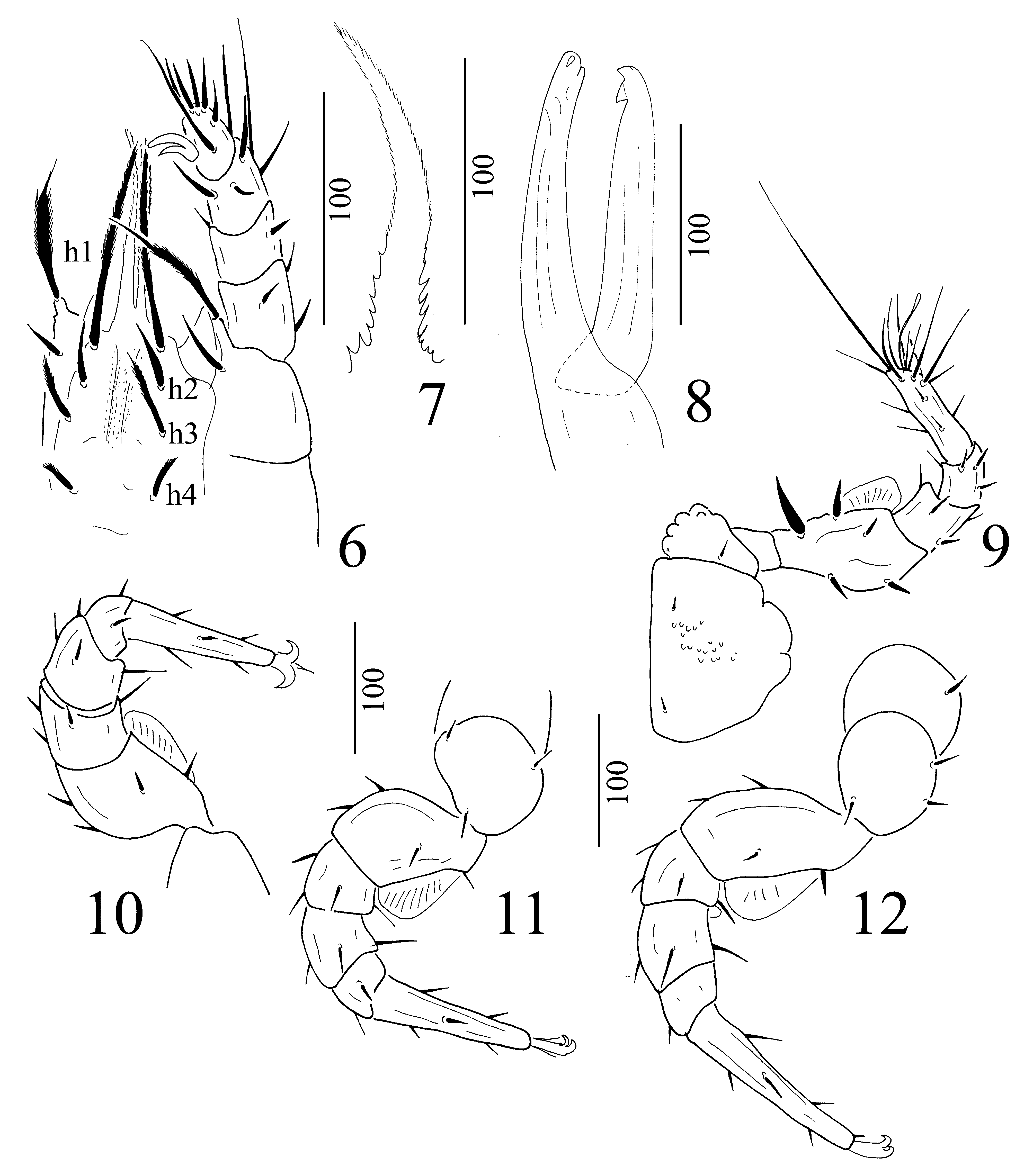
Gnathosoma ( Fig. 6 View FIGURES 6 – 12 ). Hypostomal setae h1 long and pilose (ca. 92µm), h2 short (ca. 30 µm) and smooth, h3 (ca. 38 µm) and h4 (ca. 20 µm) marginally pilose. Ventral surface of hypostome with small, spine-like structures in deutosternal groove. Palp trochanter with one long, pilose seta and one short smooth seta, other setae on palp smooth. Corniculi short and horn-shaped, internal malae longer than corniculi and marginally pilose. Epistome basally with long and narrow lateral spines, apically pilose ( Fig. 7 View FIGURES 6 – 12 ). Chelicerae long (ca. 170 µm), movable digit as long as fixed digit, movable digit bearing one large apical tooth, fixed digit with apical bulbiform sensory organ. Both digits medially smooth, without teeth, internal sclerotised node absent ( Fig. 8 View FIGURES 6 – 12 ).
Legs. Leg I without ambulacral claws ( Fig. 9 View FIGURES 6 – 12 ), coxa of leg I covered by small, oval pits, most of setae on leg I smooth and needle-like, but several setae robust. Other legs with claws, ( Figs 10–12 View FIGURES 6 – 12 ) without pits, femora of legs I–IV bearing ventral flap-shaped appendages, each segment of legs with smooth and pilose setae.
Male. Length of idiosoma 880–950 µm, width 650–680 µm (n=4). Shape and dorsal idiosoma as in female.
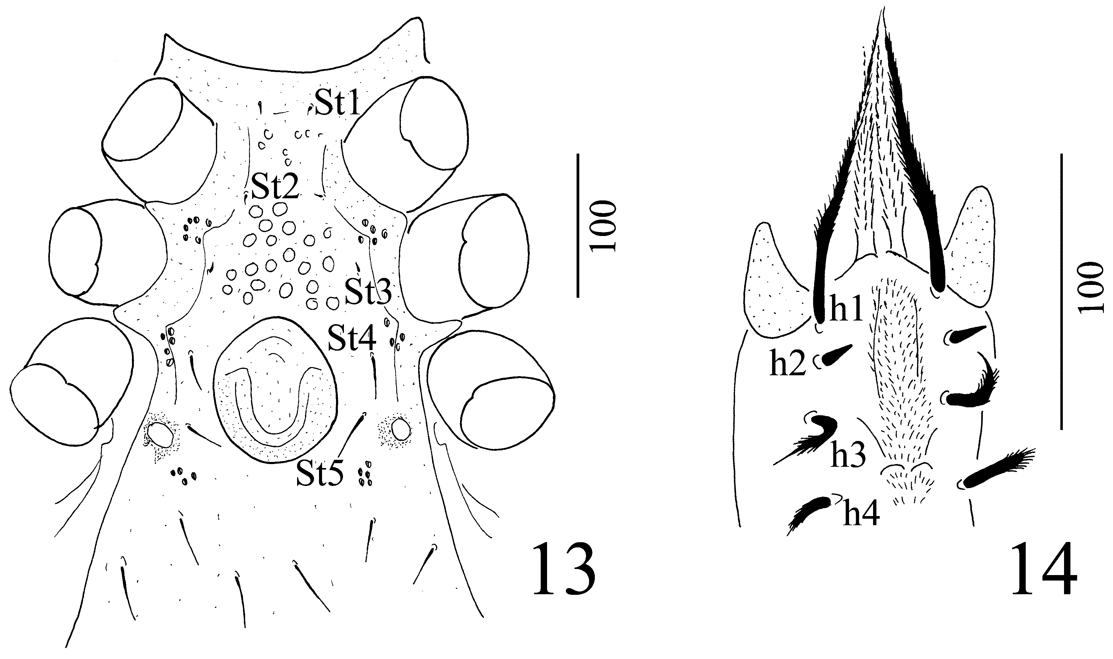
Ventral idiosoma ( Fig. 13 View FIGURES 13 – 14 ). Surface of sternal shield covered by oval pits, with five pairs of needle-like sternal setae. St1 (ca. 7 µm) placed near anterior margin of sternal shield, St2 (ca. 7 µm) at level of posterior margin of coxae II, St3 (ca. 7 µm) at level of central area of coxae III, St4 (ca. 26 µm) and St5 (ca. 27 µm) near lateral margin of genital shield. One pair of rounded depressions situated near St5. Position and shape of ventral setae and ornamentation of ventral shield as in female. Genital shield circular, without sculptural pattern, situated between coxae IV.
Gnathosoma ( Fig. 14 View FIGURES 13 – 14 ). Hypostomal setae h1 long (ca. 104 µm), pilose, h2 short (ca. 13 µm) and smooth, h3 (ca. 45–48 µm) and h4 (ca. 27 µm) long and pilose. Corniculi, internal malae, epistome, palp and chelicerae as in female.
Etymology. We dedicate the new species in honor to Dr. K. Horner, who collected these specimens in California.
Notes. Hirschmann & Zirngiebl-Nicol (1969a) described an unusual uropodine mite from North-America, which was placed into the genus Discourella ( D. sellnicki Hirschmann & Zirngiebl-Nicol, 1969 ). Later, when Hirschmann (1972) created a new subgeneric classification for Discourella , he placed this species into the Discourella caputmedusae -group on the basis of the large depression of the caudal area of dorsal shield. Hirschmann (1993) was not sure about the exact systematic position of the species; hence in his monograph this species was discussed separated from other Discourella species, similar to the species D. venusta ( Berlese, 1884) (see Kontschán, 2011).
Discourella sellnicki differs strongly from the other two species in this group on the basis of several characters [ D. caputmedusae ( Berlese & Leonardi, 1901) and D. caputmedusaesimilis Hirschmann, 1972 ] and it appears that D. sellnicki is more closely related to our new species. Hence we transfer this species into the new genus as Jedediella sellnicki ( Hirschmann & Zirngiebl-Nicol, 1969) comb. nov. Differences between the genera Jedediella and Discourella are summarised in Table 2 View TABLE 2 . and differences between the species of Jedediella and those of the Discourella caputmedusae -group are presented in Table 3 View TABLE 3 . According to our interpretation, species of the Discourella caputmedusae -group, placed in Comydinychus Berlese, 1917 in Hirschmann (1979), do not belong to the genus Discourella (this is a separate genus, which was described by Berlese (1917) earlier as Comydinychus Berlese, 1917 ), because the two species differ from other Discourella species by several characters. However, a revised placement of these two species is a topic for a future study.

No known copyright restrictions apply. See Agosti, D., Egloff, W., 2009. Taxonomic information exchange and copyright: the Plazi approach. BMC Research Notes 2009, 2:53 for further explanation.