
Phalangopsis quartzitica, Junta & Castro-Souza & Ferreira, 2020
|
publication ID |
https://doi.org/ 10.11646/zootaxa.4859.2.1 |
|
publication LSID |
lsid:zoobank.org:pub:7DFF87BA-9A4A-4773-AA2E-66A72505BC3F |
|
DOI |
https://doi.org/10.5281/zenodo.4539026 |
|
persistent identifier |
https://treatment.plazi.org/id/03DFCA0A-5B6E-5974-FF7F-FB07FA45403B |
|
treatment provided by |
Plazi |
|
scientific name |
Phalangopsis quartzitica |
| status |
sp. nov. |
Phalangopsis quartzitica n. sp.
( Figures 2–7 View FIGURES 2–7 , 8–14 View FIGURES 8–14 , 15–18 View FIGURES 15–18 , 19–23 View FIGURES 19–23 , 24–29 View FIGURES 24–29 , 148 View FIGURE 148 ; Table 1 and 4)
Material examined. Holotype ♂, code ISLA 65740, Brazil, Pará , municipality of São Geraldo do Araguaia, An- dorinhas cave (6°16’55.98”S; 48°32’33.52”O), 17.ii.2018, Sperandei, V. F., leg GoogleMaps . Holotype condition: integrate. Paratypes, 3 ♂ ♂ ( ISLA 65737; 65738; 65739) and 3 ♀ ♀ ( ISLA 65736; 65741; 65743), same data of holotype. Individuals examined , 1 ♂ ♂ ( ISLA 65742) and 1 ♀ ♀ ( ISLA 65744), same data of holotype .
Distribution. Andorinhas Cave in the municipality of São Geraldo do Araguaia, Pará, Brazil.
Etymology. The specific epithet “ quartzitica ” refers to the quartzite, the matrix rock of the cave where this specie was found.
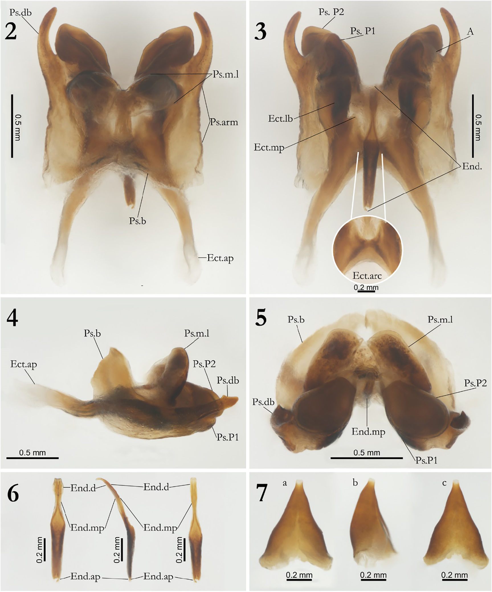
Diagnosis. Combination of the following characteristics: pseudepiphallic dorsal branch, thin and developed, projecting for the interior of the sclerite, the apex presents a visible dilation in frontal and lateral view ( Figs 2, 4 and 5 View FIGURES 2–7 ; Pd.db); pseudepiphallic paramere 2 developed, shape discoidal, projecting towards the exterior of the sclerite ( Figs 3–5 View FIGURES 2–7 , Ps.P2); pseudepiphallic arm elongated ( Fig 2 View FIGURES 2–7 , Ps.arm); pseudepiphallic median lobes developed and projecting dorsally, shape sub-quadrangular elongated towards the endophallus in frontal view and globular in dorsal view ( Figs 2, 4–5 View FIGURES 2–7 , Ps.m.1); pseudepiphallic branch little projected dorsally in dorsal view ( Figs 2, 4–5 View FIGURES 2–7 ; Ps.b); upper central and lower part of ectophallic arc horizontally curved ( Fig. 3 View FIGURES 2–7 , Ect. Arc); endophallic distal portion slightly developed in thickness ( Fig. 6 View FIGURES 2–7 , a, End.d).
Description, male holotype. Body color: general body coloration uniformly brownish, dorsal head whitish brown ( Fig. 8 View FIGURES 8–14 ); pronotum strong yellowish brown, with whitish discoloration spots ( Figs 8 and 10 View FIGURES 8–14 ); abdomen brown dorsally, white and translucent ventrally; legs yellowish brown, whitish at the start of the femur ( Figs 15–18 View FIGURES 15–18 ); cerci uniformly whitish brown. Head: slightly pubescent; elongated in front view (3.728 and 2.739 mm, length and width respectively); vertex marked with four dark vertical stripes, two starting from the eye’s region reaching the occiput, two starting from the antennas bases and reaching the occiput as two darkish spots ( Fig. 8 View FIGURES 8–14 ); gena, clypeus and labrum whitish, mandibles brownish yellow, dark brown near to the labrum; all maxillary palpomeres slightly pubescent, first and second shorter than the others and whitish, third and fourth palpomeres presenting similar size, the fourth is slightly larger, fifth palpomere lightly longer than fourth, claviform, arched and whitish at tip ( Fig. 8 View FIGURES 8–14 ); all labial palpomeres whitish, pubescent and increasing in size, third palpomere claviform ( Fig. 8 View FIGURES 8–14 ); scape pubescent, yellowish brown at the base and whitish toward the pedicel, pedicel dark yellowish brown, antennomeres uniformly yellowish brown ( Figs 8 and 9 View FIGURES 8–14 ); compound eyes black, with a small depigmented region near the scape insertion; ocelli absent ( Figs 8 and 9 View FIGURES 8–14 ). Thorax: pronotum yellowish dark brown; anterior, medial and posterior portion with spots whitish distributed along the sagittal axis in dorsal view ( Fig. 10 View FIGURES 8–14 ); dorsal disk broader than long, lateral lobes rounded, anterior and posterior margins sub-straight and with the presence of long bristles ( Fig. 10 View FIGURES 8–14 ). Legs. In general, femur, tibia and tarsus pubescent; first tarsomere serrulated; femur always smaller than tibia (μ=14.748 ± 0.587 mm; μ= 17.050 ± 0.640 mm, femur and tibia respectively, Leg III, n=4) ( Figs 15–18 View FIGURES 15–18 ). Leg I ( Figs 15 and 16 View FIGURES 15–18 ): tibia ventrally serrated and armed with two same-sized ventro apical spurs, tympanum absent; first tarsomere almost thrice longer than second and third together. Leg II ( Figs 15 and 16 View FIGURES 15–18 ): tibia ventrally serrated and armed with two same-sized ventral apical spurs; first tarsomere ventrally serrated and thrice bigger than the second and third together. Leg III: femur dilated; tibia serrulated, armed with four subapical spurs on outer side ( Fig. 17 View FIGURES 15–18 ) and three on inner side ( Fig. 18 View FIGURES 15–18 ), three apical spurs on outer ( Fig. 17 View FIGURES 15–18 ; a, b, c) and four on the inner side ( Fig. 18 View FIGURES 15–18 ; d, e, f, g), the inner being the longest; first tarsomere about thrice longer than the second and third together, armed with two apical spurs ( Figs 17–18 View FIGURES 15–18 ). Right Tegmen: pubescent, few sclerotized and underdeveloped, stridulatory file absent, poorly marked veins and with glandular thickening at the distal margin ( Fig. 11 View FIGURES 8–14 ). Abdomen: cerci pubescent and elongated, with long bristles at the base; supra-anal plate sub-quadrangular with long bristles at distal portion, base with two small lateral projections, apex slightly rounded and base curved inside ( Figs 12 and 13 View FIGURES 8–14 ); sub-genital plate sub-quadrangular, oval, short, base rounded, apex sub-triangular and lightly sharp ( Figs 13 and 14 View FIGURES 8–14 ).
Observations in Paratypes. Male phallic sclerites (paratype ISLA 65737, Figs 2–6 View FIGURES 2–7 ) Pseudepiphallus: dorsal branch well sclerotized, thin and developed, projecting for the interior of the sclerite, the apex presents a visible dilation in frontal and lateral view, ( Figs 2, 4 and 5 View FIGURES 2–7 , Pd.db), this structure is very similar to Ps.db of Phalangopsis ferratilis n. sp.; paramere 1 slightly cambered triangular, with a depression on lateral external face, connecting to the paramere two and A sclerite by a membranous tissue ( Figs 3–5 View FIGURES 2–7 , Ps.P1); paramere 2 developed, shape discoidal, projecting towards the exterior of the sclerite and more developed compared to paramere 1 ( Figs 3–5 View FIGURES 2–7 , Ps.P2); pseudepiphallic arm elongated ( Figs 2 View FIGURES 2–7 , Ps.arm); A sclerite vestigial and fused to Ps.arm, reaching paramere 1 and visible at ventral view ( Fig. 5 View FIGURES 2–7 , A); pseudepiphallic medium lobes developed and projected dorsally, shape sub-quadrangular elongated towards the endophallus in frontal view and globular in dorsal view ( Figs 2, 4–5 View FIGURES 2–7 , Ps.m.1); pseudepiphallic little projected dorsally in dorsal view, covering part of the proximal region of the ectophallic apodemes ( Figs 4–5 View FIGURES 2–7 , Ps.b). Ectophallic invagination: apodemes thin and curved dorsally, with a small break in the proximal part of the projections, followed by a inclining in dorsal and lateral view, apex little sclerotized and dilated ( Figs 2 and 4 View FIGURES 2–7 , Ect. ap.); lateral bar well developed, elongated in all its extension, internal face slightly projecting towards the Endophallus ( Fig. 3 View FIGURES 2–7 , Ect.lb); median projection undeveloped ( Fig. 3 View FIGURES 2–7 , Ect.mp); upper central and lower part of ectophallic arc horizontally curved ( Fig. 3 View FIGURES 2–7 , Ect.arc). Endophallus: endophallus partially projected dorsally in lateral view ( Fig. 6 View FIGURES 2–7 , b, End); endophallic distal portion slightly developed in thickness ( Fig. 6 View FIGURES 2–7 , a, End.d), with a small vertical groove ( Fig. 6 View FIGURES 2–7 , a–c, End.d); median portion narrow ( Fig. 6 View FIGURES 2–7 , a–c, End.mp); apodeme reduced ( Fig 6 View FIGURES 2–7 , a–c, End.ap).
Female: body size bigger than the male (♀ µ=19.976 ± 0.638mm, n=3); apterous; femur always smaller than tibia; supra-anal plate pubescent and rounded in distal portion, with long bristles, base curved inside with two small lateral projections ( Fig. 19 View FIGURES 19–23 ); sub-genital plate short, lightly pubescent, V-shaped, presenting a slight indentation ( Fig. 20 View FIGURES 19–23 ); ovipositor elongated, strong yellowish brown, sword-shaped with sharp apex (µ=11.736 ± 1.140mm, n=3) ( Figs 21–23 View FIGURES 19–23 ). Female genitalia. Copulatory papilla triangular shaped, slightly flattened dorsoventrally ( Fig. 7 View FIGURES 2–7 , a and b), the edges of the middle part are slightly bulged ( Fig. 7 View FIGURES 2–7 , a and c); presents a dorsal opening of triangular shape in the proximal portion and a small rounded orifice in the distal portion ( Fig. 7 View FIGURES 2–7 , a-c).
Ecological Remarks: Individuals of Phalangopsis quartzitica n. sp. were found in a single quartzite cave (Andorinhas cave, Fig. 24 View FIGURES 24–29 ) located in the “Serra das Andorinhas” region (São Geraldo do Araguaia municipality, Pará state). However, this cave is located at the top of the mountain, differently from the caves were Phalangopsis araguaia n. sp. were observed (that are also inserted in the same mountain). The Andorinhas cave is considerably large, with around 1 km of extension ( Auler 2019). Specimens of Phalangopsis quartzitica n. sp. are quite common along the cave, being observed in several areas, usually in considerable densities. However, they showed an obvious preference for aphotic and moistened areas, preferring the cave walls ( Figs 28 and 29 View FIGURES 24–29 ) as observed for the other species herein described. Specimens of Phalangopsis quartzitica n. sp. were mainly feeding on bat guano, especially from insectivorous bats. Among the predators that were observed feeding on the crickets, there are scorpions ( Tityus metuendus Pocock, 1897 , Fig. 25 View FIGURES 24–29 ) and whip-spiders ( Heterophrynus sp., Fig. 27 View FIGURES 24–29 ). The cave has entrances on either the side cliffs of the mountain (where occurs a typical Amazon rainforest) and the top of the mountain, which presents a savanna-like vegetation. Although the side cliffs and the top of the mountain are relatively well preserved, the landscape surrounding the mountain is strongly altered, especially by the removal of the original vegetation for agriculture. The cave receives sporadic human visitors, who seem to only go through the main conduit of the cave, thus not presenting a threat for the invertebrate community. Finally, it is important to highlight that no sampling was conducted outside caves in this study. Hence, despite the fact that specimens of P. quartzitica n. sp. were only found in one cave, they are probably not troglobitic, given the absence of any obvious troglomorphic traits. Therefore, further studies including external samplings are needed to better understand the actual distribution of this species.


| V |
Royal British Columbia Museum - Herbarium |
No known copyright restrictions apply. See Agosti, D., Egloff, W., 2009. Taxonomic information exchange and copyright: the Plazi approach. BMC Research Notes 2009, 2:53 for further explanation.
|
Kingdom |
|
|
Phylum |
|
|
Class |
|
|
Order |
|
|
Family |
|
|
Genus |