
Phalangopsis kysuia, Junta & Castro-Souza & Ferreira, 2020
|
publication ID |
https://doi.org/10.11646/zootaxa.4859.2.1 |
|
publication LSID |
lsid:zoobank.org:pub:7DFF87BA-9A4A-4773-AA2E-66A72505BC3F |
|
DOI |
https://doi.org/10.5281/zenodo.4539036 |
|
persistent identifier |
https://treatment.plazi.org/id/03DFCA0A-5B73-595C-FF7F-FF2BFEB24073 |
|
treatment provided by |
Plazi |
|
scientific name |
Phalangopsis kysuia |
| status |
sp. nov. |
Phalangopsis kysuia n. sp.
( Figures 110–115 View FIGURES 110–115 , 116–122 View FIGURES 116–122 , 123–126 View FIGURES 123–126 , 127–131 View FIGURES 127–131 , 132–135 View FIGURES 132–135 , 148 View FIGURE 148 ; Table 2 and 3 View TABLE 3 )
Material examined. Holotype ♂, code ISLA 65725, Brazil, Mato Grosso, municipality of Apiacás, Casa de Pedra da Navalha cave ( 7°28’50.70”S; 58°12’36.40”O), 09.ix.2011, Ferreira , R. L., leg GoogleMaps . Holotype condition: integrate, legs detached and stored in microtubes. Paratypes, municipality of Apiacás , Mato Grosso state, Brazil, Oncinha cave ( 8°8’49.00”S; 57°13’17.44”O), 14.ix.2011, 1 ♀ (ISLA 65726), Ferreira , R. L., leg; municipality of Apuí , Ama- zonas state, Brazil, Casa de Pedra do Pena cave ( 8°20’13.40”S; 58°19’23.20”O), 12.v.2011, 1 ♀ (ISLA 65724) and 12.ix.2011, 1 ♂ ( ISLA 65727) and 2 ♀ ♀ (ISLA 65728; 65729), Ferreira, R. L., leg GoogleMaps .
Distribution. Casa de Pedra da Navalha cave and Oncinha cave in the municipality of Apiacas, Mato Grosso Brazil; and Casa de Pedra do Pena cave in the municipality of Apuí, Amazonas, Brazil.
Etymology. The word “kysuia” means “cricket” in the native language of the Apiacá, which is one of the ethnic people from the Juruena region, where the species was found.
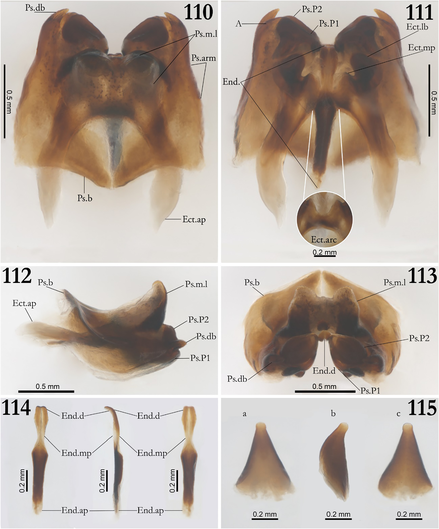
Diagnosis. Combination of the following characteristics: pseudepiphallic dorsal branch greatly reduced and curved internally toward the pseudepiphallic parameres ( Figs 110–113 View FIGURES 110–115 , Ps.db.); pseudepiphallic median lobes well rounded, dorsally projected, shape quadrangular (in front view), distal portion distant from pseudepiphallic branch (lateral view), narrow and globular in dorsal view ( Figs 110, 112–113 View FIGURES 110–115 ; Ps.m.l); pseudepiphallic branch developed, forming an semi-acute projection covering partially the endophallus (dorsal view) ( Fig. 110, 112–113 View FIGURES 110–115 ; Ps.b); pseudepiphallic arm short and inclined internally ( Fig. 110 View FIGURES 110–115 , Ps.arm); ectophallic lateral bar flattened, external face tilted out the genitalia, apex projecting inside the sclerite with a rounded apex ( Fig. 111 View FIGURES 110–115 , Ect.lb); upper central part of ectophallic arc curved in U format, lower part slightly curved horizontally ( Fig. 111 View FIGURES 110–115 , Ect. arc); proximal projections of ectophallic apodemes expanded in width (dorsal view) ( Figs 110, 112–113 View FIGURES 110–115 ; Ect. ap); endophallic distal portion slightly developed (dorsal view) ( Fig. 114 View FIGURES 110–115 , End.d).
Description, male holotype. Body color: general body coloration yellowish brown, dorsal head light yellowish brown ( Fig. 116 View FIGURES 116–122 ); pronotum yellowish brown with whitish discoloration spots ( Figs 116 and 118 View FIGURES 116–122 ); abdomen grayish white translucent ventrally and yellowish brown dorsally; brownish yellow legs, whitish at the start of the femur ( Figs 123–126 View FIGURES 123–126 ); cerci yellowish brown. Head: lightly pubescent; elongated in front view (3.721 and 2.554 mm, length and width respectively); vertex marked with two vertical dark stripes starting at the eye’s region and two starting at the antenna’s base, both reaching the occiput ( Fig. 116 View FIGURES 116–122 ); gena whitish yellow, clypeus whitish brown, labrum whitish and mandibles yellowish brown; all maxillary palpomeres slightly pubescent, first and second palpomeres whitish, and smaller than the others, third and fourth palpomeres similar size and yellowish, fifth palpomere a little longer than the forth, yellowish, whitish at the tips, claviform and curved ( Fig. 116 View FIGURES 116–122 ); all labial palpomeres whitish, pubescent and increasing in size, third palpomere claviform ( Fig. 116 View FIGURES 116–122 ); scape yellowish brown and pubescent, pedicel and antennomeres yellowish brown; compound eyes black, with a small depigmented area near the base of the scape; ocelli absent ( Figs 116 and 117 View FIGURES 116–122 ). Thorax: pronotum yellowish brown; anterior, medial and posterior portion with whitish spots distributed along the sagittal axis at dorsal view ( Fig. 118 View FIGURES 116–122 ); lateral lobes rounded, dorsal disk broader than long; anterior and posterior margins sub-straight and with long bristles ( Fig. 118 View FIGURES 116–122 ). Legs. In general, femur, tibia and tarsus pubescente; first tarsomere serrulate; femur always smaller than tibia (μ=16.911 ± 4.156 mm; μ= 19.697 ± 5.472 mm, femur and tibia respectively, Leg III, n=2) ( Figs 123–126 View FIGURES 123–126 ). Leg I ( Figs 123 and 124 View FIGURES 123–126 ): tibia serrated ventrally, armed with two same-sized apical ventral spurs, tympanum absent; first tarsomere about three times longer than the second and third together. Leg II ( Figs 123 and 124 View FIGURES 123–126 ): tibia ventrally serrulated, armed with two same-sized ventral apical spurs; first tarsomere ventrally serrated and approximately thrice bigger than the second and third tarsomeres together. Leg III ( Figs 125 and 126 View FIGURES 123–126 ): femur dilated; tibia serrulated, armed with four subapical spurs on outer ( Fig. 125 View FIGURES 123–126 ) and three on inner side ( Fig. 126 View FIGURES 123–126 ), three apical spurs on outer side ( Fig. 125 View FIGURES 123–126 ; a, b, c) and four on the inner ( Fig. 126 View FIGURES 123–126 ; d, e, f, g), the inner ones being the longest; first tarsomere about thrice longer than the second and third together, showing two apical spurs. Right Tegmen: Absent ( Fig. 119 View FIGURES 116–122 ). Abdomen: cerci slender and pubescent; supra-anal plate sub-quadrangular with long bristles at the apex, base with two reduced lateral projections, apex rounded and base curved inside ( Figs 120 and 121 View FIGURES 116–122 ); sub-genital plate sub-quadrangular, broader than long, base straight, apex sub-triangular and sharp ( Figs 121 and 122 View FIGURES 116–122 ).
Observations in paratype series. Male phallic sclerites ( paratype ISLA 65727, Fig. 110–114 View FIGURES 110–115 ) Pseudepiphallus: dorsal branch well sclerotized but greatly reduced when compared with other Phalangopsis ( P. ferratilis n. sp. e.g.), curved internally toward the pseudepiphallic parameres ( Figs 110–113 View FIGURES 110–115 , Ps.db.); paramere 1 cambered triangular, with a depression on its lateral external face, connecting to the paramere 2 and sclerite A by a membranous tissue ( Fig 111–113 View FIGURES 110–115 , Ps.P1); paramere 2 stocky discoidal (in dorsal view), same size than the paramere 1, less developed than the paramere 2 of Phalangopsis ferratilis n. sp. ( Fig 111–113 View FIGURES 110–115 , Ps.P2); pseudepiphallic arm short and inclined internally ( Fig. 110 View FIGURES 110–115 , Ps.arm); A sclerite vestigial, fused with the Ps.arm, reaching the paramere 1 and visible at ventral view ( Fig. 111 View FIGURES 110–115 , A); pseudepiphallic median lobes well rounded, dorsally projected, shape quadrangular at frontal view, distal portion distant from pseudepiphallic branch in lateral view, narrow and globular in dorsal view ( Figs 110, 112–113 View FIGURES 110–115 ; Ps.m.l); pseudepiphallic branch developed, forming an semi-acute projection covering partially the endophallus (dorsal view) ( Figs 110, 112–113 View FIGURES 110–115 ; Ps.b). Ectophallic invagination: apodeme shorter and broader than in other Phalangopsis , curved internally, apex little sclerotized and expanded in width ( Figs 110-113 View FIGURES 110–115 , Ect. ap.); ectophallic lateral bar flattened, external face tilted out the genitalia, apex projecting inside the sclerite with a rounded apex ( Fig. 111 View FIGURES 110–115 , Ect.lb); median projection undeveloped ( Fig. 111 View FIGURES 110–115 , Ect.mp); upper central part of ectophallic arc curved in U format, lower part slightly curved horizontally ( Fig. 111 View FIGURES 110–115 , Ect. Arc). Endophallus: endophallic distal portion slightly developed in thickness, apex projecting dorsally, with a small vertical groove ( Fig 114 View FIGURES 110–115 , a–c, End.d); median portion narrow ( Fig 114 View FIGURES 110–115 , a–c, End.d); apodeme reduced ( Fig 114 View FIGURES 110–115 , a–c, End.d).
Female: body size bigger than male ( ♀ µ=20.822 ± 1.446mm, n=4); apterous; femur always smaller than tibia; supra-anal plate pubescent and lightly elongated, rounded in distal portion and with long bristles, base curved inside and presence of two rounded lateral projections( Fig. 127 View FIGURES 127–131 ); sub-genital plate short, lightly pubescent, U-shaped, presenting a lightly indentation ( Fig. 128 View FIGURES 127–131 ); ovipositor elongated, yellowish brown, presenting sword-shaped with pointed apex (µ=12.452 ± 0.766mm, n=4) ( Figs 126-128 View FIGURES 123–126 View FIGURES 127–131 ). Female genitalia. Copulatory papilla triangular shaped, slightly flattened dorsoventrally ( Fig. 115 View FIGURES 110–115 , a and b); edges of middle part sub-straight (dorsal and ventral view) ( Fig. 115 View FIGURES 110–115 , a and c); presence of a triangular opening in all its extension and a small rounded orifice in the distal portion ( Fig. 115 View FIGURES 110–115 , a–c).
Ecological Remarks: Specimens of Phalangopsis kysuia n. sp. were found in three siliciclastic caves in the Juruena National park, located in the border between the Mato Grosso and Amazonas Brazilian states, in the Amazon forest. Although other nine caves were also inventoried in the region, specimens were only found in these three caves. Individuals were only observed in the aphotic, moist and deeper areas of the caves, and its non-occurrence in the other caves may be due to their small extension (thus, their inner portions were not completely aphotic). In one of the caves (Casa de Pedra da Navalha), the population was quite small, and only few specimens were observed, apparently feeding on the guano of insectivorous bats ( Peropteryx sp.). On the other hand, the Casa de Pedra do Pena cave, which comprises the largest sampled cave in the area, presented an expressive population, which only occurred in a lateral and isolated chamber of the cave. Although the main conduit of this cave is quite voluminous, the distances between the two cave entrances is relatively small, what makes this chamber lightened ( Fig. 132 View FIGURES 132–135 ). However, there is a secondary conduit, connected to the main chamber ( Fig. 133 View FIGURES 132–135 ), which has more stable and moistened conditions ( Fig. 134 View FIGURES 132–135 ). The individuals were observed feeding on bat guano, especially produced by frugivorous bats ( Carolia sp.). The area is quite remote and can only be accessed by boat or small aircraft. Thus, the whole area is well preserved, including both the caves, which showed no signs of human presence, and the forest surrounding them, which is completely preserved within a radius (from the caves) of at least 100 km. It is worth noting that samplings where not performed in the external environments, so the actual distribution of this species remains unknown.


| R |
Departamento de Geologia, Universidad de Chile |
No known copyright restrictions apply. See Agosti, D., Egloff, W., 2009. Taxonomic information exchange and copyright: the Plazi approach. BMC Research Notes 2009, 2:53 for further explanation.
|
Kingdom |
|
|
Phylum |
|
|
Class |
|
|
Order |
|
|
Family |
|
|
Genus |