
Tyttocharax metae, Román-Valencia & García-Alzate & Ruiz-C. & Taphorn B, 2012
|
publication ID |
https://doi.org/ 10.1590/S1679-62252012000300004 |
|
persistent identifier |
https://treatment.plazi.org/id/03E0305B-FFC3-FFEB-FEF5-1ECFFB6AF96A |
|
treatment provided by |
Carolina |
|
scientific name |
Tyttocharax metae |
| status |
sp. nov. |
Tyttocharax metae View in CoL , new species
Figs. 1-6 View Fig View Fig View Fig View Fig View Fig View Fig
Holotype. IUQ 2581 View Materials , 15.3 mm SL, adult male, Colombia, Meta, Vista Hermosa near Palestina, río Orinoco basin, río Güejar system, arroyo Pringamosal , tributary of arroyo Blanco 500 m below Palestina School , 03º05’22”N 73º49’27”W, 240 m a.s.l., 9 Jan 2009, C. Román-Valencia, C. García-Alzate & R. Ruiz-C. GoogleMaps
Paratypes. All from Colombia: Meta State: La Macarena mountains, río Orinoco basin, río Güejar system unless noted: AUM 50299, 2 View Materials , 15.7-18.3 mm SL, IUQ 2343 View Materials , 3 View Materials , 14.3-18.6 mm SL, Vista Hermosa , La Palestina, arroyo Palestina, 03º05’15”N 73º49’54”W, 282 m a.s.l., 8 Jul 2008, C. Román-Valencia, C. García-Alzate & R. Ruiz- C. IUQ 2344 View Materials , 4 View Materials , 13.3 View Materials -14.0 mm SL, IUQ 2345 View Materials , 3 View Materials , 13.2-14.5 mm SL, Vista Hermosa, La Palestina, creek 2 km north of Las Brisas , road to Vista Hermosa, 03º03’00”N 73º49’05”W, 264 m a.s.l., 10 Jul 2008, C. Román-Valencia, C. García-Alzate & R. Ruiz-C. MPUJ 6135 , 2 , 14.1-15.8 mm SL, Vista Hermosa, La Palestina, creek 2 km north of Las Brisas , road to Vista Hermosa, 03º03’00”N 73º49’05”W, 264 m a.s.l., 10 Jul 2008, C. Román-Valencia, C. García-Alzate & R. Ruiz- C. IUQ 2346 View Materials , 1 View Materials , 11.4 mm SL, Granada, río Ariari system, arroyo Mucuyita 03º27’05”N 73º47’49”W, 301 m a.s.l., 7 Jul 2008, C. Román-Valencia, C. García-Alzate & R. Ruiz-C. IUQ 2347 View Materials , 2 View Materials , 12.9- 14.1 mm SL, IUQ 2493 View Materials , 1 View Materials , 18.4 mm SL, collected with the holotype. IUQ 2494 View Materials , 2 View Materials , 13.2-14.3 mm SL, Vista Hermosa, Puerto Lucas, arroyo Acacias on road to Vista Hermosa, 03º05’24”N 73º45’32”W, 240 m a.s.l., 8 Jan 2009, C. Román-Valencia, C. García-Alzate & R. Ruiz- C. IUQ 2496 View Materials , 1 View Materials c&s, 12.3 mm SL, Vista Hermosa, Puerto Lucas, arroyo Luciana 100 m north of bridge in Puerto Lucas, 03º06’22”N 73º46’44”W, 253 m a.s.l., 8 Jul 2008, C. Román-Valencia, C. García- Alzate & R. Ruiz-C. IUQ 2495 View Materials , 3 View Materials c&s, 12.4-13.6 mm SL, Vista Hermosa, La Palestina, creek 1 km north of Las Brisas , 03º02’55”N 73º49’10”W, 278 m a.s.l., 10 Jul 2008, C. Román-Valencia, C. García- Alzate & R. Ruiz-C. IUQ 2755 View Materials , 4 View Materials , 15.4-17.1 mm SL, Vista Hermosa, La Palestina, arroyo Pringamosal, 03º05’29”N 73º49’47”W, 273 m a.s.l., 9 Nov 2009, M. I. González & A. M. Barrero GoogleMaps .
Diagnosis. Tyttocharax metae can be distinguished from all congeners by having bony hooks on the pectoral and caudalfin rays (vs. absent). Differs from T. madeirae and T. cochui by the absence of adipose fin (vs. adipose fin present), in having larger bony hooks on the anal fin than on the rays of the pelvic fins (vs. bony hooks of the same size on the rays of those fins). Differs from T. cochui in having i,5-6 pectoral-fin rays (vs. i,7). Tyttocharax metae can be distinguished from T. tambopatensis by absence of a sexually dimorphic color pattern in life (vs. presence of sexually dimorphic color pattern, males have bright orange abdomens), by the number of scales rows between anal-fin origin and lateral line (4 vs. 6), by the number of scale rows between pelvic-fin and lateral line (4 vs. 6), by the number of branched pectoral-fin rays (5-6 vs. 7); by the number of unbranched dorsal-fin rays (iii vs. ii) and distance between the dorsal and anal-fin origins (22.3 to 32.2% SL vs. 38.0-41.0% SL).
Description. Morphometric and meristic data for holotype and paratypes in Table 1. Body deepest between verticals through pelvic-fin and dorsal-fin origins in females; deepest between verticals through posterior margin of dorsal-fin base and middle part of anal-fin base in males. Predorsal profile of body generally convex in both sexes. Body profile slightly elevated at dorsal-fin origin then slightly concave from dorsalfin origin to procurrent caudal-fin rays; slightly convex near tips of depressed dorsal-fin rays in males. Origin of dorsal fin nearer caudal-fin base than snout. Dorsal-fin origin at vertical through anal fin in females; anterior to vertical through analfin origin in males. Tips of pelvic-fin rays reach anal-fin origin in both sexes. Ventral profile of body convex from tip of lower jaw to pelvic-fin insertion. Ventral body profile convex along anal-fin base in males, concave in females. Ventral profile of caudal peduncle convex in females, concave in sexually dimorphic males ( Figs. 1 View Fig and 2 View Fig ). Lower jaw prominent, projecting beyond upper jaw. Jaws and lips of mature males moderately thickened with specialized accommodation of prominent premaxillary teeth outside of snout. Lips thin in females. Maxilla extending anteriorly to antero-ventral border of eye in sexually dimorphic males and females.
All teeth conical and distributed over surface of mandibles in an apparently random pattern that makes them difficult to count in traditional rows. Number and distribution of teeth sexually dimorphic, with males having higher number and more prominent teeth ( Fig. 3 View Fig ). In males, premaxilla with four to five sets of teeth, each with 18 to 22 conical teeth that extend over its entire surface; fewer teeth in females, restricted to ventral half of premaxilla, with up to three sets of conical teeth, each with a maximum of 15 teeth; only two to three teeth in upper row. Maxilla in males with two to three sets of teeth that overlap dorsally; only one series of larger wellseparated teeth ventrally. Maxilla in females with only one series of non-overlapping teeth positioned dorsally ( Fig. 3 View Fig ). Dentary in males with four sets of 25 to 30 unicuspid teeth randomly distributed in front of dentary; reduced to two series laterally where they overlap with maxillary teeth. Females with only two to three sets of the teeth in front reducing to just one series laterally.
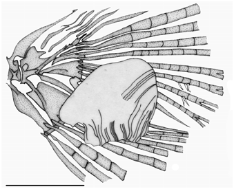
Scales cycloid, small. Lateral line complete, perforated scales 31-34 (31*, mode = 33). Scale rows between dorsal-fin origin and lateral line five (n = 28); scale rows between lateral line and anal-fin origin four (n = 28); scale rows between lateral line and pelvic-fin insertion four (n = 28). Predorsal scales 13-14, arranged in regular series (13*, mean = 13.7, mode = 13). Dorsal-fin rays ii,5-7,i (ii,5,i*, n = 28); first unbranched ray approximately one-half length of second ray, its tip reaching proximal bifurcation of first branched ray.Analfin rays ii,13-18,i (ii,14,i*; ii,13-14,i in males and ii,14-18,i in females, n = 26). Pectoral-fin rays i,5-6,i (i,5,i*; i,5-6,i in males and females). Pelvic-fin rays i,3-6,i (i,3,i*; i,3-4,i in males and i,3-6,i in females, n = 26). Pelvic-fin origin anterior to vertical through dorsal-fin origin. Caudal-fin pouch scale of irregular shape with approximately 26-28 radii ( Fig. 6 View Fig ). Total number of vertebra 40-41.
Maxilla angular, more pronounced in females in which ventral margin extends anteriorly and teeth cover anterior half of this structure. Supraneurals absent. Five infraorbitals, the first vestigial, infraorbitals two to five in typical position for genera included in Stevardiinae . The third infraorbital not in contact with preopercular sensory canal. Anterior margin of sphenotic extending over fifth infraorbital. Supraoccipital spine short, not reaching anterior margin of neural complex. Orbitosphenoid bone with an extension in anterior ventral region with bands of cartilage in anterior and posterior margins. Rhinosphenoid bone square, contacting orbitosphenoid and parasphenoids by bands of cartilage. Parasphenoid not divided, joined to ventral vomer surface by cartilage; posterior end of parasphenoid in contact with prootic and basioccipital by band of cartilage.
Dorsal margin of metapterygoid wide with a crest, foramen present in medial posterior region, with band of cartilage between it and quadrate. Ectopterygoid long and narrow, not in contact with quadrate.Mesopterygoid with entire dorsal margin in contact with ventral region of parasphenoid, band of cartilage over entire antero-ventral margin that connects to ectopterygoid.
522 A new species of Tyttocharax from the Güejar River
Nasal bone present. Basihyal cartilaginous not divided. Pharyngeal curved, an elongated plate, with thick cartilage at dorsal and ventral ends. Sixteen gill rakers on first branchial arch; 4-5 gill rakers on ceratobranchial and 11-12 gill rakers on epibranchial. Pectoral girdle with pointed dorsal process on cleithrum. Cleithrum elongated with straight posterior border; located under ventral edge of operculum. Anterior border of scapula straight. Postcleithrum 1 and 2 absent, postcleithrum 3 elongated and curved covering more than half of cleithrum. Proximal pterygiophore rays of dorsal fin inserted between neural spines 10 and 16; 17 th proximal pterygiophore of anal fin inserted between hemal spines 11 and 12.
Color in alcohol. See Figs. 1 View Fig and 2 View Fig for pigment patterns in preserved males and females. Body light yellow, dark brown on dorsum with dark spot at base of caudal peduncle. Sides of body with dark stripe that starts posterior to operculum and extends to caudal spot and widens at level of vertical through posterior tip of ventral-fin rays. Posterior margins of scales located on upper sides anterior to dorsal fin dark. Pectoral, pelvic and anal fins hyaline. Dorsal area of head dark. Humeral spot visible.
Color in life. Dorsum of body, head and post ventral area greenish yellow, with an evident absence of dark pigment. Sides of body with blue stripe, caused by presence of iridophores that generate an iridescent bluish aspect known as Rayleigh scattering. The iridophores are limited on sides to just dorsal margin of coelomic cavity. From there they extend posteriorly to the caudal peduncle. Lateral surface of coelomic cavity covered with leucophores that color this part of fish white. Bases of middle caudal-fin rays covered with narrow band of melanophores, more concentrated on dorsal lobe of caudal fin, and forming caudal spot. Posterior border of opercle covered by shiny blue iridophores. Humeral spot horizontally elongated and formed by disperse melanophores in area along dorsal margin of coelomic cavity, overlaing iridophores.Anal-fin rays with dispersed melanophores along their bases. All the fins hyaline. Color pattern identical in males and females.
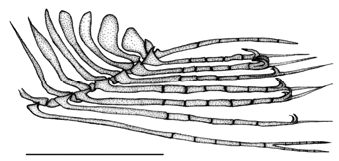
Sexual dimorphism. Neither the principal component analysis among species of Tyttocharax nor regressions comparing males and females produced significant results. In males, teeth are found along entire length of anterior margin of maxilla, which is less sharply pointed on its ventral tip than in females. In both sexes maxillary teeth diminish gradually in size ( Fig. 3 View Fig ). Posterior pterygiophores (last two or three including terminal piece) of anal fin in males of Tyttocharax metae are swollen, but in females are vestigial. Five or six penultimate anal-fin rays each have a pair of bony hooks ( Fig. 3 View Fig ) present at distal extremity. Fin-ray segment dorsal to one with hook thickened in last rays with hooks ( Fig. 4 View Fig ).
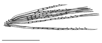
Pelvic fin of T. metae with i,3-6,i rays as in T. tambopatensis , but distribution of hooks is different: four hooks are present on simple ray and last branched ray, with one pair at terminus of each segment on their distal margins; five hooks are found on first and penultimate branched rays ( Fig. 5 View Fig ). Three or four bony hooks are present on middle portion of caudal-fin rays. Principal caudal-fin ray count 10/7. Eleventh principal caudal-fin ray (as identified in females) is transformed in males into an accessory structure coupled to caudal scale ( Fig. 6 View Fig ). Anterior anal-fin lobe larger in males (18.9% SL) than in females (15.1% SL).
Distribution. This species is known from the río Güejar system in Meta State, La Macarena Mountains, Orinoco basin, Colombia ( Fig. 7 View Fig ).
Etymology. The specific epithet refers to the Meta State, in eastern Colombia, where the new species was collected.
Ecological notes. This new species was captured in streams characterized by relatively rapid water current, running
over rocky and sandy bottoms at altitudes between 264- 282 m a.s.l. Water depth was from 0.5 to 3 m. and stream width between 1 to 4 m. Riparian vegetation was grass and trees. The transparency of the tea colored water was usually high, dissolved oxygen was also relatively high (5.7-7.1 mg /l), pH was usually around neutral (7.1-7.6). Fish species collected with T. metae are Aphyocharax alburnus , Astyanax sp., Bujurquina sp., Bryconamericus cismontanus , Charax metae , Creagrutus calai , Farlowella vittata , Hemigrammus marginatus , Hoplias malabaricus , Hyphessobrycon metae , Moenkhausia lepidura group, and Pyrrhulina brevis .
Remarks. The shape of the last anal-fin pterygiophore is reported to have an undulated margin in T. madeirae (Weitzman & Fink, 1985) and an obvious notch in T. tambopatensis (Weitzman & Ortega, 1995) . In T. metae the margin of the last anal-fin pterygiophore is concave and halfmoon shaped.
The distal extremity of principal caudal-fin ray 11 is leaf-shaped in T. metae with a straight but inclined dorsal margin and arched, protruding ventral margin vs. shaped as an elongate, thickened tubercle in T. madeirae . Three hooks on principal caudal-fin ray 7 and two on principal caudal-fin ray 8. The uroneural is subdivided in T. metae vs. uroneural continuous in T. madeirae .
Four of the five synapomorphies described for Tyttocharax (Weitzman & Fink, 1985) are present in T. metae , except for the smaller number of dentary teeth in T. metae (fewer than 50 teeth in each jaw, Fig. 3 View Fig , instead of a total of 50 to 80 or more teeth on each mandible). It is possible that this difference results from the difference in size of the fishes studied by us and by Weitzman & Fink (1985).
Comparative material. Tyttocharax madeirae : all from Colombia, Amazonas, Amazon Basin: ICNMHN 6325, 33, 10.4-13.9 mm SL, km 95 Letícia-Tarapacá road, arroyo La Arenosa; ICNMHN 10329, 6, 14.9-16.6 mm SL, Letícia, tributary of Amazon River, km 6.5, Tarapacá road, arroyo Tacana; ICNMHN 10045, 2, 15.8-17.8 mm SL, Letícia, tributary of Amazon River, arroyo Tacana; IAvH 8301, 13, 12.9-17.7 mm SL, Letícia, Matamatá Creek , 03°48’23’’S 70°15’59’’W; IAvH 8302, 4 (c&s), 16.3-20.3 mm SL, Letícia, tributary of Amazon River ; IAvH 11171, 5, 13.0- 18.4 mm SL; Letícia, arroyo Sufragio, El Zafire biological station; IAvH 11170, 23, 11.6-15.9 mm SL, Letícia, arroyo Sufragio at El Zafire biological station; IAvH 8304, 127, 15.1-20.9 mm SL, Letícia, IAvH 8304, 7 (c&s), 134, 15.4-19.8 mm SL, Letícia, río Purité , 03°41’35’’S 70°12’26’’W; IAvH 11168, 5, 16.0- 20.2 mm SL, Letícia, arroyo Gravilla at El Zafire biological station; IAvH 11169, 48, 11.6-20.3 mm SL, Letícia, IAvH 11169, 8 (c&s), 15.9-18.5 mm SL, Letícia, arroyo Sufragio at El Zafire biological station. MPUJ 3461 , 4 , 16.6 -20.0 mm SL, Letícia. Tyttocharax tambopatensis: MUSM 5087, 7 of 13 paratypes, 11.6-14.3 mm SL, Peru, Madre de Dios, Tambopata, Tambopata Reserve , río Tambopata , creek water stream at km 3, Tapir trail GoogleMaps .


| R |
Departamento de Geologia, Universidad de Chile |
| AUM |
Auburn University Museum of Natural History |
No known copyright restrictions apply. See Agosti, D., Egloff, W., 2009. Taxonomic information exchange and copyright: the Plazi approach. BMC Research Notes 2009, 2:53 for further explanation.
|
Kingdom |
|
|
Phylum |
|
|
Class |
|
|
Order |
|
|
Family |
|
|
Genus |