
Metacalanalis hakuhoae, Ohtsuka & Nishida & Machida, 2005
|
publication ID |
https://doi.org/ 10.1080/00222930500087408 |
|
persistent identifier |
https://treatment.plazi.org/id/03E0481B-FFD9-FF93-D7F4-5562FD1F2D27 |
|
treatment provided by |
Felipe |
|
scientific name |
Metacalanalis hakuhoae |
| status |
sp. nov. |
Metacalanalis hakuhoae n. sp.
( Figures 1–3 View Figure 1 View Figure 2 View Figure 3 )
Material examined
Adult female (holotype), collected from the central Sulu Sea (08 ° 52.669N, 120 ° 25.289E, 08 ° 53.979N, 120 ° 25.509E; depth 2430–2450 m) with NORPAC net set on OBT; 7 December 2002; appendages dissected on glass slides, and body proper in vial; NHM 2005.157. Copepodid V female (paratype), damaged, whole specimen; NHM 2005.158 GoogleMaps .
Description
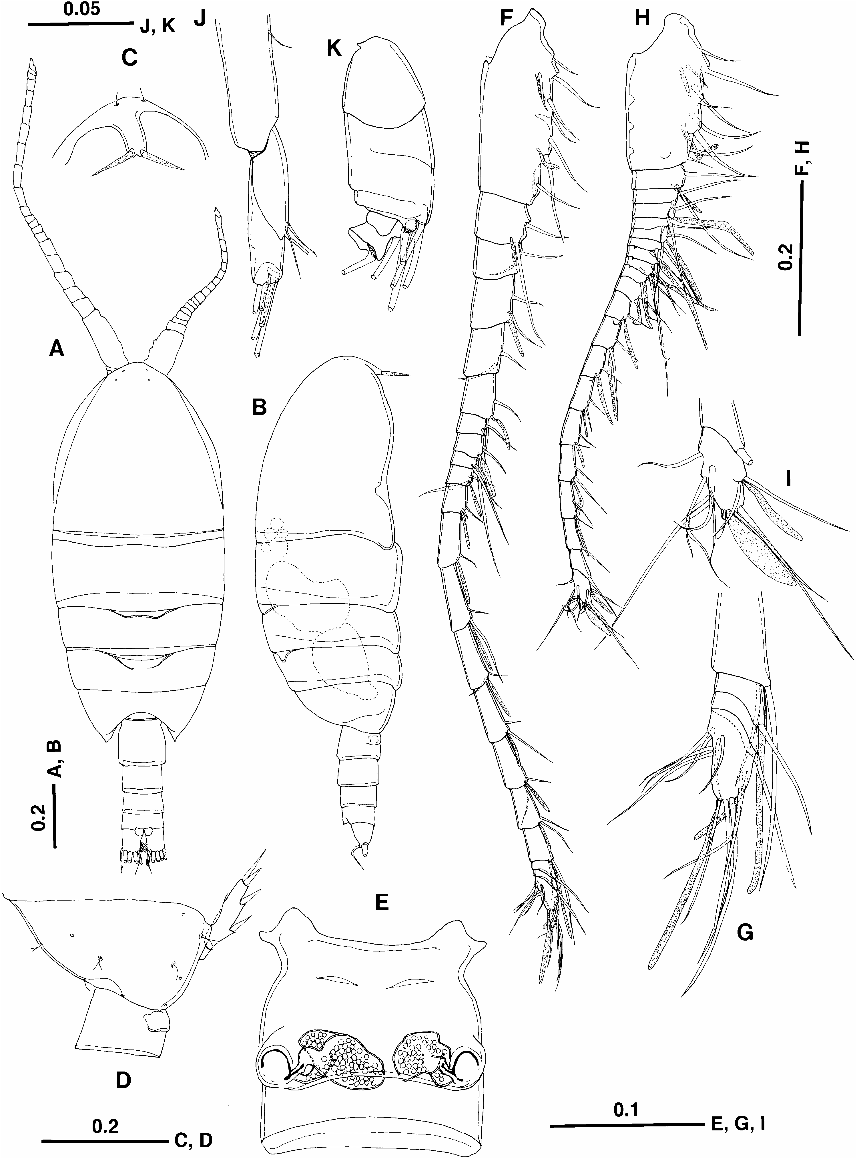
Female (holotype). Body ( Figure 1A, B View Figure 1 ) 1.55 mm, compact, plump; prosome 2.6 times as long as urosome. Cephalosome slightly asymmetrical in dorsal view; rostrum ( Figure 1B, C View Figure 1 ) strongly curved, with pair of thick filaments; ventrolateral corners expanded posteriorly to cover anterior margin of pediger 1. Pedigers 1–3 with ventrolateral corners produced posteriorly; prosomal posterior ends almost symmetrical, each produced into round lobe reaching midway along genital double-somite ( Figure 1D View Figure 1 ). Urosome four-segmented; genital double-somite wider than long, with ventral transverse ridge midway; genital system ( Figure 1E View Figure 1 ) asymmetrical, with right seminal receptacle larger than left; paired gonopores and copulatory pores located at mid-length; each copulatory pore slit-like, located at inner corner of gonopore, connected to curved, thick copulatory pore; anal somite relatively long; caudal rami symmetrical, lacking seta I; seta VII located at base of seta VI, bent at midlength. Two large, mature eggs visible within prosome ( Figure 1B View Figure 1 ).
Antennules considerably asymmetrical, with left ca 1.5 times longer than right. Left antennule ( Figure 1F, G View Figure 1 ) 21-segmented; fusion pattern and armature as follows: I–IV59+2ae, V52+ae, VI52, VII52+ae, VIII52, IX52+ae, X52 (one spiniform seta),
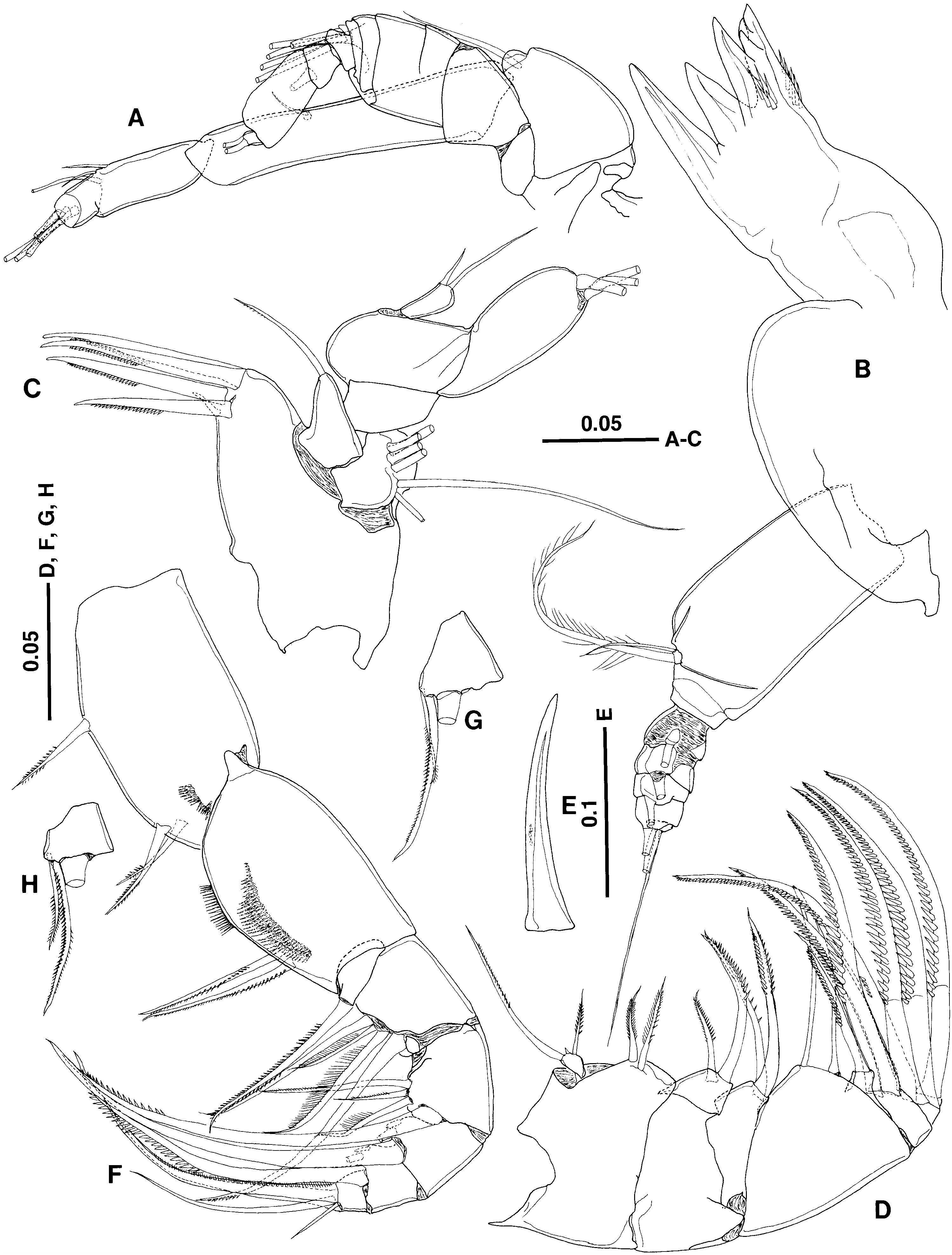
XI52+ae, XII52+ae (vestigial), XIII52+ae, XIV52 (one spiniform seta)+ae, XV52 (one missing)+ae, XVI52+ae, XVII52+ae, XVIII52+ae, XIX52+ae, XX52+ae, XXI52+ae, XXII51, XXIII51, XXIV–XXVIII512+2ae. Right antennule ( Figure 1H, I View Figure 1 ) with same fusion pattern and armature as left except for distinct aesthetasc on segment XII. Antenna ( Figures 1J, K View Figure 1 , 2A View Figure 2 ) with unarmed coxa; basis with inner seta half as long as exopod;
endopod indistinctly three-segmented, first segment with seta at two-thirds of inner margin; second and third segments almost coalescent with suture clearly visible; second segment with three unequal setae terminally; third segment with five distal setae; exopod indistinctly 10-segmented, with setal formula of 0, 0, 1, 1, 1, 1, 1, 1, 0, 2; terminal segment lacking vestigial element as seen in Metacalanus (cf. Ohtsuka et al. 1994). Mandible ( Figure 2B View Figure 2 ) without patch of long setules on gnathobase; cutting edge with two patches of minute spinules and four teeth, dorsalmost of which tricuspid; endopod rudimentary, onesegmented, with two plumose setae terminally; exopod five-segmented, with setal formula 1, 1, 1, 1, 2; terminal setae well developed. Maxillule ( Figure 2C View Figure 2 ) with one naked short, and four serrate long spines on inner distal corner of praecoxal arthrite; coxal endite with moderately developed, serrate seta; coxal epipodite bearing six setae; basal seta absent; endopod one-segmented, bulbous, with two unequal setae; exopod one-segmented, lamellar, bearing three terminal setae. Maxilla ( Figure 2D, E View Figure 2 ) stout; first praecoxal endite with two unequal spinulose setae and vestigial element; second praecoxal to second coxal endites each bearing two spinulose setae; basal spine ( Figure 2E View Figure 2 ) highly sclerotized, with short row of three or four spinules at mid-length; endopod four-segmented, with setal formula 1, 3, 2, 2; setae bearing longitudinal row of large spinules. Maxilliped ( Figure 2F– H View Figure 2 ) with syncoxa bearing one middle and two subterminal serrate setae and terminal patch of short spinules; basis as long as syncoxa, with two serrate medial setae, row of setules along proximal half of inner margin, and longitudinal patch of spinules; first endopodal segment almost separate from basis, with serrate seta; second to fifth endopodal segments bearing four, four, three, and three setae, respectively; sixth endopodal segment with setae a and b not reduced.
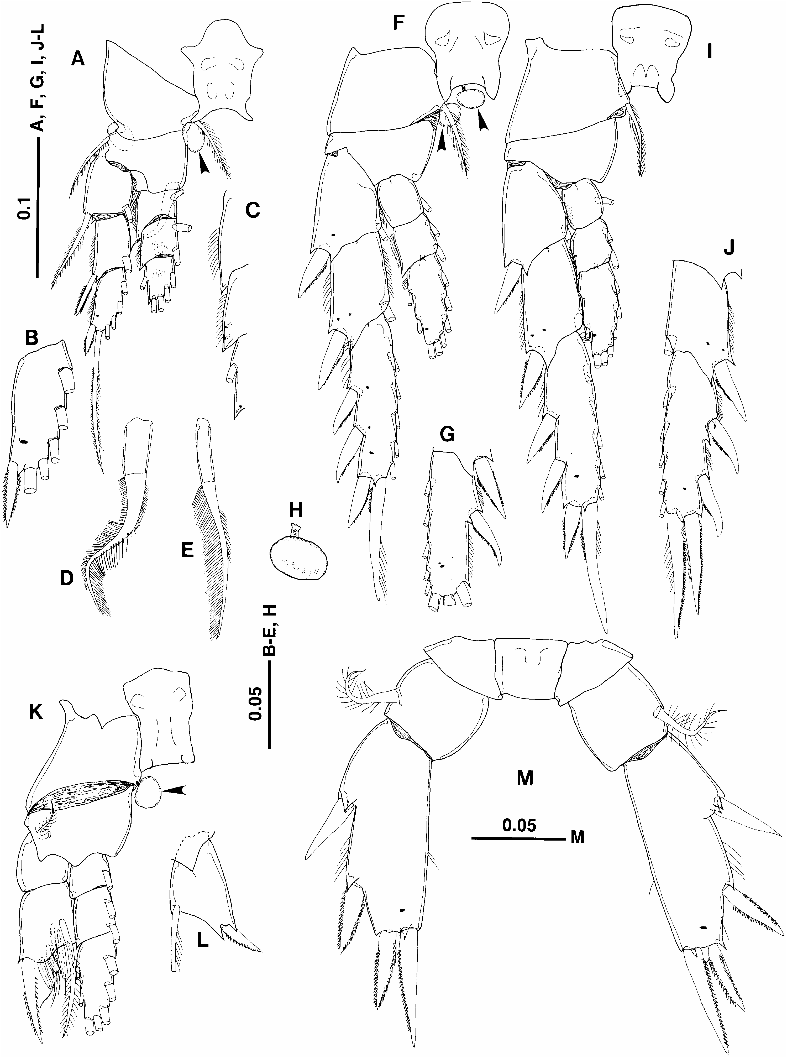
Seta and spine formula of legs 1–4 ( Figure 3A–G, I–L View Figure 3 ) shown in Table I. Both rami of legs 1–3 and endopod of leg 4 three-segmented (exopods of leg 4 broken or aberrant). Asymmetry in legs 1–4. Leg 1 ( Figure 3A–E View Figure 3 ) with inner basal setae asymmetrical, left ( Figure 3D View Figure 3 ) thicker than right ( Figure 3E View Figure 3 ); outer distal corners of three endopodal segments acutely pointed ( Figure 3C View Figure 3 ). Leg 2 ( Figure 3F, G View Figure 3 ) with outer spines on third exopodal segments asymmetrical in number, size and location. Leg 3 ( Figure 3I, J View Figure 3 ) with outer spines on second and third exopodal segments asymmetrical in length and shape. Left leg 4 ( Figure 3K View Figure 3 ) with aberrant exopod, with first segment unarmed and second bearing outer long spine and six modified setae. Terminal exopodal segments of right leg 4 missing ( Figure 3L View Figure 3 ).
Leg 5 ( Figure 3M View Figure 3 ) nearly symmetrical; coxae and intercoxal sclerite separate; basis bearing thick, subterminal seta on posterior surface; endopod lacking; exopod one-segmented, with two lateral and two terminal spines; acute process present at base of inner terminal spine; outer terminal process more acutely pointed in right leg than in left. Male. Unknown.
Remarks
Asymmetry of legs 1–3 (maybe, leg 4 also) may be related to an as yet unknown swimming behaviour of the new genus as observed in Paramisophria spp. that swim with the left lateral side parallel to the bottom and the left antennule extended anteriorly ( Fosshagen 1968; Ohtsuka and Mitsuzumi 1990). Bowman and González (1961) insisted that hyperbenthic calanoid copepods generally bear stouter outer spines on the exopods of legs in comparison with pelagic ones. Hence the longer spines on the second and third exopodal segments of left legs 2–4 suggest a possible peculiar swimming behaviour as found in Paramisophria spp. The inner basal seta of leg 1 is considered to play a role in grooming ( Vaupel Klein 1972). The thicker inner basal seta of left leg 1 of the new species is likely to be adaptive for grooming the longer left antennule.
Gut content analysis revealed that the new species fed upon copepods. Stalked cysts on legs (see Figure 3A, F, H, K View Figure 3 ) are possibly assignable to phoronts of apostome ciliates (cf. Grimes and Bradbury 1992; Ohtsuka et al. 2004).
Etymology
The new specific name, hakuhoae , refers to the RV Hokuho-maru, University of Tokyo, that carried out the survey in the Sulu Sea in 2002.
| V |
Royal British Columbia Museum - Herbarium |
No known copyright restrictions apply. See Agosti, D., Egloff, W., 2009. Taxonomic information exchange and copyright: the Plazi approach. BMC Research Notes 2009, 2:53 for further explanation.
|
Kingdom |
|
|
Phylum |
|
|
Class |
|
|
Order |
|
|
Family |
|
|
Genus |