
Metaleptobasis gabrielae, Ellenrieder, 2013
|
publication ID |
https://doi.org/10.11646/zootaxa.3738.1.1 |
|
publication LSID |
lsid:zoobank.org:pub:77D1A6F6-C320-442B-AF31-83324E5EAF3B |
|
persistent identifier |
https://treatment.plazi.org/id/03E187ED-6610-FF90-D7A8-FAB5E0A0FC5F |
|
treatment provided by |
Felipe |
|
scientific name |
Metaleptobasis gabrielae |
| status |
sp. nov. |
Metaleptobasis gabrielae View in CoL new species
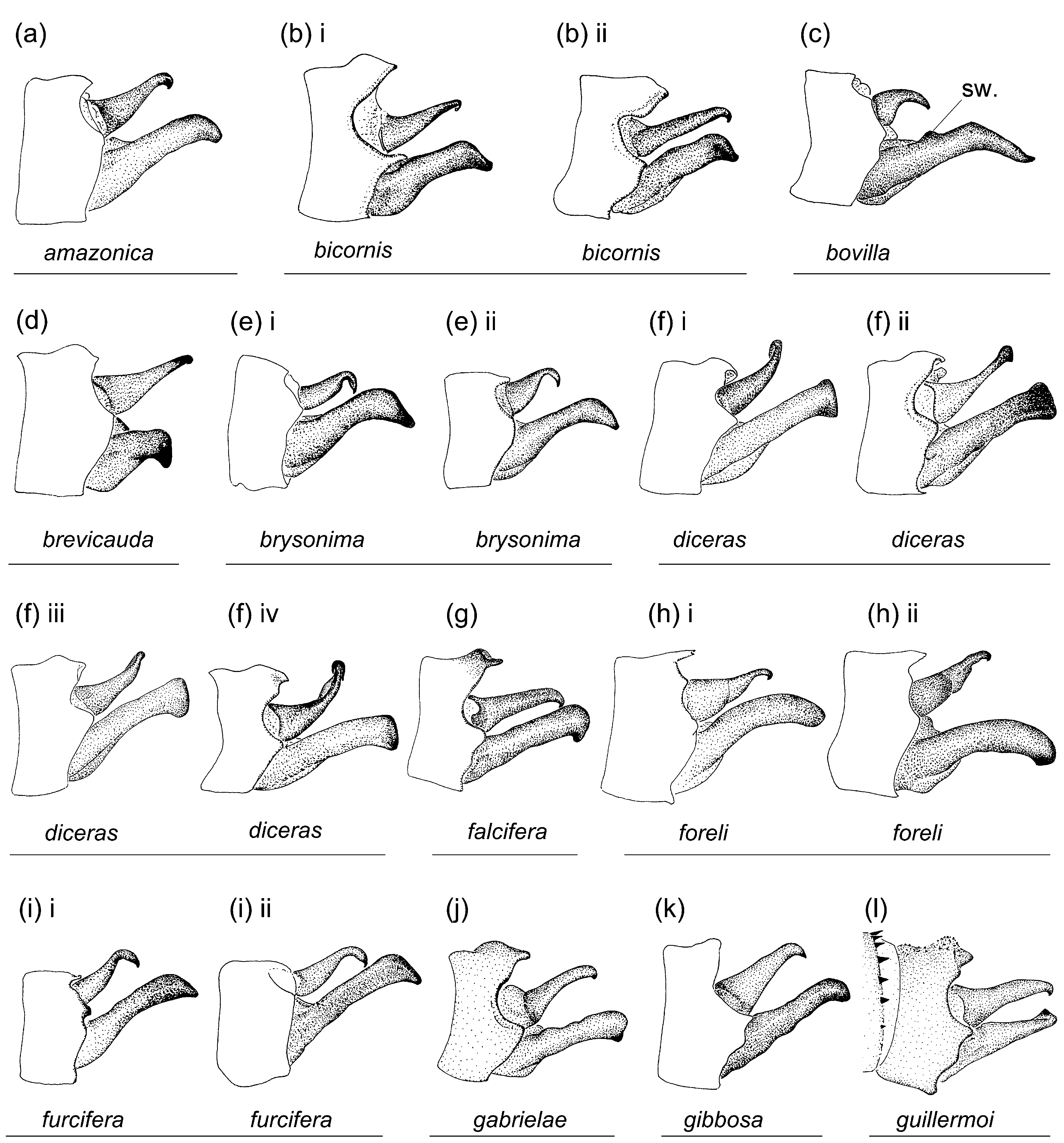
Figs. 1j View FIGURE 1 ; 3j View FIGURE 3 ; 4j View FIGURE 4 ; 5j View FIGURE 5 ; 8j View FIGURE 8 ; 9j View FIGURE 9 ; 10j View FIGURE 10 ; 11j View FIGURE 11 ; 12j View FIGURE 12 ; 13e View FIGURE 13 ; 14g
Etymology. I name this species gabrielae (noun in the genitive case) after my dear mother, who always encouraged my creativity and provided support and advice for whatever endeavors I would embark on.
Types. ( all *)— Holotype ♂: PERU, LoretoDep., Tamshiyacu-Tahuayo Reserve , forest interior ( 4°23'40''S, 73°14'56''W), 27 vii 2009, TF leg. [ RMNH]; 1 ♀ paratype, in copula with holotype [ TF]; 2 ♂, 1 ♀ paratypes, same data as holotype [ TF]; 2 ♂, 2 ♀ paratypes, same data as holotype but ( 4°23'47''S, 73°15'13''W), 23 ii 2010 [ TF]; 3 ♂, 1 ♀ paratypes, same data as holotype but ( 4°23'49''S, 73°14'57''W), 25 ii 2010 [ TF]; 1 ♂, 2 ♀, 27 ii 2010 [ TF]. GoogleMaps
Specimens examined. 9 ♂, 7 ♀.
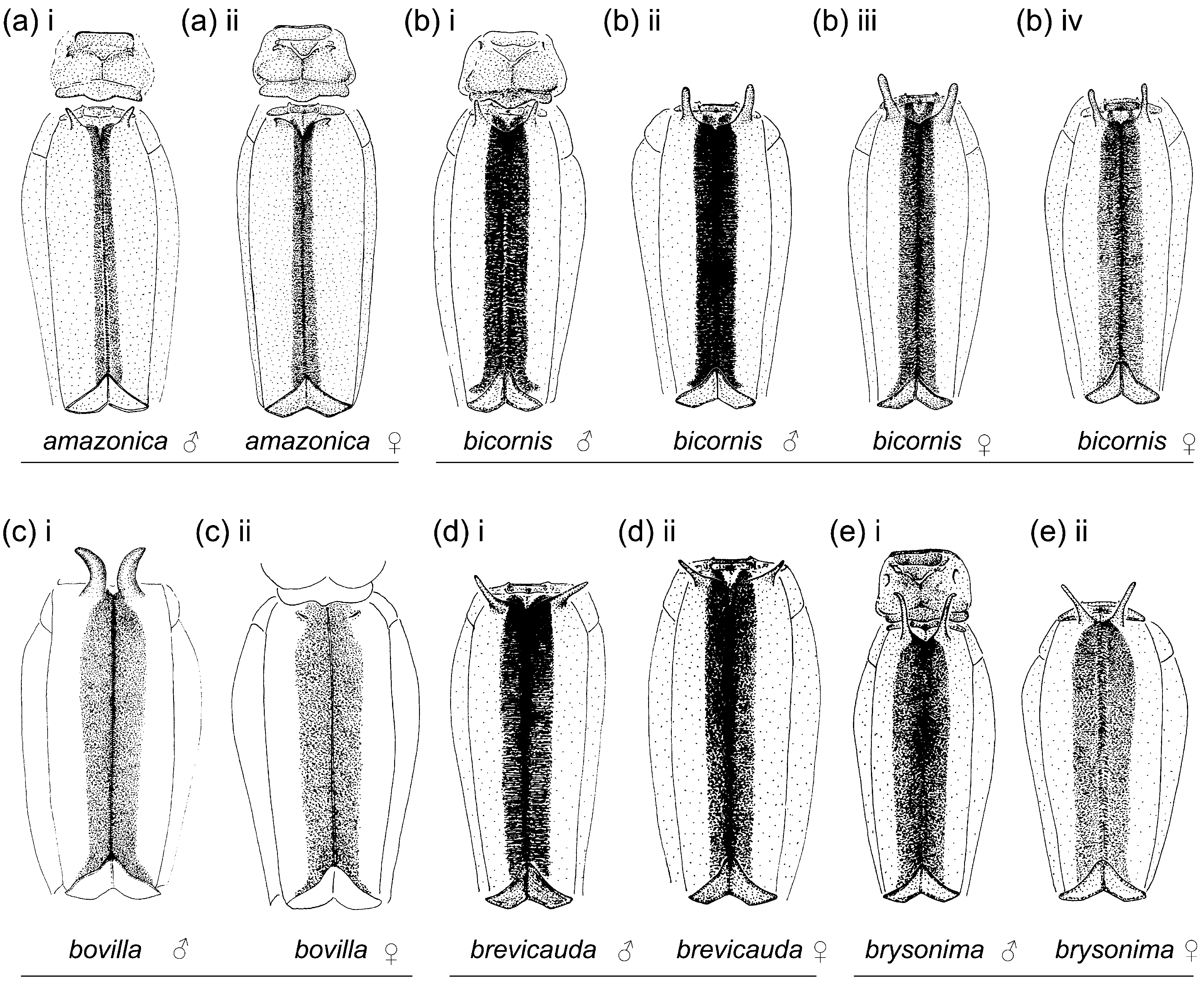
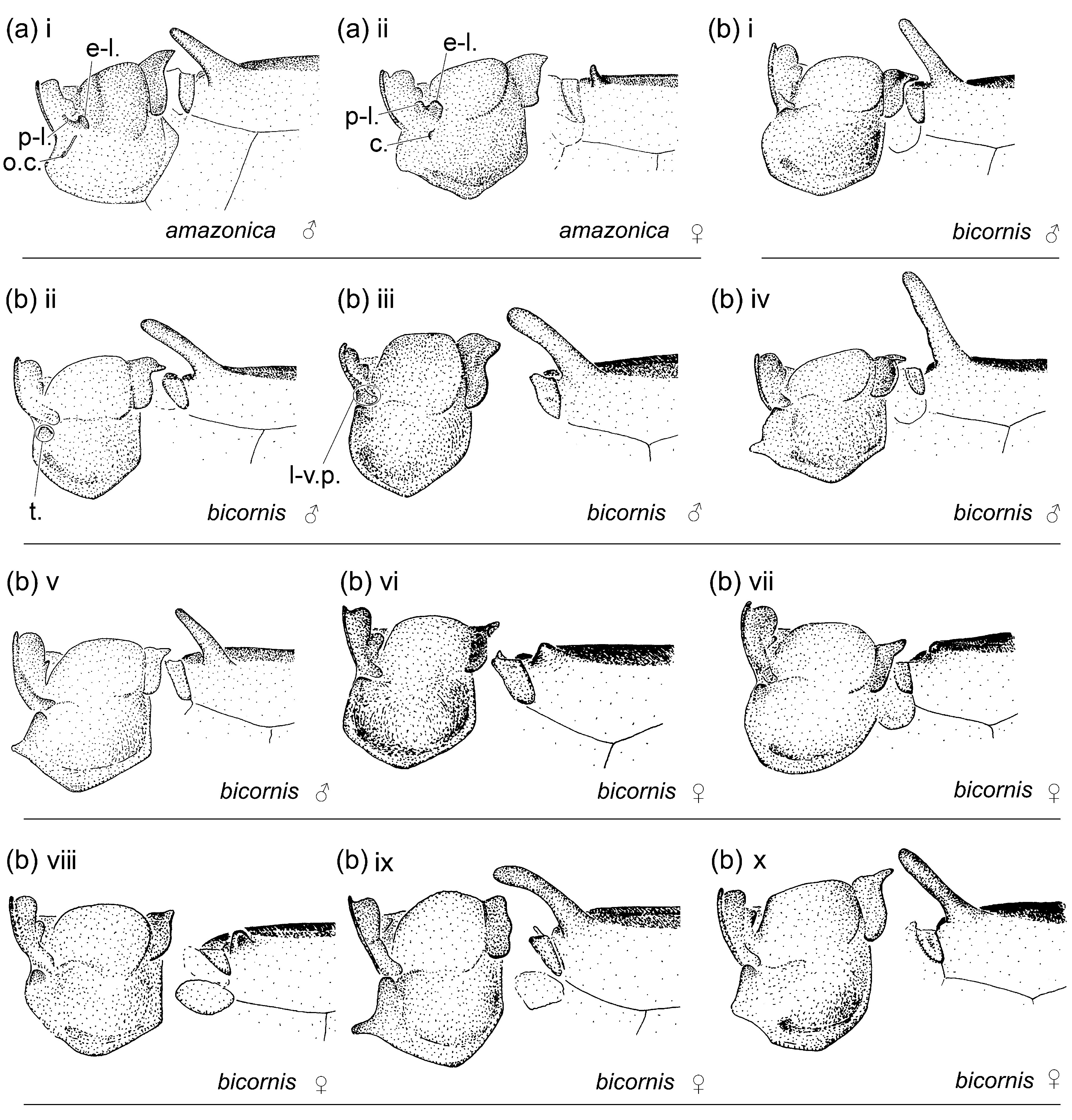
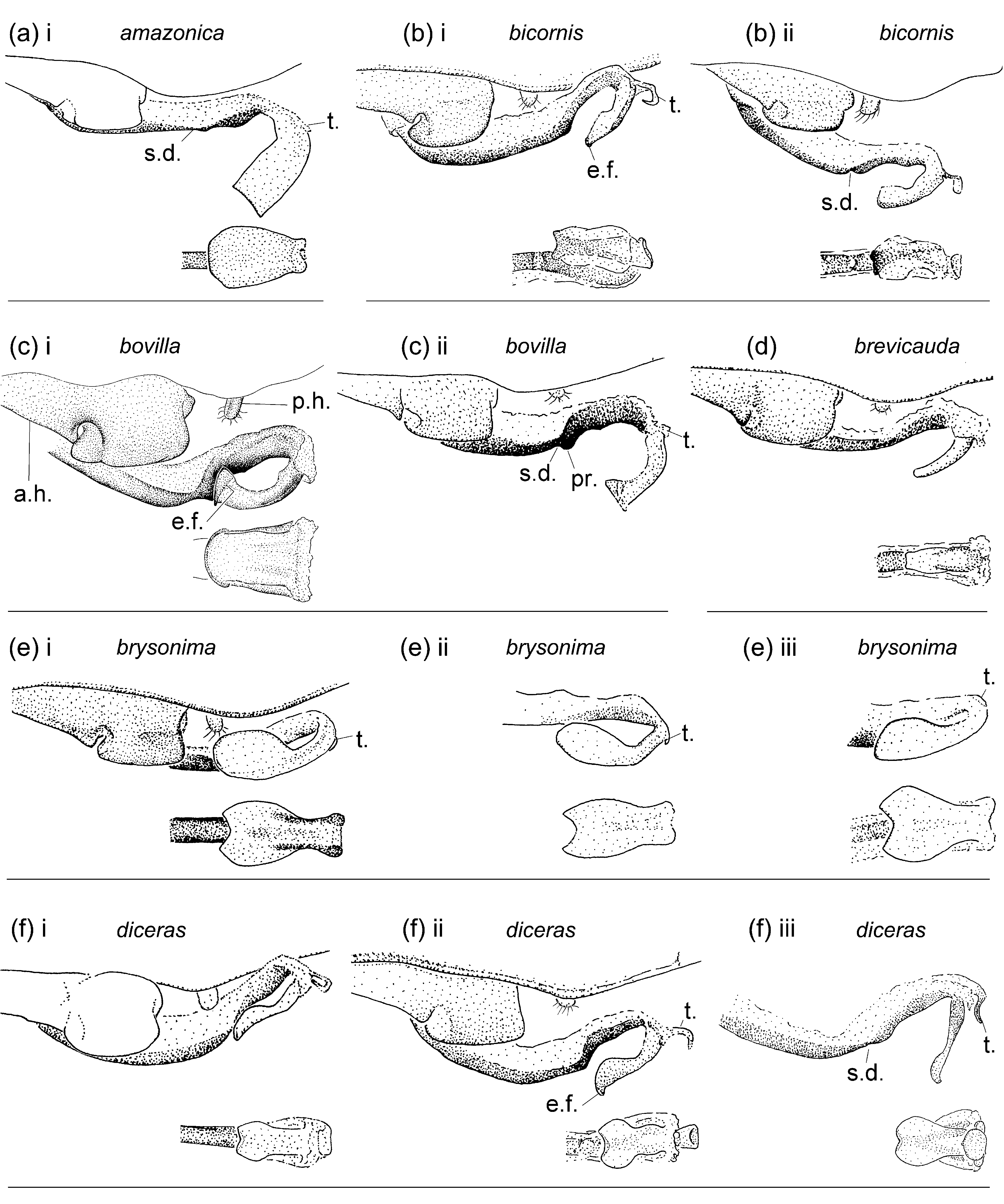
Description of male holotype. Labium and rear of head ivory; base of mandible pale bluish grey with a brown spot; labrum yellow with black line along latero-basal margin; gena pale bluish grey with a brown spot to the side of antefrons; anteclypeus pale bluish grey with a pair of medio-lateral brown spots; postclypeus distal half black, basal half pale bluish grey; antefrons pale bluish grey; postfrons and epicranium black with bluish grey pale spots and stripes as depicted in Fig. 1j i View FIGURE 1 ; postocular lobes posterior to vertex pale bluish grey; eyes brown, in life bright green with a dorso-medial orange area (as in Fig. 13e i View FIGURE 13 ); postocular lobes rounded. Thorax. Color as described for genus, with mid-dorsal dark stripe on pterothorax black with metallic greenish-blue reflections, wider than interlaminal sinus, maximum width at about 0.30 of pterothoracic length of about 0.50 of mesanepisterna width, slightly narrowing posteriorly, extended along sides of antealar sinus ( Fig. 3j View FIGURE 3 ). Pronotum anterior lobe ( Figs. 4j i View FIGURE 4 ; 5j i View FIGURE 5 ) smooth; anterior and middle lobes of pronotum separated dorso-laterally by a groove; anterior area of propleuron with a prominent rounded tubercle (t.); dorsum and sides of middle lobe of pronotum smooth; pronotum posterior lobe slightly trilobed, with medial lobe smoothly convex and lateral lobes narrowing to free angled lateral corners, slightly shorter than 0.50 of medial lobe. Mesanepisternal horns with bases separated, horns as long as 1.7 times mesostigmal plate width, thin, cylindrical, diverging from each other in posterior view, directed anterodorsally at an angle of 30° with dorsum in lateral view, ending on a bluntly pointed tip. Wings hyaline, veins reddish brown; Pt sub-rectangular, with anterior and posterior sides longer than distal side, and membrane pale brown margined by pale yellow; 12 (right) and 13 (left) pnx in Fw, 11 (right) and 12 (left) in Hw. Abdomen. Postmortem background color pale yellow on S1–7, yellowish orange on S8–10; S1 with a dark reddish brown dorso-posterior spot and latero-ventral margin of terga margined in black; dorsum of S2 reddish brown and of S3– 7 dark reddish brown, with a basal pale incomplete ring at anterior edge interrupted by dorso-longitudinal dark line, on S2–4 a small diffuse pale transverse spot near posterior margin on each side, and dark dorsal color posterior to pale transverse spot extended ventrally along sides of lateral terga on S3–6; intersegmental membrane on dorsum and sides of S2–6 dark reddish brown; dorsum of S8 reddish brown along basal 0.50, turning into yellowish orange distally; dorsum of S9–10 yellowish orange; medio-longitudinal carina on sterna brown; caudal appendages yellowish orange with black apex. Genital lobe ( Fig. 8j i View FIGURE 8 ) short, less than 0.50 of anterior hamule height, smoothly convex in lateral view; posterior hamule laminar and large, clearly surpassing ventral margin of genital fossa, slightly higher than 0.50 of anterior hamule, sickle-shaped, of about uniform width, with posterior margin smoothly curved, with tip curved medially gradually and rounded in ventral view; curvature of basal segment of genital ligula marked by a deep concave depression, followed by a prominent convex protuberance; genital ligula distal segment pear-shaped, distinctly widened sub-apically, with ratio maximum width/length of 0.55, apex slightly concave with a narrow ectal fold (e.f.). Medial portion of S10 postero-dorsal margin ( Figs. 10j i View FIGURE 10 ; 11j i View FIGURE 11 ; 12j i View FIGURE 12 ) projected posteriorly, lacking a medial incision, with a dorsal prominence represented by an entire transverse sub-apical swelling. Cercus sub-cylindrical, slightly narrowing distally in lateral view ( Fig. 12j i View FIGURE 12 ), with a longitudinal medial concavity, in dorsal view ( Fig. 10j i View FIGURE 10 ) about straight along medial portion, with tip curved medio-ventrally gradually, wider than medial third of cercus, and ending on a single point directed medio-ventrally; ratio of cercus length to S10 maximum length in lateral view 1.4; ratio of cercus length to paraproct length in lateral view 0.95; paraproct in lateral view sub-cylindrical, of uniform width along medial portion and slightly widening distally, in dorsal view with a medio-longitudinal concavity extended almost to tip, tip curved medially at about 90°, ending on a single medio-ventral tooth.
Dimensions. Hw 23; abdomen 39.8; total length 46.8.
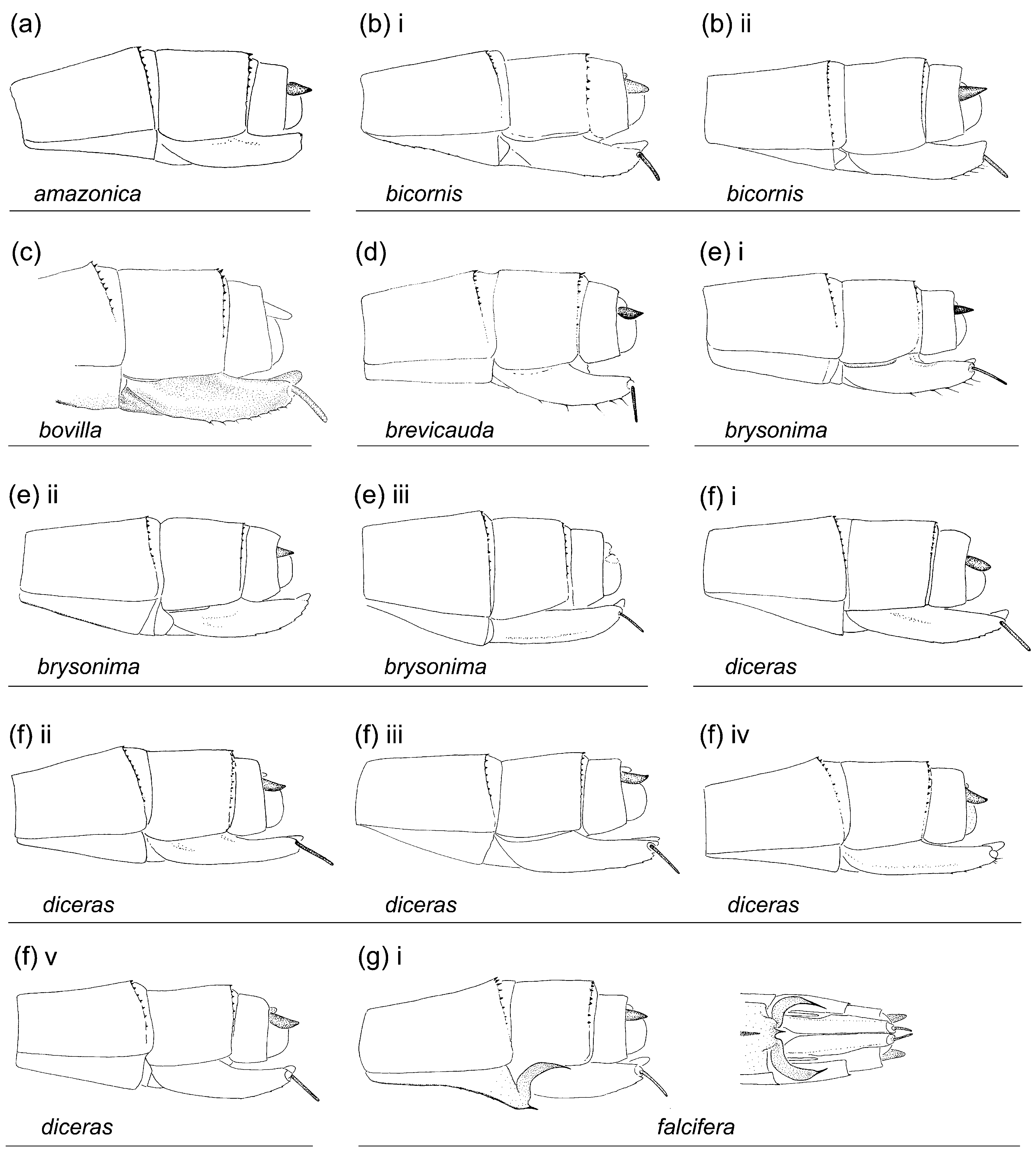
Female paratype (in copula with holotype). Head. As in holotype but pale color of labrum pale brown and of remainder of head pale yellow; eyes brown, in life bright green (as in Fig. 13d i View FIGURE 13 ); pattern of epicranium as in Fig. 3j View FIGURE 3 ii.— Thorax. As in holotype but lateral lobes of posterior lobe of pronotum projected latero-dorsally, and posterior margin of medial lobe projected dorso-posteriorly ( Figs. 4j View FIGURE 4 ii; 5j ii); mesanepisternal horns represented by very low blunt tubercles; 13 pnx in Fw, 11 (right) and 12 (left) in Hw.— Abdomen. Color pattern as in holotype but pale color on S8–10 reddish orange; posterior margin of S8 sternum with two strong spines, with base bulbous and distal portion needle-shaped, about parallel to each other and separated at their bases by a distance greater than their individual width, extending along ventro-lateral margin of ovipositor external valve, as long as about 0.66 of S9 and 0.33 of ovipositor external valve ( Fig. 9j View FIGURE 9 ); ovipositor surpassing slightly level of tip of cercus; caudal appendages and ovipositor external valves reddish orange; stylus dark brown.
Variation in paratypes. Paratypes as holotype but postfrons and epicranium in teneral specimens metallic green, eyes pale green, and thorax and distal abdominal segment pale yellow; mesanepisternal horns in males as long as about 1.5–1.7 of mesostigmal plate width; Pt pale yellow to pale brown; 12–14 pnx in Fw and 11–13 in Hw; small diffuse pale transverse spot near posterior margin on each side of dorsum of S2 only in some specimens; pale color of abdomen S8–10 pale yellow in teneral specimens; pronounced prominence following deep depression on basal segment of genital ligula triangular to rounded in lateral view; spines on posterior margin of S8 sternum as long as about 0.50–0.66 of S9 and about parallel sided to each other to slightly diverging; ovipositor reaching level of tip of paraproct to level of tip of cercus.
Dimensions. Males ( n 9, including holotype): Hw 22.3 ± 0.7 [21–23]; abdomen 38 ± 1.4 [36–39.8]; total length 45 ± 1.5 [43–46.8]. Females ( n 7): Hw 23.1 ± 0.9 [22–24.3]; abdomen 36.7 ± 1.1 [35.4–38.6]; total length 43.4 ± 1.3 [42–45.7].
Diagnosis. Among the species with large laminar male posterior hamule clearly surpassing ventral margin of genital fossa in lateral view and posterior margin of female S8 sternum with denticles, spines, or processes, M. gabrielae shares male posterior hamule not surpassing anterior hamule ventrally and propleuron with an anterior prominent rounded tubercle only with M. gibbosa , M. guillermoi , M. mauffrayi , and M. panguanae , and middle lobe of female pronotum smooth only with M. gibbosa ; in the other species of this group male posterior hamule is as high or slightly higher than anterior hamule, propleuron lacks an anterior prominent tubercle, and there are dorsal or lateral ear-like lobes on anterior portion of middle lobe of female pronotum. It differs from M. gibbosa , M. guillermoi , and M. mauffrayi by the distal segment of male genital ligula pear-shaped and by female S8 with two strong straight sub-cylindrical ventro-lateral spines ( Figs. 8j View FIGURE 8 ; 9j View FIGURE 9 ; vs. distal segment of male genital ligula subrectangular and female sternum S8 with denticles or small spines, Figs. 8k–l, r View FIGURE 8 ; 9k–l, q View FIGURE 9 ). The male of M. gabrielae differs further from M. mauffrayi and M. panguanae by distal portion of cercus wider than medial third ( Fig. 10j View FIGURE 10 ; vs. about as wide as medial third width, Figs. 10r, v View FIGURE 10 ), from M. gibbosa by posterior margin of S10 with a mediodorsal posterior projection (p-m.p., Figs. 10j View FIGURE 10 ; 11j View FIGURE 11 ; lacking a medio-dorsal posterior projection in M. gibbosa , Figs. 10k View FIGURE 10 ; 11k View FIGURE 11 ), and from M. guillermoi by paraproct ending on a single ventral tooth (st., 10j; 11j; paraproct ending on a distal carina with dorsal and ventral teeth in M. guillermoi , te., Figs. 10l View FIGURE 10 ; 11l View FIGURE 11 ). Female of M. gabrielae shares the two strong straight sub-cylindrical ventro-lateral spines on posterior margin of sternum S8 with M. panguanae , but they are separated at their bases by a distance greater that their individual width ( Fig. 9j View FIGURE 9 ), whereas in M. panguanae they are separated at their bases by a distance shorter than their individual width ( Fig. 9u View FIGURE 9 ).
All known females are dimorphic, differing from males in the development of mesanepisternal horns and shape of pronotum posterior lobe.
Habitat. Forest interior.
Distribution. Loreto Dep. in Peru ( Fig. 14g).


| RMNH |
National Museum of Natural History, Naturalis |
| TF |
Department of Mineral Resources |
No known copyright restrictions apply. See Agosti, D., Egloff, W., 2009. Taxonomic information exchange and copyright: the Plazi approach. BMC Research Notes 2009, 2:53 for further explanation.
|
Kingdom |
|
|
Phylum |
|
|
Class |
|
|
Order |
|
|
Family |
|
|
Genus |