
Metaleptobasis Calvert, 1907
|
publication ID |
https://doi.org/10.11646/zootaxa.3738.1.1 |
|
publication LSID |
lsid:zoobank.org:pub:77D1A6F6-C320-442B-AF31-83324E5EAF3B |
|
persistent identifier |
https://treatment.plazi.org/id/03E187ED-6630-FFA8-D7A8-FAEBE7FEFEA7 |
|
treatment provided by |
Felipe |
|
scientific name |
Metaleptobasis Calvert, 1907 |
| status |
|
Metaleptobasis Calvert, 1907 View in CoL
Type species: Metaleptobasis bovilla Calvert, 1907: 386 , by original designation.
Other species included: Metaleptobasis amazonica Sjöstedt, 1918 ; M. bicornis ( Selys, 1877) [ = M. mauritia Williamson, 1915 syn. nov.]; M. brevicauda sp. nov.; M. brysonima Williamson, 1915 [ = M. tetragena Calvert, 1948 syn. nov.; = M. weibezahni Rácenis, 1955 syn. nov.; = M. incisula De Marmels, 1989 syn. nov.]; M. diceras ( Selys, 1877) [= M. manicaria Williamson, 1915 ; = M. fernandezi Rácenis, 1955 syn. nov.]; M. falcifera sp. nov.; M. foreli Ris, 1918 [= M. westfalli Cumming, 1954 syn. nov.]; M. furcifera sp. nov.; M. gabrielae sp. nov.; M. gibbosa Tennessen, 2012 ; M. guillermoi sp. nov.; M. inermis sp. nov.; M. knopfi Tennessen, 2012 ; M. leniloba sp. nov.; M. longicauda sp. nov.; M. lillianae Daigle, 2004 ; M. mauffrayi Daigle, 2000 ; M. minteri Daigle, 2003 ; M. orthogonia sp. nov.; M. paludicola sp. nov.; M. panguanae sp. nov.; M. peltata sp. nov.; M. prostrata sp. nov.; M. quadricornis ( Selys, 1877) ; M. selysi Santos, 1956 ; M. silvicola sp. nov.; M. spatulata sp. nov.; M. tridentigera sp. nov.; M. truncata sp. nov.; M. turbinata sp. nov.
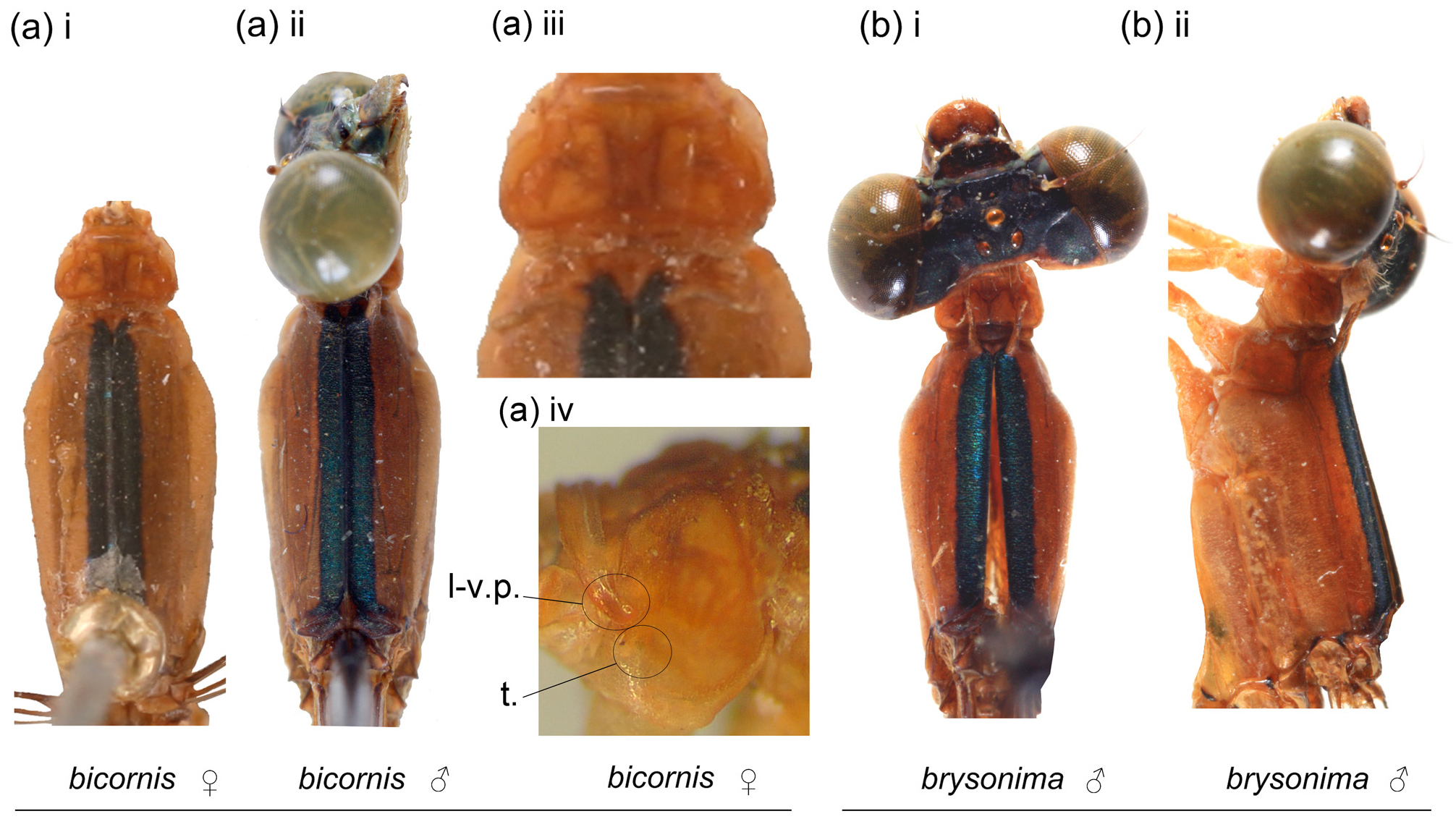
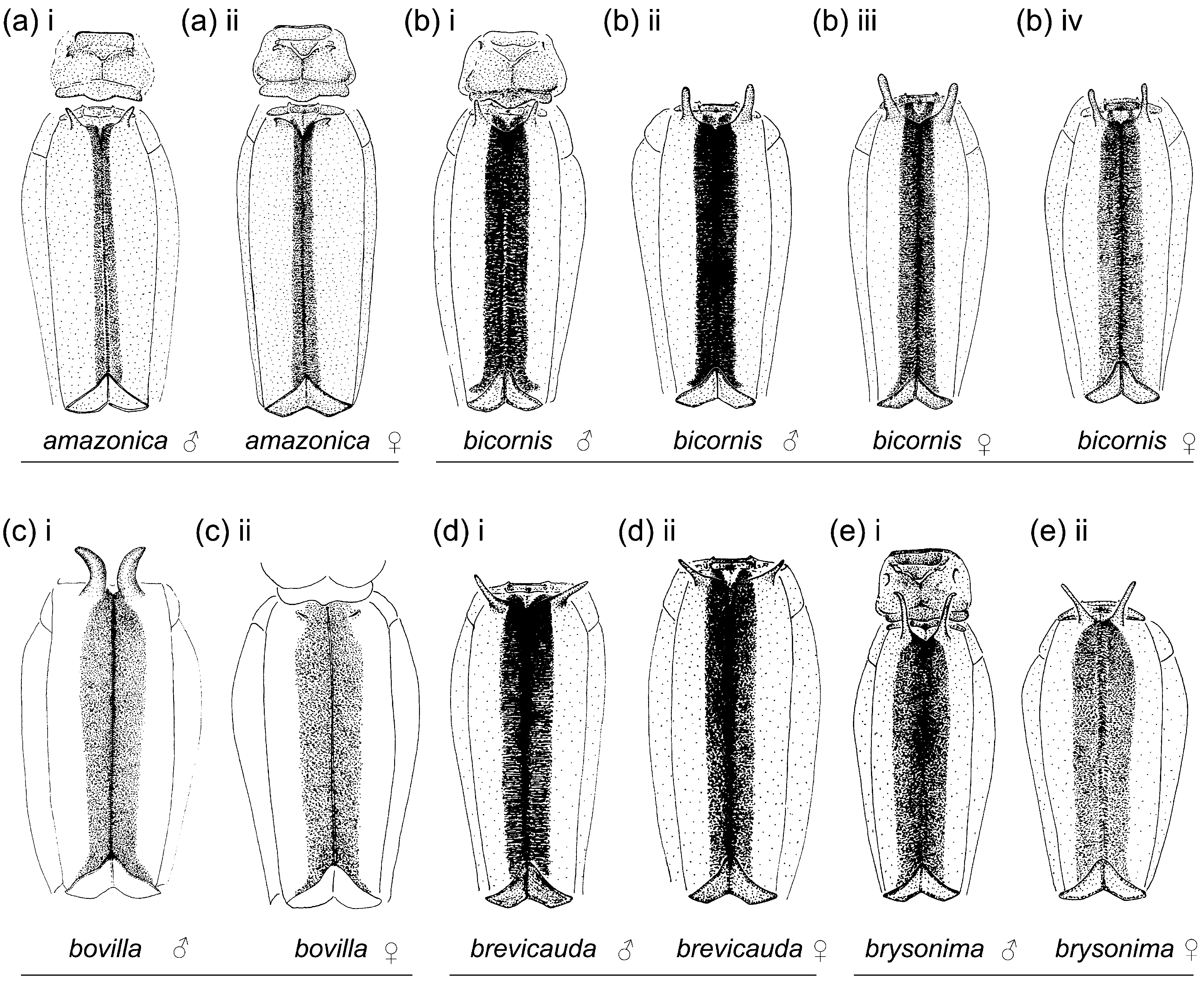
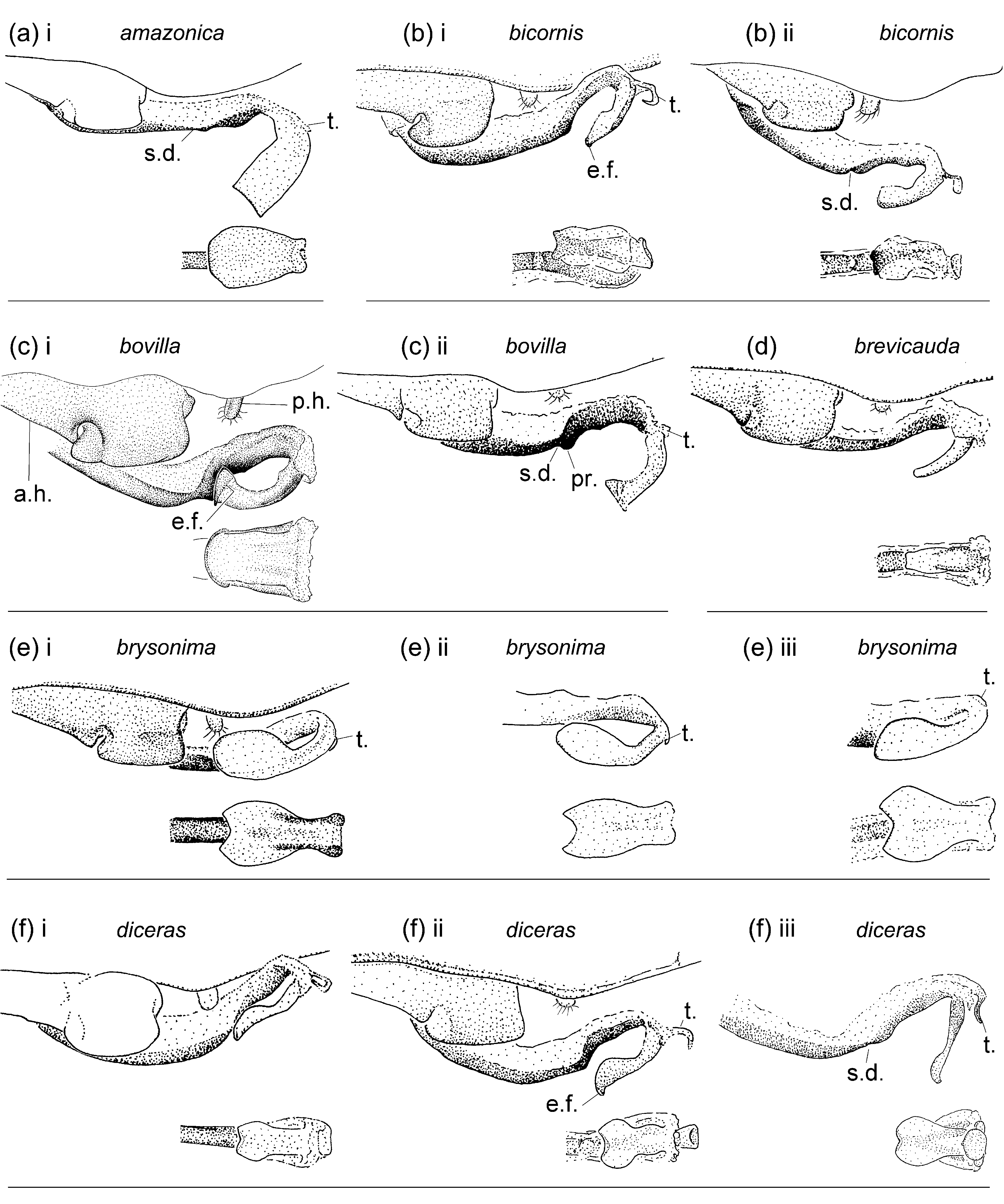
Generic characterization. Medium to large sized coenagrionids (total length 38–53; Hw length 19–29; abdomen length 32–44). Head pale orange or yellow to pale or dark blue or green, variable intraspecifically and usually paler in females than in males, dorsum with some black markings to mostly black ( Figs. 1–2 View FIGURE 1 View FIGURE 2 , 13 View FIGURE 13 ); black areas usually with metallic reflections; frons angulate. Pronotum pale orange, sometimes with a dark spot on medial area of middle and/or posterior lobes, of variable extension to entirely absent in the same species, bearing a variety of diagnostic lobes, tubercles, and/or crests on anterior and/or middle lobes ( Figs. 4–5 View FIGURE 4 View FIGURE 5 ); middle lobe with a shallow medio-dorsal depression on each side, and posterior lobe posterior margin of various shapes, from entire to trilobed with lobes inflated and corners strongly projected postero-dorsally ( Figs. 4–5 View FIGURE 4 View FIGURE 5 ). Pterothorax usually pale orange, but it can also be yellowish, brownish, greenish or bluish in life ( Fig. 13 View FIGURE 13 ), with venter pale yellow and bearing powdery white pruinescence in both males and females, and dark areas limited to black spurs on legs, reddish brown apical 0.25–0.33 of pretarsus, an oval black spot on prosternellum, a small black spot on dorsoanterior corner of mesepimeron and metepimeron, on dorso-anterior corner of metepimeron postero-dorsal triangular section, and mid-dorsal black stripe, with green, bluish, or purple metallic reflections, the particular color of the metallic shine being variable within the same species, probably according to preservation ( Figs. 2–3 View FIGURE 2 View FIGURE 3 , 13 View FIGURE 13 ). Anterior portion of mesanepisterna with a pair of smooth horns, in several females shorter than in conspecific males or reduced to bases or low protuberances ( Figs. 4–5 View FIGURE 4 View FIGURE 5 ). Metatibial spurs shorter than twice intervening spaces; pretarsus with vestigial supplementary tooth represented by an obtuse low prominence, which may be very small but is always visible under high magnification. Fw CuA extending for more than six cells distal to vein descending from subnodus; CuP linking CuA to CuP&AA and not reaching posterior wing margin; vein descending from quadrangle forming an unbroken line to wing margin (v.q., Fig. 6 View FIGURE 6 ). Abdomen orange to yellow, light blue, or green, with brown to black areas as follows ( Fig. 13 View FIGURE 13 ): S1 with a brown to reddish brown dorso-posterior spot and ventral carina of lateral tergum usually margined with brown or black; S2–7 with reddish-brown to black dorsum, with a basal pale incomplete ring on anterior edge interrupted by dorso-longitudinal dark line, and with a small diffuse pale transverse subapical spot near posterior margin on each side, which can be present or absent on some or all segments, on S3–6 dark dorsal marking posterior to pale transverse spot usually extending ventrally along sides of lateral terga; dorsum of S8–10 ranging from dark reddish brown to yellow or reddish-orange; antero-basal corner of female S8 with a black spot; medio-longitudinal carina on sterna yellow to brown to black; S2–9 with denticles along postero-lateral margin, very small on S2–6, larger on S7–9. Genital ligula distal segment lacking inner fold and lateral lobes, with a membranous terminal fold of variable length, its apparent length related to degree of hydration at preservation, sometimes barely visible but always present under high magnification (t in Fig 8 View FIGURE 8 ). Posterior margin of female sternum S8 ( Fig. 9 View FIGURE 9 ) smooth, with some denticles or small spines, or with strong spines or a bifid or trifid process. Male cercus entire and sub-cylindrical in lateral view; male paraproct entire, subcylindrical and directed dorso-posteriorly in lateral view, with a wide squarish base in ventral view ( Figs. 10–12 View FIGURE 10 View FIGURE 11 View FIGURE 12 ). Ovipositor valves smooth, bearing only some sparse long hairs along ventral margin, surpassing posterior margin of S10 and reaching level between paraproct tip to slightly longer than level of cercus tip ( Fig. 9 View FIGURE 9 ). Larva unknown.
Generic diagnosis. Among New World coenagrionids, Metaleptobasis shares the vein descending from quadrangle forming an unbroken line to wing margin (v.q., Fig. 6 View FIGURE 6 ) with only four other genera: Aceratobasis , Calvertagrion St. Quentin , Mesoleptobasis , and Tukanobasis Machado. It differs from all four by the pair of smooth horns on anterior portion of mesepisterna present in most males and some females, and when reduced, represented by at least a pair of low protuberances ( Figs. 4–5 View FIGURE 4 View FIGURE 5 ), and by its pterothoracic color pattern, pale with dark color limited to a mid-dorsal stripe ( Figs. 3 View FIGURE 3 , 13 View FIGURE 13 ); in the other four genera the anterior portion of mesanepisterna is smooth, and the pterothorax either lacks dark stripes ( Mesoleptobasis ) or includes additional dark stripes or areas (other three genera). Among other characters, it further differs from Aceratobasis by head dorsum including at least some small pale areas ( Fig. 1 View FIGURE 1 ) and postero-dorsal margin of S9 ( Fig. 9 View FIGURE 9 ) with denticles (dorsum entirely black and denticles on postero-dorsal margin of S9 absent in Aceratobasis ), from Calvertagrion by supplementary tooth of pretarsal claw vestigial and three cells between quadrangle and vein descending from subnodus in Hw (well developed pretarsal tooth and two cells between quadrangle and vein descending from subnodus in Hw in Calvertagrion ), from Mesoleptobasis by its angulate frons, pterothoracic dark mid-dorsal stripe, and Fw CuA extending for seven or more cells distal to vein descending from subnodus (frons rounded, no dark mid-dorsal stripe, and Fw CuA extending for six or less cells distal to vein descending from subnodus in Mesoleptobasis ), and from Tukanobasis by head dorsum including some pale areas, frons angulate, venter of pterothorax flat, and genital ligula lacking inner fold and lateral lobes (dorsum entirely black, frons rounded, venter of pterothorax with a mound-like medial tubercle, and genital ligula with inner fold and lateral lobes in Tukanobasis ).
Characters analyzed. Characters listed below were found to be useful for species recognition. Character states used in species diagnoses and descriptions throughout the text, with variability noted and illustrated when observed, are as follows:
Head.—Labrum color: mostly pale ( Figs. 1a–b, d View FIGURE 1 –ae); mostly black ( Fig. 1c View FIGURE 1 ).
Extension of black on dorsum of head: limited to isolated spots and stripes covering less than 0.50 of dorsum ( Figs. 1a–b, d, f, m–q, s, x, z View FIGURE 1 –aa, ad); extensive, covering about 0.50 to most of dorsum ( Figs. 1c, e, g–l, r, t–w, y View FIGURE 1 , ab–ac, ae).
Postocular lobes: rounded ( Figs. 1a–b, g–h, j–p, r–t, v–x View FIGURE 1 , ab–ac, ae) to slightly or markedly angled medially ( Figs. 1c–f, i, q, u, y View FIGURE 1 –aa, ad); in some species intermediates between two states occur, i.e. rounded to slightly angled in M. inermis and M. paludicola , and slightly to markedly angled in M. selysi .
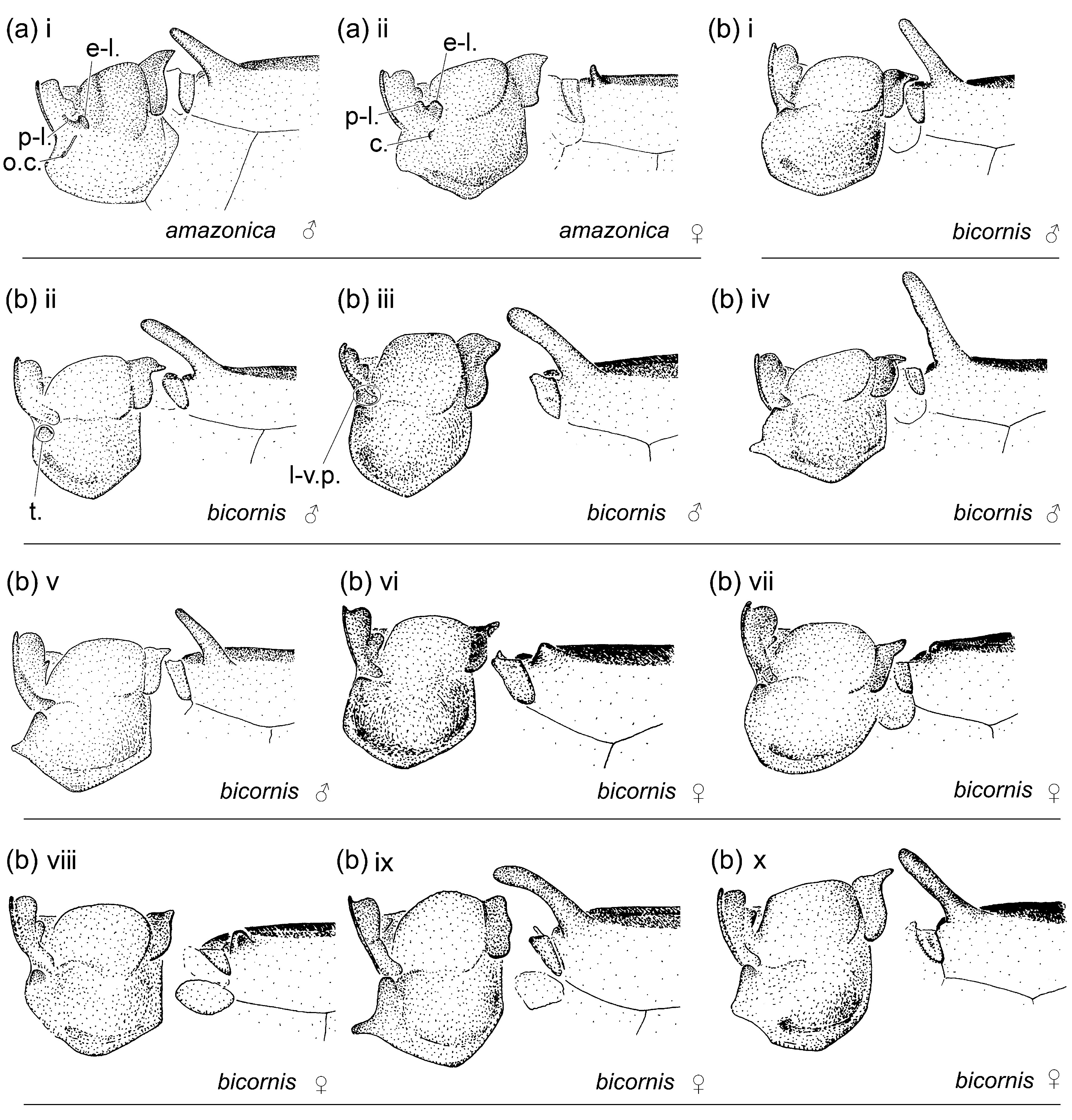
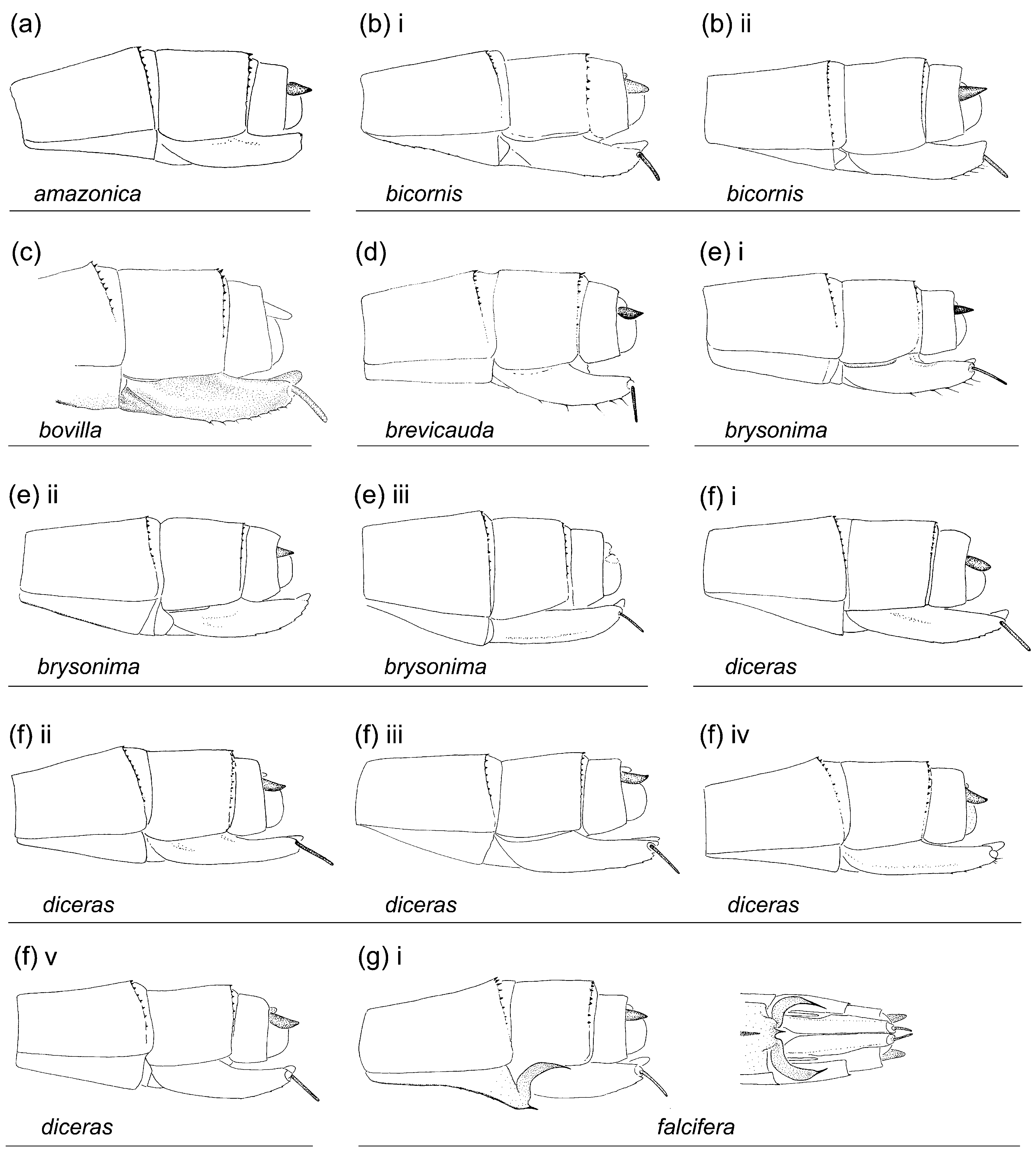
Thorax.—Pronotum anterior lobe dorsally [NOTE: to properly see the ultrastructures of pronotum high microscopy magnification (at least 50 x) and good illumination are necessary, since small relief structures usually have the same color as the surface and are not necessarily evident; softening of specimens to rotate the head might also be necessary in order to properly view these structures]: inflated laterally (in., Figs. 4z View FIGURE 4 , 5z View FIGURE 5 ); with medio-lateral portions of posterior margin with a marginal crest (cr., Fig. 4b View FIGURE 4 ); with a postero-lateral projection on each side, free (po., Fig. 4h View FIGURE 4 iii–iv), or fused to middle lobe of pronotum (p-l., Fig. 4a View FIGURE 4 ); with lateral margin projected posteroventrally forming a smooth L-shaped ridge (l-v. p., Figs. 4b, x View FIGURE 4 ); with lateral margin projected ventrally forming a denticulated ridge ending on a rounded tip (d.r., Fig. 4k View FIGURE 4 ii); smooth ( Figs. 4c–g, h i View FIGURE 4 –ii, i–j, k i, l–p, r–w, y–ae).
Anterior and middle lobes of pronotum dorso-laterally (best seen in dorso-lateral view examined from outer side): separated by an evident groove (g., Figs. 5a–o, r–t, v–z View FIGURE 5 , ab–ac, ae); almost touching, separated by a narrow fissure (f., Figs. 5q, u View FIGURE 5 , aa, ad).
Propleuron: smooth ( Figs. 5c, g, t, w View FIGURE 5 , ac); with a denticulated anterior oblique crest (c., Fig. 5a View FIGURE 5 ); with a longitudinal crest (lo., Figs. 5h, n, y–z View FIGURE 5 ); with sub-vertical crest/s or ridge/s (l.c., Figs. 5f, o–q View FIGURE 5 , aa, ad); with a cshaped crest (c., Figs. 5e, i, m, u, y View FIGURE 5 ); with a rounded tubercle or plate, low or prominent (t., Figs. 5b, d–e, h–l, n, r, u–v, x–z View FIGURE 5 , ab, ae).
Anterior margin of middle lobe of pronotum: smooth ( Figs. 4a–g, i–x, z View FIGURE 4 –ad); forming a crest (cr.), which can be entire ( Figs. 4h, y View FIGURE 4 ; 5h, y View FIGURE 5 ) or bilobate ( Fig. 4 View FIGURE 4 ae; 5ae).
Dorsum of middle lobe of pronotum: with a latero-dorsal c-shaped longitudinal depression (de., Fig. 5c View FIGURE 5 ); lacking a latero-dorsal c-shaped longitudinal depression ( Figs. 5. a–b, d View FIGURE 5 –ae).
Paired ear-lobe projections on anterior portion of middle lobe of pronotum: digit like or tongue-shaped, oriented laterally or postero-laterally ( Figs. 4a View FIGURE 4 iii, g, t, v–w, ac); flap-like with a dorsal concavity, oriented dorso-ventrally ( Fig. 4l View FIGURE 4 ); flap-like and upright, wide and sinuous ( Fig. 4r View FIGURE 4 iii–iv, vi, viii–ix) or narrow ( Fig. 4r v View FIGURE 4 , vii); absent ( Figs. 4b–f, h–k, m–q, s, u, x View FIGURE 4 –ab, ad–ae).
Shape of pronotum posterior lobe: entire, smoothly convex ( Figs. 4k, l i, s View FIGURE 4 iii, y ii, ab iii–iv, vi) or with a medial point ( Figs. 4l View FIGURE 4 ii–iii, ab x); bilobed, with a slight medial concavity shallower than 0.30 of posterior lobe length ( Figs. 4b View FIGURE 4 xiv, d iv, f iv–vii, p viii–x), with a medial u- or v-shaped incision about as deep or deeper than 0.50 of posterior lobe length ( Figs. 4e v View FIGURE 4 –xiii, i iii–vi), and with lateral lobes entire ( Figs. 4b View FIGURE 4 xiv, d iv, e vi–x, i iii–vi, p viii, x) or bilobed ( Figs. 4b View FIGURE 4 vi, e v, ix, xi–xiii, f iv–vii, p ix); trilobed, with medial lobe bilobed ( Figs. 4b View FIGURE 4 iii, vii–viii, x–xi, g i, p v, s ii, iv–v, u iii, x v, y iii, vi–vii, ab i–ii, ix, ac iv), smoothly convex ( Figs. 4a–b i View FIGURE 4 –ii, iv–v, ix, xii–xiii, c v, d ii–iii, f i–iii, viii–ix, g ii, h ii–iv, j, m–o, p ii–vi, xi, q i, r i–iii, s i, vi–viii, t, u i, iv, vi, y i, iv–v, z i–iii, aa i, iii, ab v, vii–viii, ac i–iii, ad i–ii, ae), with a central point ( Figs. 4c i View FIGURE 4 , iii, d i, e i–iv, h i, i i–ii, p i, vii, q ii, r iv–ix, u ii, v, v–w, x i–ii, z iv, aa ii, ad iii), with a central posterior or postero-dorsal projection ( Figs. 4c View FIGURE 4 vi–vii, r iv–v, ix), and lateral lobes entire ( Figs. 4a i View FIGURE 4 –ii, b i–v, vii–xiii, c v–vii, f i–iii, h, i i–ii, m–o, p ii–vi, xi, r, t i–iii, v–ad, ae i) or bilobed ( Figs. 4a View FIGURE 4 iii, t iv, ae ii) [in some species shape is variable, especially in females, presenting two or more possible states].
Lateral lobes: shorter ( Figs. 4a–b, c View FIGURE 4 vi–vii, e iii, v, ix, xi–xiii, f–g, h i, i i, iii–iv, j–m, n i–ii, o i, iii–iv, p i–vii, q i, r–x, y i–ii, z, ab i–v, vii–viii, x, ac, ad i–ii, ae i), as long as (4c i, iii, v, e i–ii, iv, vi–viii, x, h ii–iv, i ii, v–vi, n iii, o ii, p viii, q ii, y, ab vi, ix), or longer ( Figs. 4y View FIGURE 4 iii–vii, ad iii, ae ii) than medial lobe or central area; smoothly convex ( Figs. 4a–f, h–v, w i, x View FIGURE 4 –ab, ac i–iii, ad–ae) or dorsally globose ( Figs. 4g View FIGURE 4 ii, w ii, ac iv); not projected ( Figs. 4a–f, g i, h–i, j i, k, l i, m–q, s, t i View FIGURE 4 –ii, u, v i, w–ae) or projected dorsally ( Figs. 4g View FIGURE 4 ii, j ii, l ii–iii, t iii–iv, v ii).
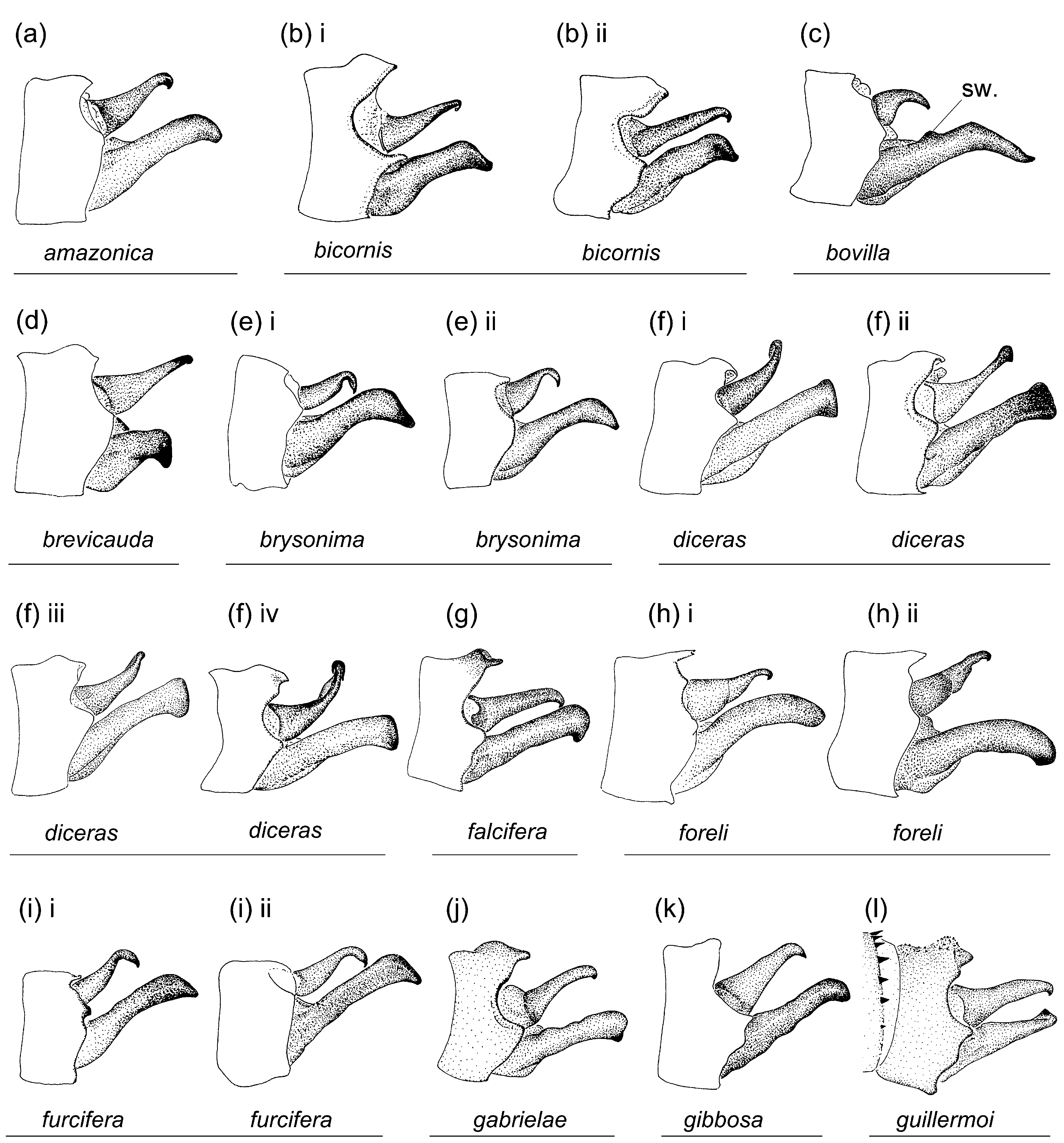
Mesanepisternal horns ( Figs. 4–5 View FIGURE 4 View FIGURE 5 ) length: about as long as 0.30–0.50 of mesostigmal plate width ( Figs. 4a View FIGURE 4 ii, b vi–viii, c iii–iv, f vi–ix, g ii, h iii, j ii–iii, k ii, m i, q ii, r ii–iii, s i–iii, t iii, v ii, y iv–v, aa ii,) to well developed, from about equal to mesostigmal plate width to as long as 4–5 times mesostigmal plate width [even though length can be variable range of values can aid in diagnosis].
Relative width of mesanepisternal horns (width can be variable, but approximate ranges can aid in diagnosis): thin ( Figs. 4d–e, g–k, n, s–t, v–x, y View FIGURE 4 iii–v, z iii–iv, ac i) to thick ( Figs. 4c View FIGURE 4 ).
Orientation of free portion of mesanepisternal horns in dorsal view (orientation can be variable, but approximate ranges can aid in diagnosis): lateral ( Figs. 4p, s View FIGURE 4 ) to antero-lateral ( Figs. 4a, b View FIGURE 4 vi–vii, xiii–xiv, d, e v–xiii, f–g, i iii, vi, j, l i, n i, o ii–iii, q–r, t, v, x, z iii–iv, aa, ab i–iv, ac ii–iii, ad–ae), antero-dorsal ( Figs. 4b i View FIGURE 4 –iv, e i–ii, k, l ii, n ii, o, v, u, w, y, z, ab v–x, ac i), and dorsal ( Figs. 4h View FIGURE 4 ).
i, iv
i–ii
Angle of mesanepisternal horns to mesepisternal surface in lateral view (angle can be variable, but range of values can aid in diagnosis): parallel to surface ( Figs. 5b View FIGURE 5 vii–vii, d, p, s) or at an angle with surface.
Bases of mesanepisternal horns (both states can occur in some species, i.e. M. bicornis , M. leniloba ): adjacent or almost touching ( Figs. 4e–f, p–q View FIGURE 4 , aa, ad) to separated.
Shape of tip of mesanepisternal horns: rounded ( Figs. 4a–b, l–m, n View FIGURE 4 ii–iii, z); pointed or bluntly pointed ( Figs. 4c– d, e–h, n i, o–y View FIGURE 4 , ac); with a transverse dorsal slit ( Fig. 4c v View FIGURE 4 ); antero-posteriorly compressed ( Figs. 4 View FIGURE 4 ab, ae).
Mid-dorsal dark stripe ( Fig. 3 View FIGURE 3 ): parallel sided; widening medially; widening posteriorly; narrowing posteriorly.
Pt shape: rectangular, with anterior and posterior sides longer than distal side ( Fig. 6a View FIGURE 6 ii–iii); squarish, with anterior and posterior sides about equal to distal side; trapezoidal, with anterior side shorter than distal side ( Figs. 6b, c View FIGURE 6 ) [in some species intermediates between two states occur].
Abdomen.—Male genital lobe height: distinctly shorter than 0.50 of anterior hamule height ( Figs. 8a–f, h–v, x View FIGURE 8 – ab, ad–ae); about as high as 0.50 of anterior hamule height and shorter than 0.50 of genital lobe’s basal width ( Fig. 8g View FIGURE 8 , ac); about as high as anterior hamule and as high as 0.50 of genital lobe’s basal width ( Fig. 8w View FIGURE 8 ).
Male genital lobe contour: smoothly curved ( Figs. 8a–s, u View FIGURE 8 –ae); with a distinct anterior angle ( Fig. 8t View FIGURE 8 ).
Male posterior hamule: digit-like and small, with at most only tip surpassing ventral margin of genital fossa in lateral view ( Figs. 8a–f, h–i, m–q, s, u, x View FIGURE 8 –ab, ad–ae); laminar and large, clearly surpassing ventral margin of genital fossa in lateral view ( Figs. 8g, j–l, r, t, v–w View FIGURE 8 , ac), and shorter than ( Figs. 8j–l, v View FIGURE 8 ) or as high as or higher than anterior hamule ( Figs. 8g, r, t, w View FIGURE 8 , ac), with posterior side smoothly convex ( Figs. 8g, j–l, r, w View FIGURE 8 , ac) or angled ( Figs. 8t, v View FIGURE 8 ).
Tip of posterior hamule: not differentiated ( Figs. 8a–f, h–i, m–q, s, u, x View FIGURE 8 –ab, ad–ae); bluntly pointed ( Figs. 8j–l, r, v View FIGURE 8 ); or bent at about 90° and triangular ( Figs. 8g, t View FIGURE 8 , ac) or rounded ( Figs. 8w View FIGURE 8 ).
Curvature of basal segment of genital ligula: marked by a slight depression followed by a slight convex prominence ( Figs. 8a–f, h–i, m–q, s, u, x View FIGURE 8 –ab, ad–ae); marked by a slight depression followed by a pronounced prominence ( Fig. 8c View FIGURE 8 ii), with tip of ligula laying between depression and prominence when folded ( Fig. 8c i View FIGURE 8 ); marked by a deep depression, followed by a pronounced prominence ( Figs. 8g, j–l, r, t, v–w View FIGURE 8 , ac), with tip of ligula laying in the depression when folded. Shape of genital ligula distal segment: sub-quadrate ( Figs. 8a, c, m, o View FIGURE 8 ); sub-rectangular ( Figs. 8b, d, k–l, n, p–s, x, z View FIGURE 8 –aa, ad–ae); pear-shaped, distinctly widened sub-apically ( Figs. 8e–f, g–j, t–w, y View FIGURE 8 , ab– ac).
Terminal fold (ectal fold at base of genital ligula distal segment): short to long (t., Fig. 8 View FIGURE 8 ) [the length of this fold can be variable according to the degree of hydration of the membrane, which can extend considerably when placed in water (i.e. Figs. 8c i View FIGURE 8 – ii), and should therefore not be used as a diagnostic feature].
Base of genital ligula distal segment: with a lateral sclerotized pointed tubercle (p.t., Fig. 8h View FIGURE 8 ); smooth ( Figs. 8a–g, i View FIGURE 8 – ae).
Shape of apex of genital ligula distal segment: convex ( Figs. 8c, h View FIGURE 8 ); transverse or with a shallow concavity ( Figs. 8a–b, d–g, i–r, t View FIGURE 8 –aa, ac–ad); with a deep concavity or cleft, about as deep as wide or deeper ( Figs. 8s View FIGURE 8 , ab); with lateral corners projected into long coiled flagella ( Fig. 8 View FIGURE 8 ae).
Ectal fold on apex of genital ligula distal segment: present, narrow ( Figs. 8b, f–g, j–l, p, r, t–w View FIGURE 8 ) or wide ( Figs. 8c, h View FIGURE 8 ); absent ( Figs. 8a, i, m–o, q, s, x View FIGURE 8 –ae).
Posterior margin of female S8 sternum: smooth, lacking any denticles, spines or processes ( Figs. 9a–f, h–i, m–p, r–s, w–z View FIGURE 9 , ab–ac); with 1 to 6 denticles or small spines ( Figs. 9k–l, q View FIGURE 9 ); with strong spines ( Figs. 9j, u View FIGURE 9 ); with a two ( Figs. 9g, t, v View FIGURE 9 ) or three ( Fig. 9 View FIGURE 9 aa) pronged process, which can be parallel to the sternum ( Figs. 9t View FIGURE 9 , aa), or to the sides of the lateral terga ( Figs. 9g, v View FIGURE 9 ).
Distal end of ovipositor reaching ( Fig. 9 View FIGURE 9 ): apex of paraproct to level slightly distal to cercus [length of ovipositor is variable].
Male S10 postero-dorsal margin: with a small medial u-shaped incision or longitudinal slit ( Figs. 10a–b, d–f, i, m–q, s, u, x View FIGURE 10 –aa, ad); smoothly convex ( Figs. 10c, g–h, j–l, r, t, v–w View FIGURE 10 , ab–ac, ae).
Male S10 postero-dorsal surface: with a medial prominence, which can be entire ( Figs. 11a–d, f–g, j, l–r, t–x, z View FIGURE 11 – ae; 12a–d, f–g, j, l–r, t–x, z–ae) or divided in two lateral portions ( Figs. 11k View FIGURE 11 ; 12k View FIGURE 12 ); lacking a prominence ( Figs. 11e, h–i, s, y View FIGURE 11 ; 12e, h–i, s, y View FIGURE 12 ).
Male S10 postero-dorsal margin: with a medial projection ( Figs. 10a–b, d–j, l–r, t View FIGURE 10 –ad; 11a–b, d–j, l–r, t–ad); lacking a projection ( Figs. 10c, k, s View FIGURE 10 , ae; 11c, k, s, ae).
Male cercus shape in dorsal view: curved medially ( Figs. 10a–d, f–g, j–o, q–x, z View FIGURE 10 –ae); about straight ( Figs. 10e, h–i, p, y View FIGURE 10 ).
Male cercus width in dorsal view: about uniform ( Figs. 10q, r, t, w, z View FIGURE 10 , ad–ae); narrowing to tip ( Figs. 10a, c–e, h–i, m–p, s, u–v, x–y View FIGURE 10 , aa–ac); widening sub-apically ( Figs. 10b, f–g, j–l View FIGURE 10 ).
Shape of male cercus tip: pointed ( Figs. 10a–e, g–n, p, r–y View FIGURE 10 , aa–ac); rounded or blunt ( Figs. 10f, o, q, z View FIGURE 10 , ad–ae).
Ratio of male cercus length to S10 maximum length in lateral view ( Fig. 12 View FIGURE 12 ): from about 0.5 to about 2.
Ratio of male cercus length to paraproct length in lateral view ( Fig. 12 View FIGURE 12 ): from about 0.5 to about 1.5.
Width of male paraproct distal third in lateral view: (0) narrower than medial third ( Figs. 12a, c, m–n, q–r, z View FIGURE 12 ); about as wide as medial third ( Figs. 12b, h, k–l, o, s, x–y View FIGURE 12 , ab, ae); wider than medial third ( Figs. 12d–g, i–j, p, t–w View FIGURE 12 , aa, ac–ad).
Male paraproct tip: with smooth medial surface and no apical tooth ( Figs. 11 View FIGURE 11 aa, ae); with smooth medial surface and ending on an apical tooth ( Figs. 11a, c, g–h, j–k, m, o, q–w, y View FIGURE 11 , aa, ac–ad); with a ridge on medial surface ending on an apical tooth ( Figs. 11b, d–f, n, x, z View FIGURE 11 ); with a ridge on medial surface with basal and apical teeth ( Figs. 11i, l, p View FIGURE 11 ).
Remarks. Several male characters are species specific: shape of genital ligula distal segment, posterior hamule in species where it is well developed, dorsum of S10, cerci, and paraprocts. Conspecific males and females share the same ultrastructural features of the pronotal and propleural surface (presence and position of various crests, ridges, tubercles, and projections), the position of the bases of the mesanepisternal horns, and the basic black pattern of head and width and shape of dark mid-dorsal stripe of pterothorax. However, the latter two characters show considerable intraspecific plasticity in a few species, i.e. the extension of the black pattern on head dorsum observed in M. paludicola and M. spatulata ( Figs. 1u View FIGURE 1 , ab) and on the back of the head in M. minteri , and the width of the mid-dorsal stripe in M. minteri ( Fig. 3s View FIGURE 3 ) vary considerably even among specimens within the same population.
The shape of the posterior lobe of the pronotum and the exact length, width, orientation, and shape of mesanepisternal horns show considerable intraspecific variability, especially in females. In addition, in females one or both of these structures can be similar to those of conspecific males (species with no secondary sexual dimorphism: M. leniloba , M. minteri , and M. paludicola ) or different (species with secondary female dimorphism: M. bovilla , M. brysonima , M. falcifera , M. furcifera , M. gabrielae , M. gibbosa , M. knopfi , M. lillianae , M. mauffrayi , M. longicauda , M. panguanae , M. peltata , M. quadricornis , M. selysi , M. tridentigera , M. truncata , and M. turbinata ), and several species include both dimorphic and not dimorphic females (species with andromorphic and heteromorphic females: M. bicornis , M. brevicauda , M. diceras , M. foreli , M. guillermoi , M. orthogonia , M. silvicola , and M. spatulata ). Female dimorphism can include just the mesanepisternal horns (with shape of posterior lobe of pronotum the same as in male: M. amazonica , M. foreli , M. knopfi , M. truncata ), just the shape of posterior lobe of pronotum (with mesanepisternal horns similar to those of male: M. brevicauda , M. lillianae , M. prostrata ), both mesanepisternal horns and/or posterior lobe of pronotum ( M. bicornis , M. diceras , M. guillermoi , M. orthogonia , M. spatulata , M. truncata ), or involve other structures of pronotum (i.e. antero-dorsal ear-like lobes on middle lobe of pronotum in M. mauffrayi ). Dimorphic females may display more than one distinct morph in terms of the characters in which they differ from their males (i.e. the upright ear-like lobe on dorsum of middle lobe of pronotum in females of M. mauffrayi , absent in males, can be either narrow or wide, with no gradation between these two states). Species that were represented by small series of available females in this study (see Table 2) might be found to present more morphs when more specimens become available in the future.
I was able to reliably confirm conspecifity of males and dimorphic or polymorphic females in species for which pairs in copula and/or tandem were available, from which I was able to identify diagnostic characters shared by males and females, i.e. ultrastructural features on propleural and pronotal surface, position of bases of mesanepisternal horns, basic black pattern of head, and width and shape of dark mid-dorsal stripe of pterothorax. Conspecificity was reinforced by the examination of series of specimens from the same locality that included males and different female morphs. After discovering the existence of female dimorphism and polymorphism in some species and recognizing which characters of diagnostic value are shared by conspecific males and females, I was accordingly able to associate males and females of species not found in copula but which share states of these diagnostic characters.
structures. Polymorphic species including both andromorphic and heteromorphic females are indicated in bold, and dimorphic species including more than one distinct female morph are underscored.
Key to species of Metaleptobasis View in CoL
1. Male posterior hamule (p.h.) digit-like, small, with at most only tip surpassing ventral margin of genital fossa in lateral view ( Figs. 8a–f, h–i, m–q, s, u, x View FIGURE 8 –ab, ad–ae); curvature of basal segment of genital ligula marked by a slight concave depression (s.d., Figs. 8a–f, h–i, m–q, s, u, x View FIGURE 8 –ab, ad–ae); posterior margin of female S8 sternum smooth ( Figs. 9a–f, h–i, m–p, r–s, w–z View FIGURE 9 , aa, ac–ad)........................................................................................ 2
1’. Male posterior hamule (p.h.) laminar and curved posteriorly or postero-medially, large, clearly surpassing ventral margin of genital fossa in lateral view ( Figs. 8g, j–l, r, t, v–w View FIGURE 8 , ac); curvature of basal segment of genital ligula marked by a deep concave depression (d.d., Figs. 8g, j–l, r, t, v–w View FIGURE 8 , ac); posterior margin of female S8 sternum with either denticles or small spines ( Figs. 9 View FIGURE 9 k-l, q), strong spines ( Figs. 9j, u View FIGURE 9 ), or a bifid or trifid process ( Figs. 9g, t, v View FIGURE 9 , ab)............................ 23
2(1). Black on head dorsum covering half to most of dorsal surface (1c, e, g–l, r, t–w, y, ab–ac, ae)....................... 3
2’. Black on head dorsum covering less than half of dorsum, limited to isolated spots and stripes ( Figs. 1a–b, d, f, m–q, s, x, z View FIGURE 1 – aa, ad) .............................................................................................10
3(2). Labrum mostly black, with only distal margin narrowly pale ( Figs. 1c View FIGURE 1 ); lateral surface of middle lobe of pronotum with a cshaped latero-dorsal longitudinal depression (de.), shallow in males ( Figs. 4c I View FIGURE 4 , 5c I View FIGURE 5 –II), deep in females ( Figs. 4c V View FIGURE 4 –VI, 5 c III–IV); mesanepisternal horns thick in male ( Figs. 4c View FIGURE 4 II–IV, 5c I–II), reduced to tubercles with a dorsal transverse slit in females ( Figs. 4c V View FIGURE 4 , 5c View FIGURE 5 III–IV); male cercus about as long as 0.33 of paraproct ( Figs. 10c View FIGURE 10 , 12c View FIGURE 12 ); male paraproct with a dorsal swelling at basal 0.40 of its length (sw., Fig. 12c View FIGURE 12 ); Guatemala to Costa Rica ( Fig. 14a)........................................ M. bovilla View in CoL
3’. Labrum mostly pale, with marginal black stripe along base and sides ( Figs. 1e, h–i, u, y View FIGURE 1 , ab, ae); lateral surface of middle lobe of pronotum smooth ( Figs. 4e, h–i, u, y View FIGURE 4 , ab, ae; 5e, h–i, u, y, ab, ae); mesanepisternal horns thin in males, and never with a dorsal transverse slit in females ( Figs. 4e, h–i, u, y View FIGURE 4 , ab, ae; 5e, h–i, u, y, ab, ae); male cercus about as long as 0.50 of paraproct or longer; male paraproct lacking a dorsal swelling at basal 0.40 of its length ( Figs. 12e, h–i, u, y View FIGURE 12 , ab, ae)........ 4
4(3’). Anterior margin of middle lobe of pronotum forming a distinct crest (cr., Figs. 4h, y View FIGURE 4 , ae; 5h, y, ae)............... 5
4’. Anterior margin of middle lobe of pronotum smooth ( Figs. 4e, i, u View FIGURE 4 , ab; 5e, i, u, ab)............................. 7
5(4’). Black on head dorsum covering about half of dorsal surface ( Figs. 1 View FIGURE 1 ae); crest on anterior margin of middle lobe of pronotum bilobate ( Figs. 4 View FIGURE 4 ae; 5ae); distal segment of genital ligula with long paired coiled flagella (fl., Fig. 8 View FIGURE 8 ae); N Peru ( Fig. 14a)..................................................................................... M. turbinata View in CoL
5’. Black on head dorsum covering most of dorsal surface ( Figs. 1h, y View FIGURE 1 ); crest on anterior margin of middle lobe of pronotum entire (cr., Figs. 4h, y View FIGURE 4 ; 5h, y View FIGURE 5 ); distal segment of genital ligula lacking flagella ( Figs. 8h, y View FIGURE 8 )........................ 6
6(4’). Anterior lobe of female pronotum with a lateral digit-shaped projection directed posteriorly (po., Figs. 4h View FIGURE 4 III–IV, 5h V), rarely reduced ( Fig. 5h View FIGURE 5 VI), absent (as in Fig. 5h I View FIGURE 5 , IV), or fused to middle-lobe of pronotum ( Fig. 5h View FIGURE 5 VII); posterior lobe of female pronotum slightly trilobed with lateral lobes shorter than medial lobe length ( Figs. 4h View FIGURE 4 III–IV); distal segment of genital ligula with a pair of sclerotized pointed tubercles at base (p.t.) and with a wide ectal fold (e.f., Fig. 8h View FIGURE 8 ); male cercus basal half globose, distal half sub-cylindrical, with only extreme tip pointed ventrally; male paraproct curved ventrally gradually, with distal end rounded in lateral view ( Figs. 12h I View FIGURE 12 –II); Costa Rica to N Colombia and Venezuela ( Fig. 14b).................... M. foreli View in CoL
6’. Anterior lobe of female pronotum smooth ( Figs. 4y View FIGURE 4 IV, 5y IV–VII); posterior lobe of female pronotum deeply trilobed with lateral lobes rounded and longer than medial lobe length ( Figs. 4y View FIGURE 4 III–VII); distal segment of genital ligula lacking paired sclerotized pointed tubercles at base and lacking an ectal fold ( Fig. 8y View FIGURE 8 ); male cercus sub-cylindrical, gradually narrowing distally, with tip curved ventrally forming a hook; male paraproct curved ventrally at an angle, with tip pointed in lateral view ( Figs. 12y I View FIGURE 12 –II); Venezuela, Surinam, French Guiana, and N Brazil ( Fig. 14b)................................... M. quadricornis View in CoL
7(4’). Propleuron with a short triangular anterior laminar ridge (t., Fig. 5 View FIGURE 5 ab); mesanepisternal horns wide at base and compressed antero-posteriorly ( Figs. 4 View FIGURE 4 ab I–VI,VIII, 5ab I–II,IV–V) or reduced to low blunt prominences ( Figs. 4 View FIGURE 4 ab VII, 5ab III); Ecuador, Peru, and W Brazil ( Fig. 14a)......................................................................... M. spatulata View in CoL
7’. Propleuron lacking a laminar ridge, with an anterior rounded tubercle (t., Figs. 4e, i, u View FIGURE 4 ; 5e, i, u View FIGURE 5 ); mesanepisternal horns sub-cylindrical ( Figs. 4e, i, u View FIGURE 4 ; 5e, i, u View FIGURE 5 ).................................................................. 8
8(7’). Female pronotum trilobed with smoothly convex medial lobe ( Figs. 4u V View FIGURE 4 –VI); dark mid-dorsal pterothoracic stripe about as wide as 0.33 of mesepisterna width ( Fig. 3u View FIGURE 3 ); male S 10 in lateral view with a well developed medial prominence on postero-dorsal margin ( Fig. 12u View FIGURE 12 ); Amazon forest of N Peru and N Brazil ( Fig. 14e).............................. M. paludicola View in CoL
8’. Female pronotum unlobed to slightly trilobed always with a pronounced medial u or v-shaped incision ( Figs. 4e V View FIGURE 4 –XIII, i III–VI); dark mid-dorsal pterothoracic stripe about as wide as 0.50 of mesepisterna width ( Figs. 3e, i View FIGURE 3 ); male S 10 in lateral view flat, lacking a medial prominence on postero-dorsal margin ( Figs. 12e, i View FIGURE 12 ).......................................... 9
9(8’). Apex of male genital ligula distal segment with a u-shaped incision ( Fig. 8e View FIGURE 8 ), embracing sides of basal segment when ligula is folded; tip of male paraproct with a poorly developed longitudinal crest on inner surface, terminating as a medially directed single apical tooth (s.t., Figs. 10e View FIGURE 10 , 11e View FIGURE 11 ); Trinidad and Venezuela to E Peru and N Bolivia ( Fig. 14b)....... M. brysonima View in CoL
9’ Apex of male genital ligula distal segment transverse ( Fig. 8i View FIGURE 8 ), only touching ventral surface of basal segment without extending over its sides when ligula is folded; tip of male paraproct with a well developed longitudinal crest on inner surface, with two medially directed teeth (te.), one on its basal end (may be small) and one on its apical end ( Figs. 10i View FIGURE 10 , 11i View FIGURE 11 ); N Peru ( Fig. 14b)......................................................................... M. furcifera View in CoL
10(2’). Middle lobe of pronotum with an antero-dorsal rounded thumb-like process (e-l., Figs. 4a View FIGURE 4 ; 5a View FIGURE 5 ) directed laterally on each side; Pará State in Brazil ( Fig. 14e)......................................................... M. amazonica View in CoL
10’. Middle lobe of pronotum lacking an antero-dorsal thumb-like process directed laterally on each side ( Figs. 4b, d, f, m–q, s, x, z View FIGURE 4 –aa, ad; 5b, d, f, m–q, s, x, z–aa, ad).............................................................. 11
11(10’). Lateral margin of anterior lobe of pronotum extended postero-ventrally forming a ridge (l-v. p.) opposite an anterior tubercle (t.) on propleuron ( Figs. 4b, x View FIGURE 4 ; 5b, x View FIGURE 5 )............................................................ 12
11’. Lateral margin of anterior lobe of pronotum not extended postero-ventrally forming a ridge ( Figs. 4d, f, m–q, s, z View FIGURE 4 –aa, ad; 5d, f, m–q, s, z–aa, ad)......................................................................... 13
12(11). Lateral portions of posterior margin of pronotum anterior lobe with a marginal crest (cr., Fig. 4b View FIGURE 4 ); mesanepisternal horns oriented antero-dorsally at an angle of about 45° with dorsum ( Figs. 5b I–V View FIGURE 5 ,IX–X) or represented by bases oriented laterally ( Figs. 5b View FIGURE 5 VI–VIII); distal portion of male cercus depressed dorso-ventrally in lateral view ( Fig. 12b View FIGURE 12 ) and slightly widened sub-apically in dorsal view ( Fig. 10b View FIGURE 10 ); Trinidad, Venezuela, Guyana, Surinam, French Guiana, and N Brazil ( Fig. 14c)... M. bicornis View in CoL
12’. Lateral portions of posterior margin of pronotum anterior lobe lacking a crest ( Fig. 4x View FIGURE 4 ); mesanepisternal horns directed antero-laterally at an angle of about 15° with dorsum ( Fig. 5x View FIGURE 5 ); distal portion of male cercus sub-cylindrical in lateral view ( Fig. 12x View FIGURE 12 ) and gradually narrowing distally in dorsal view ( Fig. 10x View FIGURE 10 ); Junín Dep. in Peru ( Fig. 14c)....... M. prostrata View in CoL
13(11’). Mesanepisternal horns directed antero-laterally or laterally forming an angle of 0°–15° with dorsum ( Figs. 4d, p, s View FIGURE 4 ; 5d, p, s View FIGURE 5 ), and if limited to horn bases these arising at level of mesostigmal plates bases or more externally ( Figs. 4s I View FIGURE 4 , III,V,VIII)...... 14
13’. Mesanepisternal horns directed antero-dorsally forming an angle of 35°–90° with dorsum ( Figs. 4f, m–o, q, z View FIGURE 4 –aa, ad; 5f, m– o, q, z–aa, ad), and if limited to horn bases these arising medially to level of mesostigmal plates bases ( Figs. 4f View FIGURE 4 IV–VIII, m, q II, aa III, ad II–III)....................................................................................... 16
14(11). Mesanepisternal horns distinctly separated at bases, arising at level of base of mesostigmal plates or more externally ( Fig. 4s View FIGURE 4 ); male S10 lacking a postero-dorsal prominence and a postero-medial projection ( Fig. 12s View FIGURE 12 ); male paraproct sub-apically about as wide as at medial third ( Fig. 12s View FIGURE 12 ) and lacking an apical ridge on medial surface ( Figs. 10s View FIGURE 10 ; 11s View FIGURE 11 ); S Colombia, Ecuador, Peru, and W Brazil ( Fig. 14e)............................................................ M. minteri View in CoL
14’. Mesanepisternal horns adjacent at base ( Fig. 4p View FIGURE 4 ) or if separated, arising medially to level of mesostigmal plates bases ( Fig. 4d View FIGURE 4 ); male S10 with a postero-dorsal prominence and a postero-medial projection ( Figs. 12d, p View FIGURE 12 ); male paraproct sub-apically wider than at medial third ( Figs. 12d, p View FIGURE 12 ) and with an apical ridge on medial surface (r., Figs. 10d, p View FIGURE 10 ; 11d, p View FIGURE 11 ).......... 15
15 (14’). Bases of mesanepisternal horns separated ( Fig. 4d View FIGURE 4 ); male paraproct shorter than 0.50 of cercus length ( Figs. 10d View FIGURE 10 ; 12d View FIGURE 12 ); Huánuco Dep. in Peru ( Fig. 14c)............................................................ M. brevicauda View in CoL
15’. Bases of mesanepisternal horns adjacent ( Fig. 4p View FIGURE 4 ); male paraproct about as long as twice cercus length ( Figs. 10p View FIGURE 10 ; 12p View FIGURE 12 ); Ecuador, Peru, Bolivia, and W Brazil ( Fig. 14d)................................................. M. lillianae View in CoL
16(15’). Anterior area of propleuron with two sub-vertical crests (l.c., Figs. 5f, q View FIGURE 5 , aa, ad; CAUTION: anterior crest can be low and rounded as in Figs. 5f View FIGURE 5 IV,IX).............................................................................17
16’. Anterior area of propleuron with only one crest, sub-vertical, transverse, or c-shaped (c., l.c., lo., Figs. 5m –n, o, z View FIGURE 5 )..... 20
17(16’). Pt trapezoidal, with anterior side shorter than distal side ( Figs. 6b–c View FIGURE 6 ); tip of male cercus sub-cylindrical ( Figs. 11q View FIGURE 11 , ad; 12q, ad)..............................................................................................18
17’. Pt sub-rectangular, with anterior and posterior sides longer than distal side ( Fig. 6a View FIGURE 6 ); tip of male cercus depressed dorso-ventrally ( Figs. 11f View FIGURE 11 , aa; 12f, aa)......................................................................... 19
18(17’). Mid-dorsal dark stripe on pterothorax at mid-length as wide as about 0.20 of mesepisterna width ( Fig. 3q View FIGURE 3 ); male cercus about as long as 2 times maximum length of S 10 in lateral view, about as long as paraproct, with tip rounded ( Fig. 12q View FIGURE 12 ); Mato Grosso and Goias States in Brazil ( Fig. 14d).............................................. M. longicauda View in CoL
18’. Mid-dorsal dark stripe on pterothorax at mid-length about as wide as 0.25 of mesepisterna width ( Fig. 3 View FIGURE 3 ad); male cercus about as long as 1.5 times maximum length of S 10 in lateral view, shorter than paraproct, with tip truncate posteriorly ( Fig. 12 View FIGURE 12 ad); Pará State in Brazil ( Fig. 14d)..................................................... M. truncata View in CoL
19(17’). Anterior and middle lobes of pronotum separated dorso-laterally by a groove (g., Fig. 5f View FIGURE 5 ); mesanepisternal horns of males and andromorphic females about as long as 1.5 times the width of a mesostigmal plate ( Figs. 4f View FIGURE 4 ; 5f View FIGURE 5 ); pterothoracic mid-dorsal stripe about as wide as 0.25 of mesepisterna width ( Fig. 3f View FIGURE 3 ); distal segment of genital ligula widened from base to tip ( Fig. 8f View FIGURE 8 ); male cercus in dorsal view slightly widened distally and constricted sub-apically ( Fig. 10f View FIGURE 10 ); tip of male paraproct with ventral bicuspidate tooth pointed medially (b.t.), and with an inner transverse sub-apical ridge (r., Figs. 10f View FIGURE 10 ; 11f View FIGURE 11 ); Trinidad, Venezuela, Surinam, Guyana, Brazil, and Peru ( Fig. 14d)....................................... M. diceras View in CoL
19’. Anterior and middle lobes of pronotum almost touching dorsally, separated by a fissure (f., Fig. 5 View FIGURE 5 aa); mesanepisternal horns of males and andromorphic females about as long as two times the width of a mesostigmal plate ( Figs. 4 View FIGURE 4 aa; 5aa); pterothoracic mid-dorsal stripe about as wide as 0.18 of mesepisterna ( Fig. 3 View FIGURE 3 aa); distal segment of genital ligula sub-rectangular, with sides almost straight, slightly narrowing from base to tip ( Fig. 8 View FIGURE 8 aa); male cercus in dorsal view of uniform width sub-apically ( Fig. 10 View FIGURE 10 aa); tip of male paraproct with a single triangular medio-ventral tooth (s.t.), and with two inner longitudinal sub-apical ridges (rs., Figs. 10 View FIGURE 10 aa; 11aa); SE Peru ( Fig. 14d)................................... M. silvicola View in CoL
20(16’). Latero-ventral area of middle lobe of pronotum with a short c-shaped crest, located opposite a short c-shaped crest on antero-dorsal area of propleuron (c., Fig. 5m View FIGURE 5 ); male mesanepisternal horns sub-triangular, about as long as 0.50 of mesostigmal plate width ( Figs. 4m View FIGURE 4 ; 5m View FIGURE 5 ); Pará State in Brazil ( Fig. 14e) [female unknown]................ M. inermis View in CoL
20’. Latero-ventral area of middle lobe of pronotum lacking any crest or ridge ( Figs. 5n, o, z View FIGURE 5 ); male mesanepisternal horns sub-cylindrical, about as long as mesostigmal plate width or longer ( Figs. 5n, o, z View FIGURE 5 )...............................21
21(20’). Pterothoracic mid-dorsal dark stripe narrower than 0.20 of mesepisterna width, slightly widening posteriorly ( Fig. 3o View FIGURE 3 ); anterior area of propleuron with a short comma-shaped vertical crest (l.c., Fig. 5o View FIGURE 5 ); distal segment of genital ligula sub-quadrate ( Fig. 8o View FIGURE 8 ); N Peru and N Brazil ( Fig. 14e)....................................................... M. leniloba View in CoL
21’. Pterothoracic mid-dorsal dark stripe as wide or wider than 0.33 of mesepisterna width, about parallel sided ( Figs. 3n, z View FIGURE 3 ); anterior area of propleuron with a short longitudinal ridge (lo., Figs. 5n, z View FIGURE 5 ); distal segment of genital ligula sub-rectangular ( Figs. 8n, z View FIGURE 8 ).......................................................................................22
22(21’). Lateral margin of pronotum anterior lobe not inflated ( Figs. 4n View FIGURE 4 ; 5n View FIGURE 5 ); mesanepisternal horns thin, narrowing from base to bluntly pointed tip, forming an angle of 80°–95° with dorsum in lateral view ( Fig. 5n View FIGURE 5 ); male cercus in dorsal view only slightly curved medially, sub-cylindrical, gradually narrowing to tip ( Figs. 10n View FIGURE 10 ; 11n View FIGURE 11 ), in lateral view about as long as 0.50 of paraproct length ( Fig. 12n View FIGURE 12 ); Ecuador ( Fig. 14c).................................................... M. knopfi View in CoL
22’. Lateral margin of pronotum anterior lobe inflated (in., Figs. 4z View FIGURE 4 ; 5z View FIGURE 5 ); mesanepisternal horns of medium thickness, subcylindrical with rounded tips, forming an angle of 35°–45° with dorsum in lateral view ( Fig. 5z View FIGURE 5 ); male cercus in dorsal view abruptly bent medially at about mid-length, with distal half about parallel-sided ( Fig. 10z View FIGURE 10 ), and depressed dorso-ventrally ( Figs. 11z View FIGURE 11 ; 12z View FIGURE 12 ), in lateral view distinctly longer than 0.50 of paraproct length ( Fig. 12z View FIGURE 12 ); E Brazil and NE Argentina ( Fig. 14c)....................................................................................... M. selysi View in CoL
23(1’). Males........................................................................................... 24 23’. Females......................................................................................... 32
24(23’). Medially bent distal portion of cercus widened, wider than medial third of cercus in dorsal view ( Figs. 10j–l View FIGURE 10 )........ 25
24’. Medially bent distal portion of cercus not widened, about as wide as or narrower than medial third of cercus in dorsal view ( Figs. 10g, r, t, v–w View FIGURE 10 , ac)............................................................................ 27
25(24). Cercus about as long as paraproct in lateral view ( Fig. 12l View FIGURE 12 ); paraproct ending on apical dorsal and ventral teeth ( Figs. 10l View FIGURE 10 ; 11l View FIGURE 11 ); N Peru ( Fig. 14g)................................................................... M. guillermoi View in CoL
25’. Cercus shorter than paraproct in lateral view ( Figs. 12j–k View FIGURE 12 ); paraproct ending on a single medio-ventral apical tooth ( Figs. 10j–k View FIGURE 10 ; 11j–k View FIGURE 11 ) .............. .. ........................... ............. ......... .. ................. 26
26(25’). Posterior hamule tip in same plane as hamule axis; axis narrow, about as wide as width of medial sclerotized portion of genital ligula basal segment in ventral view ( Fig. 8k View FIGURE 8 ); genital ligula distal segment sub-rectangular in ventral view ( Fig. 8k View FIGURE 8 ); dorsum of S10 with sub-apical prominence (pr.) divided into two lateral mounds, and lacking a postero-medial projection ( Figs. 10k View FIGURE 10 ; 11k View FIGURE 11 ); Ecuador ( Fig. 14g)........................................................ M. gibbosa View in CoL
26’. Posterior hamule tip bent medially at an angle of about 90° with hamule axis; axis wide, about as wide as 1.5 times width of medial sclerotized portion of genital ligula basal segment in ventral view ( Fig. 8j View FIGURE 8 ); dorsum of S10 with sub-apical prominence (pr.) entire and about transverse, and with a postero-medial convex projection (p-m.p., Figs. 10j View FIGURE 10 ; 11j View FIGURE 11 ); Loreto Dep. in Peru ( Fig. 14g).................................................................... M. gabrielae View in CoL
27(24’). Genital lobe (g.l.) as tall as or taller than 0.50 of anterior hamule (a.h., Figs. 8g, w View FIGURE 8 , ac)........................... 28
27’. Genital lobe shorter (g.l.) than 0.50 of anterior hamule (a.h., Figs. 8r, t, v View FIGURE 8 )..................................... 30
28(27). Genital lobe (g.l.) as tall as anterior hamule (a.h.), and slightly taller than 0.50 of genital lobe basal width ( Fig. 8w View FIGURE 8 ); N Peru ( Fig. 14f)....................................................................... M. peltata View in CoL
28’. Genital lobe (g.l.) as tall as 0.50 of anterior hamule (a.h.), and shorter than 0.50 of genital lobe basal width ( Figs. 8g View FIGURE 8 , ac) 29
29(28’). Male posterior hamule (p.h.) about as wide as 1.5 times width of medial sclerotized portion of genital ligula basal segment in ventral view ( Fig. 8g View FIGURE 8 ); distal portion of male cercus about as wide as medial portion ( Fig. 10g View FIGURE 10 ); S Peru and W Brazil ( Fig. 14f).................................................................................... M. falcifera View in CoL
29’. Male posterior hamule (p.h.) about as wide as width of medial sclerotized portion of genital ligula basal segment in ventral view ( Fig. 8 View FIGURE 8 ac); distal portion of male cercus narrower than medial portion ( Fig. 10 View FIGURE 10 ac); N and W Brazil ( Fig. 14f)............................................................................................. M. tridentigera View in CoL
30(27’). Posterior hamule slightly (p.h.) taller than 0.50 of anterior hamule (a.h.), with anterior margin smoothly curved and posterior margin widened into a heel, with tip curved gradually medially and rounded in ventral view ( Fig. 8v View FIGURE 8 ); Huánuco Dep. in Peru ( Fig. 14g)............................................................................. M. panguanae View in CoL
30’. Posterior hamule (p.h.) about as tall as anterior hamule (a.h.) or slightly taller, about semicircular with both anterior and posterior margins smoothly curved and tip in same plane as axis ( Figs. 8r View FIGURE 8 ), or with anterior margin straight and posterior margin angled near base and hamule tip bent medially at about 90° with hamule axis and triangular in ventral view ( Figs. 8t View FIGURE 8 ).. 31
31(30’). Posterior hamule (p.h.) elongated crescent shaped with both anterior and posterior margins smoothly curved and laminar, with tip in same plane as axis ( Fig. 8r View FIGURE 8 ); distal segment of male genital ligula rectangular in ventral view ( Fig. 8r View FIGURE 8 ); N Peru, Ecuador, and S Colombia ( Fig. 14g)......................................................... M. mauffrayi View in CoL
31’. Posterior hamule (p.h.) with posterior margin angled near base, and hamule tip bent medially at about 90° with hamule axis and triangular in ventral view ( Fig. 8t View FIGURE 8 ); distal segment of male genital ligula pear-shaped, distinctly widened sub-apically in ventral view ( Fig. 8t View FIGURE 8 ); N Peru and Ecuador ( Fig. 14f)...................................... M. orthogonia View in CoL
32(23’). Posterior margin of sternum S8 with 1–6 denticles or small spines ( Figs. 9k–l, q View FIGURE 9 )............................... 33
32’. Posterior margin of sternum S8 with strong spines or processes ( Figs. 9g, j, t, u–v View FIGURE 9 , ab)........................... 35
33(32’). Latero-ventral corners of anterior lobe of pronotum projected ventrally forming a ridge with rounded tip (d.r., Fig. 5k View FIGURE 5 II); middle lobe of pronotum smooth; hind lobe of pronotum entire, smoothly convex with lateral corners fused to middle lobe of pronotum ( Figs. 4k View FIGURE 4 ; 5k View FIGURE 5 ); Ecuador ( Fig. 14g)................................................... M. gibbosa View in CoL
33’. Latero-ventral corners of anterior lobe of pronotum not projected ventro-laterally ( Figs. 5l, r View FIGURE 5 ); middle lobe of pronotum with antero-dorsal ear-like lobes (e.l., Figs. 4l View FIGURE 4 ; 5l, r View FIGURE 5 ); female hind lobe of pronotum more or less trilobed, with lateral corners angled ( Figs. 4l View FIGURE 4 ; 5l, r View FIGURE 5 ).......................................................................... 34
34(33’). Middle lobe of pronotum with antero-dorsal ear-like lobes (e.l.) directed laterally, with surface oriented dorso-ventrally and with a dorso-basal concave depression ( Figs. 4l View FIGURE 4 ; 5l View FIGURE 5 ); posterior margin of female sternum S8 with 5 small denticles ( Fig. 9l View FIGURE 9 ); N Peru ( Fig. 14g)................................................................ M. guillermoi View in CoL
34’. Middle lobe of pronotum with antero-dorsal ear-like lobes (e.l.) upright, narrow or wide and sinuous ( Figs. 4r View FIGURE 4 ; 5r View FIGURE 5 ); posterior margin of female sternum S8 with 1–3 denticles or spines ( Fig. 9q View FIGURE 9 ); N Peru, Ecuador, and S Colombia ( Fig. 14g)................................................................................. M. mauffrayi View in CoL
35(32’). Posterior margin of sternum S8 with two strong straight sub-cylindrical spines separated at their bases ( Figs. 9j, u View FIGURE 9 ).... 36
35’. Posterior margin of sternum S8 with two or three prongs joined at a common base ( Figs. 9g, t, v View FIGURE 9 , ab)................ 37
36(35). Middle lobe of female pronotum with tongue-shaped antero-lateral lobes (e.l.) directed latero-ventrally ( Figs. 4v View FIGURE 4 ; 5v View FIGURE 5 ); spines of distal margin of female sternum S8 separated by a distance less than their individual width at their base ( Fig. 9u View FIGURE 9 ); Huánuco Dep. in Peru ( Fig. 14g)........................................................... M. panguanae View in CoL
36’. Middle lobe of female pronotum smooth ( Figs. 4j View FIGURE 4 ; 5j View FIGURE 5 ); spines of distal margin of female sternum S8 separated by a distance greater than their individual width at their base ( Fig. 9j View FIGURE 9 ); Loreto Dep. in Peru ( Fig. 14g)................. M. gabrielae View in CoL
37(35’). Posterior margin of sternum S8 projected into a bifid process with lateral prongs sub-cylindrical and parallel to sternum ( Fig. 9t View FIGURE 9 ); N Peru and Ecuador ( Fig. 14f).......................................................... M. orthogonia View in CoL
37’. Posterior margin of sternum S8 projected into a trifid process with a central spine and laminar lateral prongs ( Figs. 9g, v View FIGURE 9 , ab) ..................... .. ....................... .. .. .. ............ .................... ............. 38
38(37’). Lateral laminar prongs oriented parallel to sternum, semicircular with medial margin straight, narrowing into slender posterior spines, and central spine about as long as 0.33 of lateral prongs ( Fig. 9 View FIGURE 9 ab); N and W Brazil ( Fig. 14f)................................................................................................... M. tridentigera View in CoL
38’. Lateral laminar prongs oriented parallel to sides of ovipositor, scythe-shaped, and central spine about as long as 0.10–0.12 of lateral prongs ( Figs. 9g, v View FIGURE 9 ).......................................................................... 39
39(38’). N Peru ( Fig. 14f)........................................................................... M. peltata View in CoL
39’. S Peru and W Brazil ( Fig. 14f)............................................................... M. falcifera View in CoL


No known copyright restrictions apply. See Agosti, D., Egloff, W., 2009. Taxonomic information exchange and copyright: the Plazi approach. BMC Research Notes 2009, 2:53 for further explanation.