
Epistylis plicatilis Ehrenberg, 1831
|
publication ID |
https://doi.org/10.11646/zootaxa.1454.1.4 |
|
DOI |
https://doi.org/10.5281/zenodo.5077555 |
|
persistent identifier |
https://treatment.plazi.org/id/03E1B34E-FFF9-FFEA-E0AE-F8E736D9F8A6 |
|
treatment provided by |
Felipe |
|
scientific name |
Epistylis plicatilis Ehrenberg, 1831 |
| status |
|
Epistylis plicatilis Ehrenberg, 1831
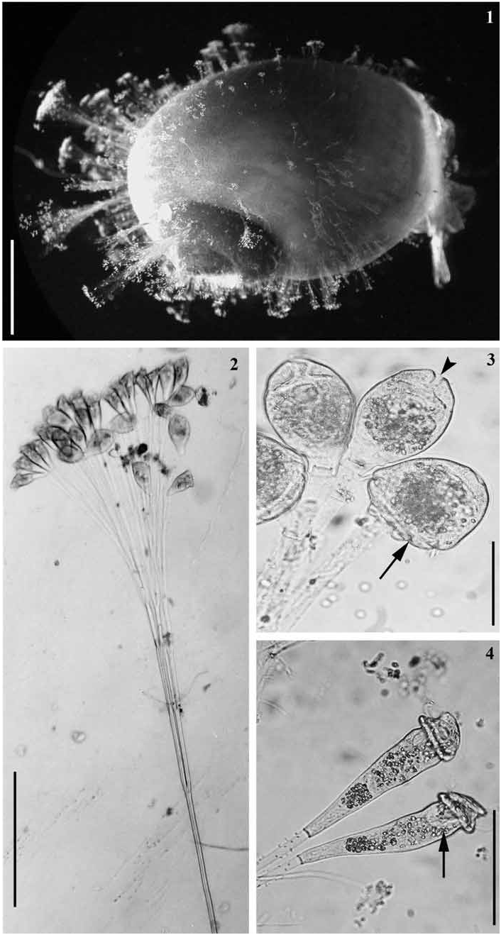
Morphology of live specimens: Colonies were dichotomously branched with alternate branches terminating approximately at the same level ( Fig. 2 View FIGURES 1–4. 1 ). Neither the main stalk nor its branches were contractile. The main basal stalk was short, with a smooth surface, without protuberances or ridges. Primary branches were the longest in the colony, and similar in width to the main stalk with no difference found between collection sites ( Table 1). Colonies had up to 100 zooids, but usually consisted of 10–20 zooids that were similar in size. Zooids were elongate and ranged from 105.0 to 187.5 µm in length and from 25.0 to 52.5 µm in width ( Table 1, Fig. 2 View FIGURES 1–4. 1 ). When contracted, the zooid was transversely folded in the region near to the scopula ( Fig. 3 View FIGURES 1–4. 1 ). The peristomial lip was wider than the body and folded transversally in the fully extended zooid ( Fig. 4 View FIGURES 1–4. 1 ). It also presented a snout-like protuberance when the zooid was contracted ( Fig. 3 View FIGURES 1–4. 1 ). The epistomial disk was highly elevated, and was ~ ¾ as wide as the peristomial lip. A single “C-shaped” macronucleus was observed in the oral half of the cell, lying transversely below the peristomial lip ( Fig. 4 View FIGURES 1–4. 1 ).
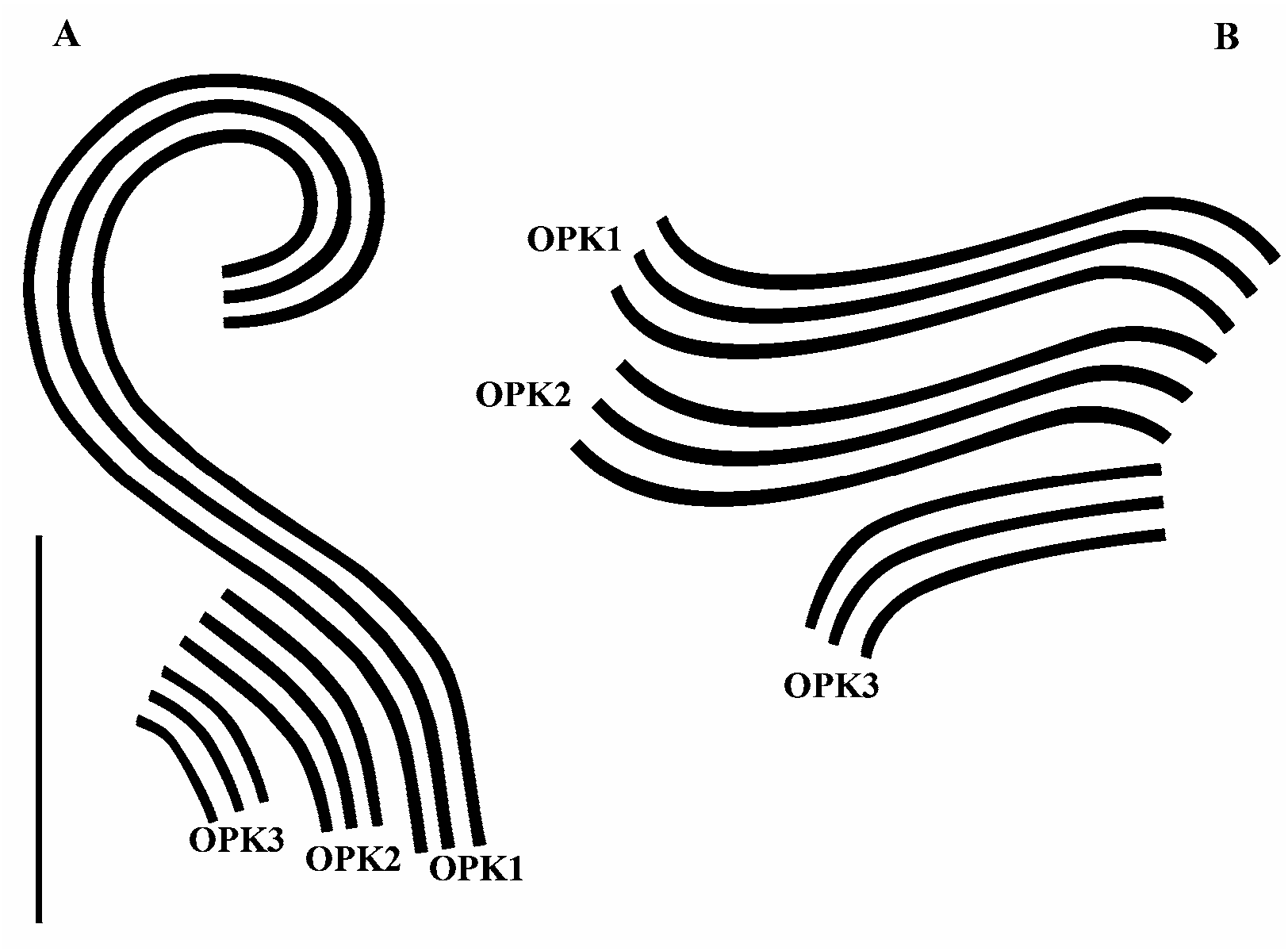
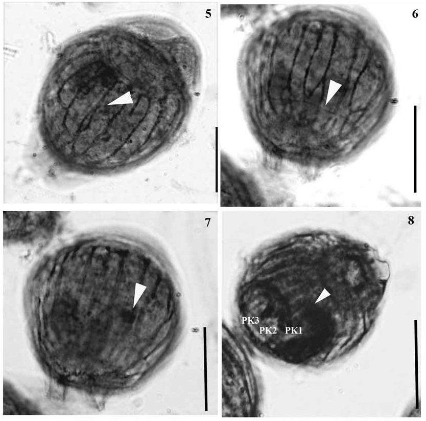
Morphology of stained specimens: Infraciliary and nuclear characteristics of E. plicatilis were easily recognized in protargol stained colonies. A total of 22 somatic myonemes extended from the scopula to the epistomial disk of the zooid ( Fig. 5 View FIGURES 5–8. 5 ). In the trochal band, only one row of kinetosomes was present ( Fig. 6 View FIGURES 5–8. 5 ). A spherical micronucleus was located close to the C-shaped macronucleus ( Figs. 7 and 8 View FIGURES 5–8. 5 ). The oral infraciliature is typical of sessile peritrichs and presents an outer haplokinety and an inner polykinety (PK1) that describes approximately 1 ½ turns around the peristomial disk before entering the infundibulum ( Figs. 8 View FIGURES 5–8. 5 and 9a View FIGURE 9 ). In addition to PK1, the infundibular polykineties PK2 and PK3 were also identified ( Figs. 8 View FIGURES 5–8. 5 and 9b View FIGURE 9 ): PK1 consisted of three rows of kinetosomes equal in length. PK2 also presents three rows of kinetosomes equal in length that terminate at the adstomal curvature of PK1; only a narrow gap existed between PK1 and PK2. PK3 consists of three short rows of kinetosomes that lie in a right angle to PK2 ( Figs. 8 View FIGURES 5–8. 5 and 9b View FIGURE 9 ). PK3 terminates adstomally, close to the adstomal end of PK2.
No known copyright restrictions apply. See Agosti, D., Egloff, W., 2009. Taxonomic information exchange and copyright: the Plazi approach. BMC Research Notes 2009, 2:53 for further explanation.
|
Kingdom |
|
|
Phylum |
|
|
Class |
|
|
Order |
|
|
Family |
|
|
Genus |