
Episcepsis andina, Mantilla & Grados, 2021
|
publication ID |
https://doi.org/ 10.11646/zootaxa.5020.2.7 |
|
publication LSID |
lsid:zoobank.org:pub:AD98C54D-6F78-4461-B4A6-C93F4D995F97 |
|
persistent identifier |
https://treatment.plazi.org/id/1FFB58DF-9F0B-483F-9F47-F9A49172C4EB |
|
taxon LSID |
lsid:zoobank.org:act:1FFB58DF-9F0B-483F-9F47-F9A49172C4EB |
|
treatment provided by |
Plazi |
|
scientific name |
Episcepsis andina |
| status |
sp. nov. |
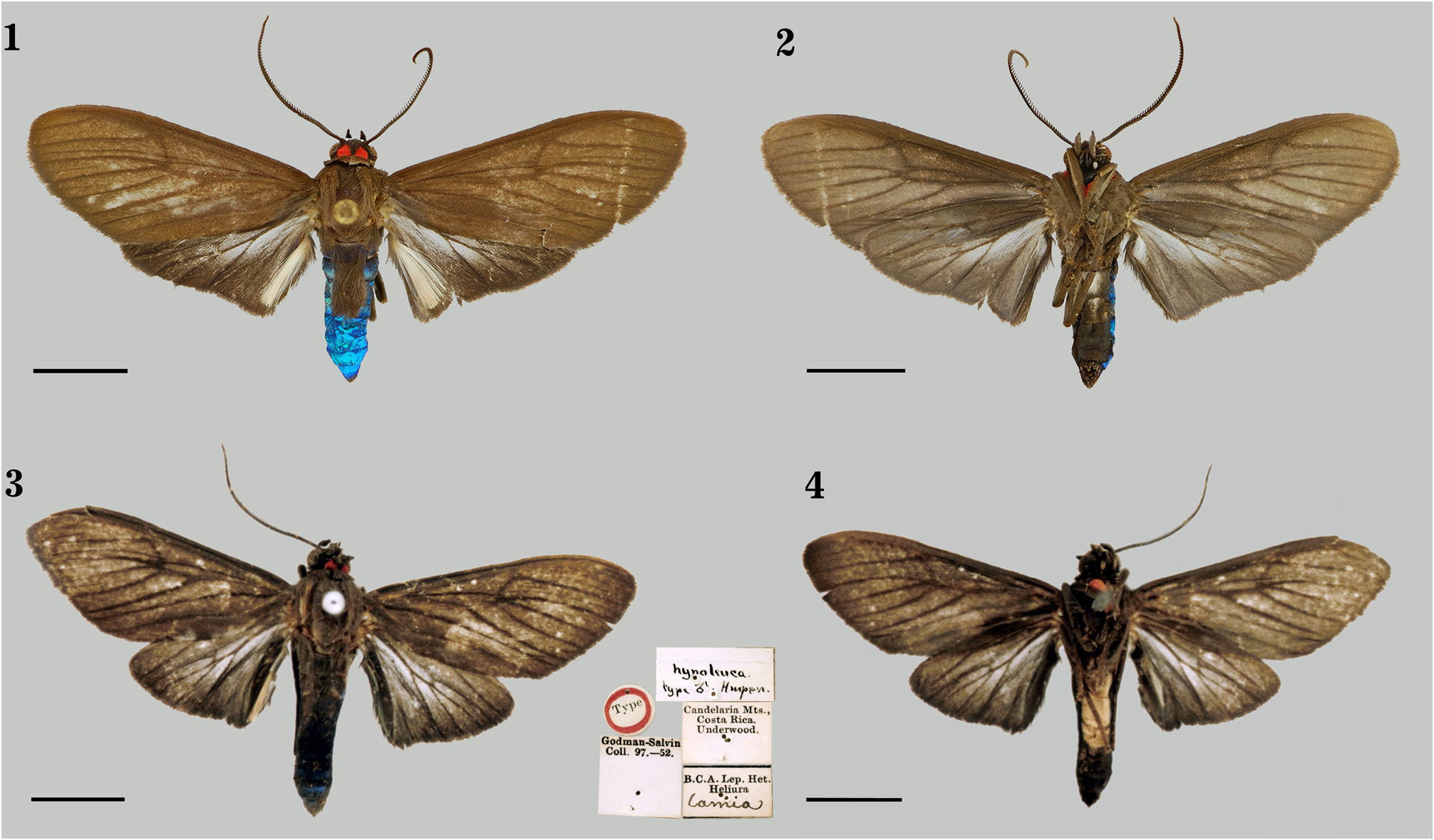
Episcepsis andina sp. nov.
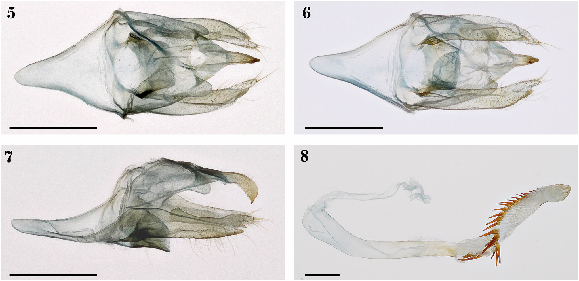
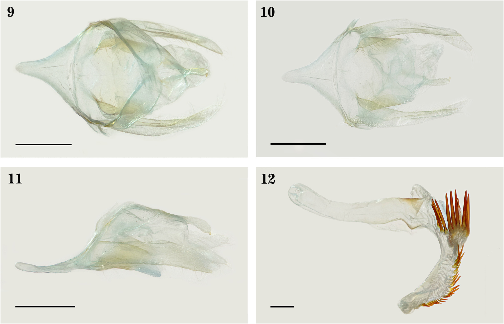
( Figs. 1–2 View FIGURES 1–4 , 5–8 View FIGURES 5–8 )
Holotype male ( Figs. 1–2 View FIGURES 1–4 ): PERU, Cuzco. San Pedro, 13°03’S / 71°33’W, 1400 m, 04.xi.2001, J. Grados ( MUSM). 15 paratypes (all in MUSM). PERU, Amazonas, 1 male, Qda. Condorpuquio, 06°27’S / 77°25’W, 1500 m, 22.viii.98, J. Grados (Genitalia # JGA- 620, MUSM). San Martín. 1 male, Carretera Tarapoto-Yurimaguas, 06°28’S / 76°19’W, 950 m, 16.xi.98, J. Grados. Pasco, 1 male, La Suiza Alta, 10°38’S / 75°27’W, 2189 m, 15.i.2004, J. Grados. Junín. 1 male, 1.2 Km O de San Miguel de Autiki, 10°48’19’’S / 74°49’31’’W, 1418 m, 06.vi.2014, E. Rázuri (Voucher DNA-Artc#00814- JGA MUSM); 1 male, 1-3 Km S Mina Pichita , 11°05’S / 75°25’W, 2100 m, 25.viii.88, G. Lamas (Genitalia # JGA- 623, MUSM). Cuzco. 1 male, 3.5 km ONO de Monte Carmelo, Echarate , 12°26’21.0’’S / 72°59’21.1’’W, 1349 m, 21.ii.2011, M. Alvarado & E. Rázuri (Voucher DNA-Artc#00815- JGA MUSM); 1 male, La Convención, Echarate, CC. Ochigoteni, 12°39’31.36’’S / 73°08’57.71’’W, 1449 m, 17.x.2009, C. Carranza & C. Rossi, Light Trap; 1 male, Campamento Comerciato, 12°47’S / 73°22’W, 1350 m, 18.xi.2002, J. Grados; 2 males, idem except 25.xi.2002; 3 males, Quebrada (Puente) Santa Isabel, 13°02’S / 71°31’W, 1194 m, 13.v.2018, J. Grados; 1 male, idem except (Genitalia # JGA- 1024, MUSM); 1 male, San Pedro, 13°03’S / 71°33’W, 1400 m, 15.viii.2001, J. Grados. GoogleMaps
Diagnosis. Similar to Episcepsis hypoleuca Hampson, 1898 ( Figs. 3–4 View FIGURES 1–4 , 9–12 View FIGURES 9–12 ) (Slide USNM 115,960) and distinguished from it by having a white spot on the ventral side of the hindwing, at the 1A+2A-3A while spot absent in E. hypoleuca ; whitish area of the hindwing slightly wider in E. andina sp. nov. than in E. hypoleuca . The IV abdominal tergite of E. andina sp. nov. presenting two black androconial submedial patches whereas absent in E. hypoleuca . Tegumen of E. andina sp. nov. with posterolateral projections while absent in E. hypoleuca .
Description. Male ( Figs. 1–2 View FIGURES 1–4 ). Head. Brown. Labial palps curved and reaching the vertex. First palpomere white ventrally; second with white scales scattered anteriorly; third, about one third of the length of the second. Frontoclypeus with numerous white scales on the latero-superior surfaces. Occiput with red spots on the lateral surfaces. Antenna bipectinate brown; scapus and pedicel with few white spots anteriorly. Red spot on the lateral surfaces of the cervical region. Thorax. Brown. Tymbal organ present. Legs. Brown. Foreleg coxae red anteriorly. Trochanters with white spots. Anterior and middle legs with femora whitish on posterodistal and external surfaces, the latter less evident on the middle legs; tibiae slightly whitish on external surfaces. Forewing. Length (15–16.5 mm) (n= 16). Brown. Dorsal side, with dark brown veins. Ventral side, similar to the dorsal one, slightly more greyish. Hindwing. Brown. Dorsal side, notorious dark brown veins and a whitish area less prominent, with few brown scales on proximal region. The whitish area comprises the posterior portion of the discal cell, proximal portion of M 2 -M 3, M 3 -Cu 1 and Cu 1 -Cu 2, towards the half of the Cu 2 -1A+2A. 1A+2A-3A modified, with an enlarged invagination, deeper than the proximal portion; white hair-pencils originating from the basal portion of the cell. Ventral side, similar to the dorsal one, except for the presence of whitish scales on its proximal region; white scales on the proximal region of Cu 2 -CuP. 1A+2A-3A with no hair-pencils, with a white spot at the proximal half.
Abdomen. Brown. Tergites with an iridescent blue hue. Four anteriormost tergites with piliform scales. Tergite IV with two black androconial submedial patches covered by piliform scales of the tergite III. Sternites II-IV with white spots on the median region. Male genitalia ( Figs. 5–8 View FIGURES 5–8 ) (Genitalia # JGA, 1024-MUSM). Anterior portion of tegumen V-shaped with a cup-shape posterolateral portion; posterolateral surfaces with short membranous projections, lobulated shape. Uncus and tegumen united by a membrane. Uncus with a bifurcated base; dorsal view with a broad proximal half and a narrow distal half; lateral view, globose and hooklike with a sharp apex. Saccus V-shaped. Juxta semicylindric, as wide as long. Valva nearly straight, proximal third wider; ventral margin with long setae; close to the middorsal region with a process until the internal part, which fuses distally with the dorsal process; dorsal process membranous, proximally wide and tapering distally with numerous setae; ventral process sclerotized and narrow; dorsal process about twice the length of the ventral one. Coecum slightly rounded. Aedeagus elongate, somewhat sinusoid, distal portion slightly curved toward the right side; two distinct areas short and sclerotized distally: a rectangular one ventrally and a longer one oblique on the right side. Vesica everted dorsally, almost as long as the aedeagus, helicoidal and directed toward the left side. Cornuti distributed helicoidally to their length: from the ventroproximal portion to the distodorsal one. Cornuti enlarged, decreasing in length towards the distal part. Ventral portion of the vesica with an enlarged sclerite.
Female: Unknown.
Etymology. andina is a feminine adjective in the nominative singular that refers to the species region of occurrence, the Andes.
Distribution. Montane forests of Amazonas, San Martín, Pasco, Junín and Cuzco departments in Peru.
Remarks. This species is very similar to E. hypoleuca ( Figs. 3–4 View FIGURES 1–4 , 9–12 View FIGURES 9–12 ) and both species can be distinguished by the following characters: ventral side of the hindwing of E. andina sp. nov. with a white spot on the proximal half of 1A+2A-3A and with no androconial organ (white hair-pencils), while in E. hypoleuca 1A+2A-3A is brown and an androconial organ is found. The whitish area of the hindwing in E. andina sp. nov. is more evident than in E. hypoleuca . The IV abdominal tergite of E. andina sp. nov. presents two black androconial submedial patches, while in E. hypoleuca no patch is found. Tegumen of E. andina sp. nov. presents projections on its posterolateral portions which are absent in E. hypoleuca .
Episcepsis hypoleuca was described from two females and one male specimen from Candelaria ( Costa Rica) (syntypes), collected by Godman and Salvin ( Hampson 1898). The male specimen is designated herein as the lectotype, which is deposited in NHMUK ( Figs. 3–4 View FIGURES 1–4 ) .
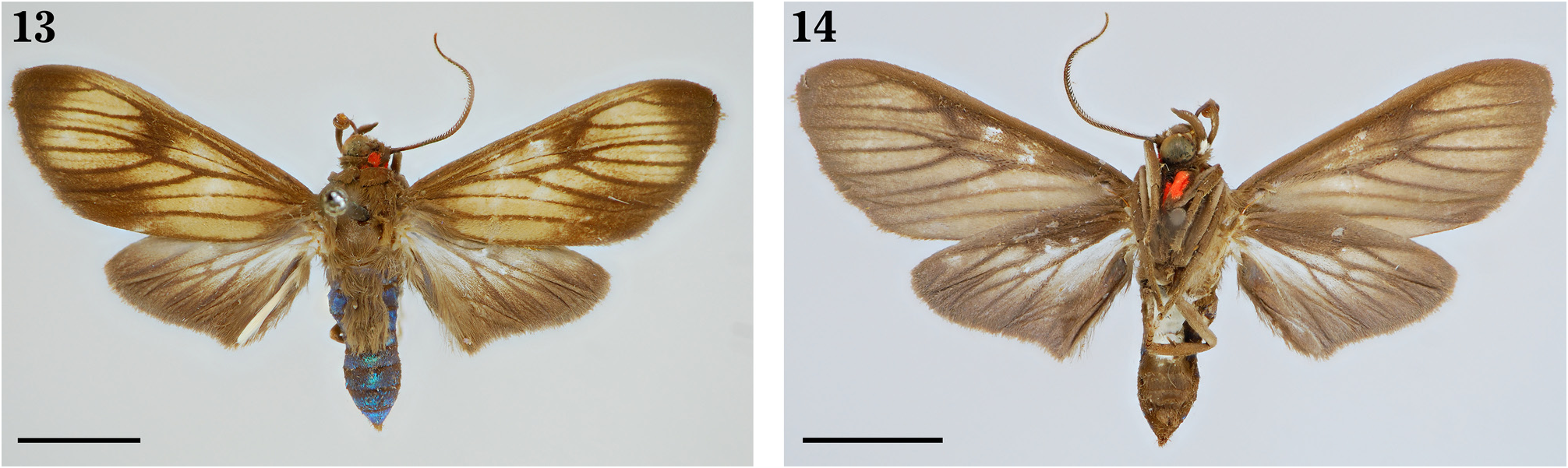
Episcepsis diversa sp. nov.
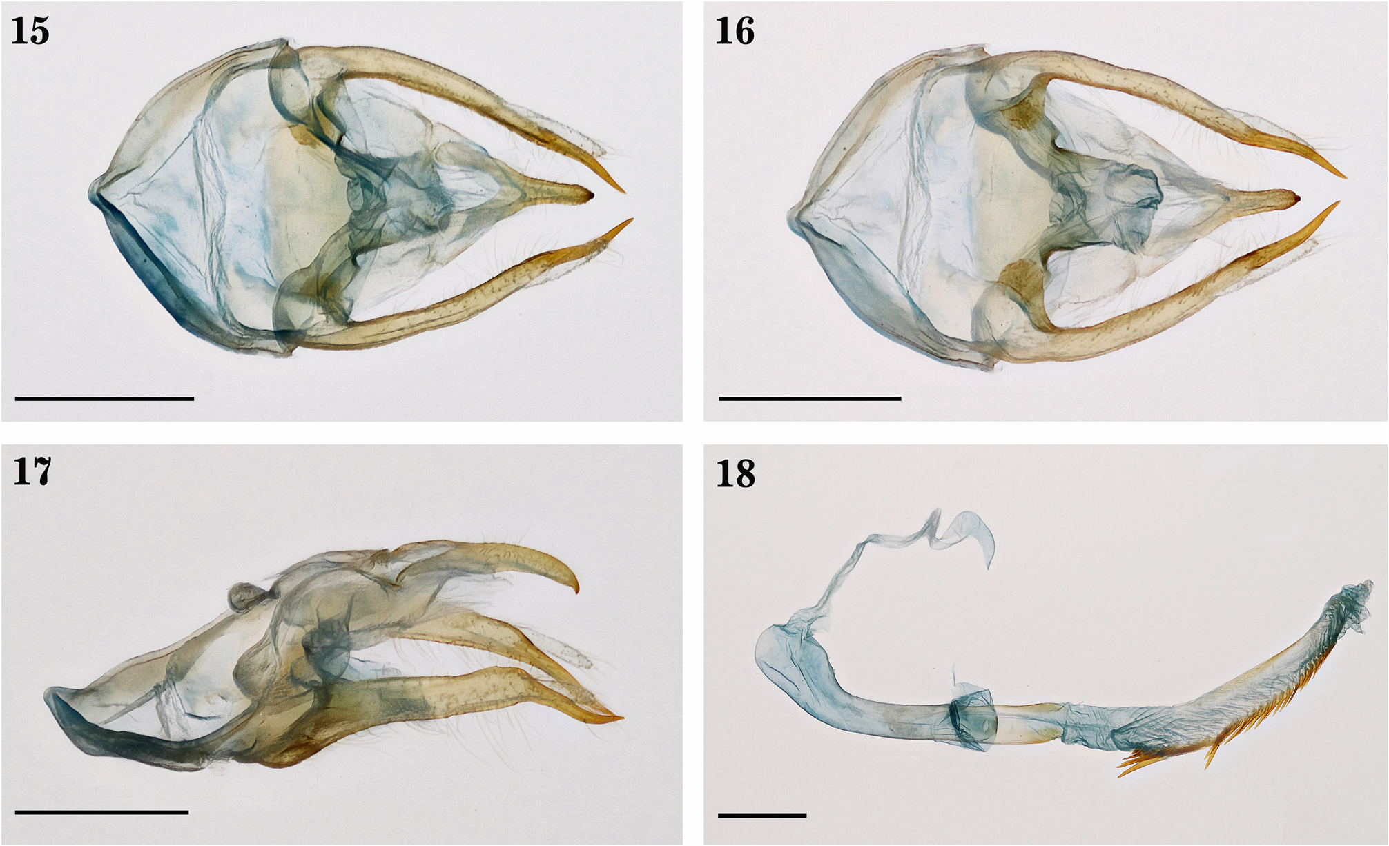
( Figs. 13 – 18 View FIGURES 13–14 View FIGURES 15–18 )
Holotype male ( Figs. 13–18 View FIGURES 13–14 View FIGURES 15–18 ): [ Brazil, Pará, Santarém], Taperinha Amazones Brésil, Dognin Collection ( Slide
USNM 115,967 About USNM ) ( USNM). 1 male paratype . [ Brazil, Pará, Santarém], Unt. Amaz. Taperinha b[ei]. Santarém, 1- 10.VIII.27, Zerny, Auf Heliotropium indicum L. ( NHMW) .
Diagnosis: Similar to Episcepsis venata ( Butler, 1877) ( Figs. 19–30 View FIGURES 19–24 View FIGURES 25–26 View FIGURES 27–30 ) and distinguished from it in the forewing is wide and has a large brown spot on the tornus (dorsal side) whereas in E. venata is the opposite. The IV abdominal tergite of E. diversa sp. nov. presents two black androconial submedial patches while two invaginations with light brown androconial patches in E. venata ( Figs. 25 – 26 View FIGURES 25–26 ). Ventral process of valva of E. diversa sp. nov. about two times greater the length than the dorsal process whereas one-third of the length in E. venata .
Description. Male ( Figs. 13–14 View FIGURES 13–14 ). Head. Brown. Labial palps curved and reaching the vertex. First palpomere white ventrally; second with few white scales scattered anteriorly; third, almost one-third the length of the second. Frontoclypeus white on latero-superior surfaces. Occiput red on lateral surfaces. Antenna bipectinate brown with white scapus anteriorly. A red spot on the lateral surfaces of the cervical region. Thorax. Brown. Tymbal organ present. Legs. Brown. Foreleg coxae red anteriorly. Trochanters with a white spot, less evident on first pair. Femora with white scales on external sides, and on the first pair with whitish scales on the posterodistal surfaces. Tibiae with whitish scales on external sides, less evident on the third pair. Forewing. Length (12.5–13 mm) (n=2). Dorsal side, light brown ground color with dark brown spots; veins, dark brown. Margins of the wing dark brown, except on the subapical portion of the costal margin. Presence of three notorious dark brown spots: the first one on the apex, which extends to almost all R 3 -R 4, R 4 -R 5, distal part of R 5 -M 1 y M 1 -M 2 decreasing towards the M 2 -M 3 y M 3 - Cu 1; second, on the distal portion of the discal cell; third, on the Cu 1 -Cu 2, Cu 2 -1A+2A and anal cell distally. CuP covered by dark brown scales; basal part of anal cell dark brown. Ventral side, greyish brown with dark brown veins. Hindwing. Brown. Dorsal side, with a whitish area covered by few brown scales; veins, dark brown. Whitish area comprising the posterior portion of the discal cell, proximal part of M 2 -M 3, M 3 -Cu 1, Cu 1 -Cu 2 and Cu 2 -CuP, decreasing in area towards the 1A+2A. 1A+2A-3A modified in an enlarged invagination, deeper than the proximal portion; hair-pencils cream originating from the basal portion of the cell. Ventral side, similar to dorsal one, slightly white on the proximal area, except for, Rs-M 1 slightly whitish; white scales on the proximal portion of M 1 -M 2 and median portion of CuP-1A+ 2A. Abdomen. Brown. Tergites with an iridescent blue hue. Four anteriormost tergites with piliform scales. Tergite IV with two black androconial submedial patches covered by piliform scales of the tergite III. Sternites II-V with white scales in almost all its area, scarce on the VI. Male genitalia ( Figs. 15 – 18 View FIGURES 15–18 ) (Slide USNM 115,967). Anterior portion of the tegument V-shaped; posteromedial part cup-shape. Uncus and tegumen united by a membrane. Uncus with a base bifurcated; in lateral view, enlarged with a sharp apex. Saccus poorly developed but wider than long, almost cordiform. Juxta, subrectangular proximally and cylindrical distally. Valva elongate, nearly straight with numerous setae along the ventral part; in ventral view, sacculus short with a sclerotized internal process, flattened dorsoventrally with nearly rounded margin; dorsal process membranous, somewhat straight, with numerous setae and rounded apex; ventral process sclerotized and narrow, tapering towards the apex, curved downward ventrally, and almost two times the length than the dorsal process. Coecum slightly rounded.Aedeagus elongate, somewhat sinusoid and curved distally towards the right side; distally with two distinct sclerotized areas: one broad, dorsally located; another, similar in length to the dorsal one, with an oval apex on the ventral side. Vesica dorsally everted, almost as wide as the aedeagus. Cornuti distributed along the vesica on the ventral side that decrease in size distally. Dorsal portion of the vesica with an enlarged sclerite.
Female: Unknown.
Etymology. diversa is a feminine adjective in the nominative singular, used in the sense of being different.
Distribution. From the eastern part of the Amazon basin, in the humid forests of the Tapajós-Xingu on Taperinha (Santarém, Pará, Brazil); in the Caura valley, Venezuela ( Pinheiro & Araujo 2017) and French Guiana ( Cerda 2017).
Remarks. The genus Episcepsis presents some species groups quite similar to each other. One of these, the E. venata group, is composed of the following species: E. aelia (Schaus, 1889) , E. melanoneura Zerny, 1931 , E. atlantica Pinheiro & Araujo, 2017 , and E. diversa sp. nov. These species are very similar regarding external morphology, and have been easily confused and misidentified ( Schaus 1892; Hampson 1898; Zerny 1912; Draudt 1915; Fleming 1959; Hernández-Baz & Grados 2004). Some characters help in their distinction, but in many cases, internal morphology is needed.
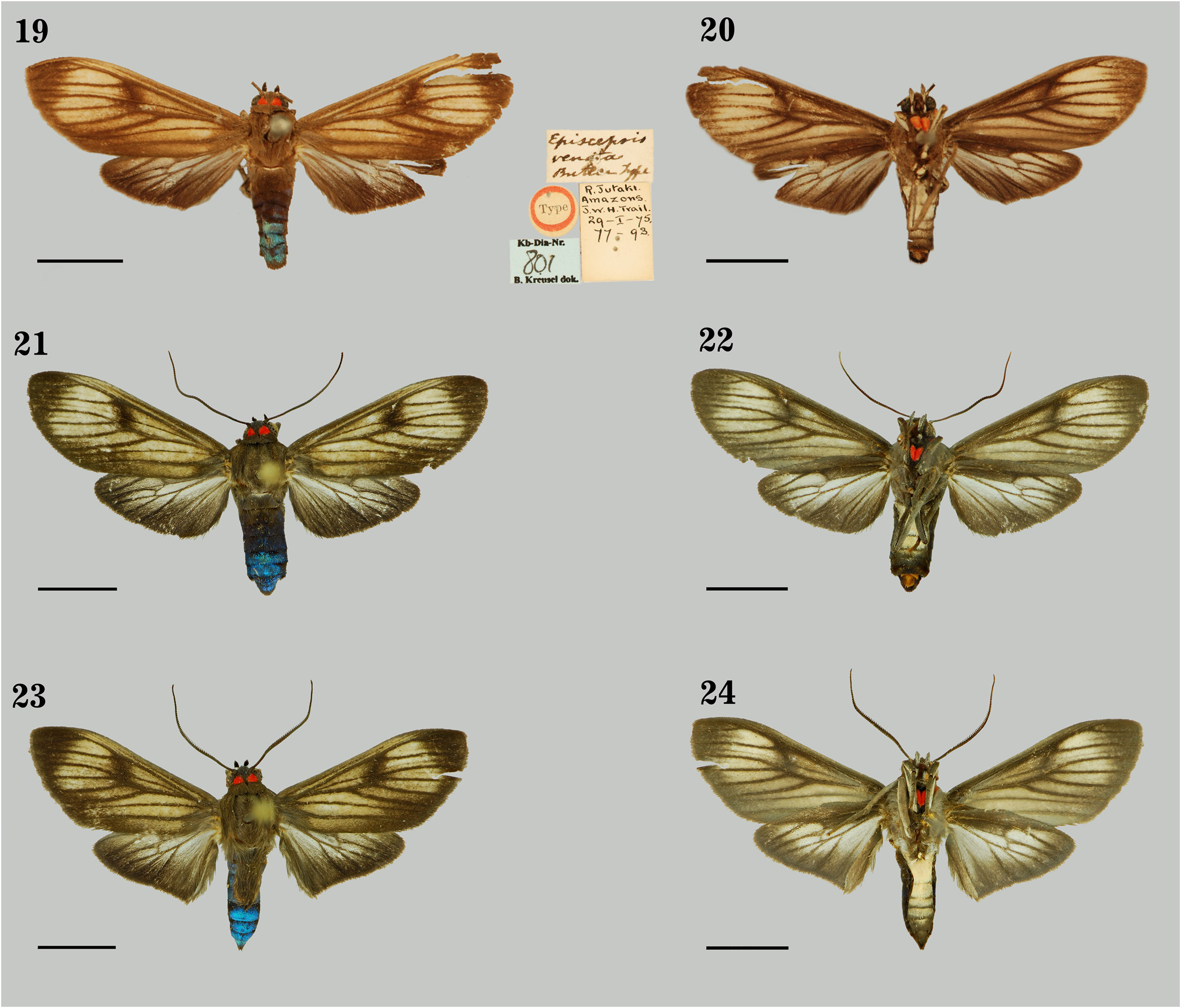
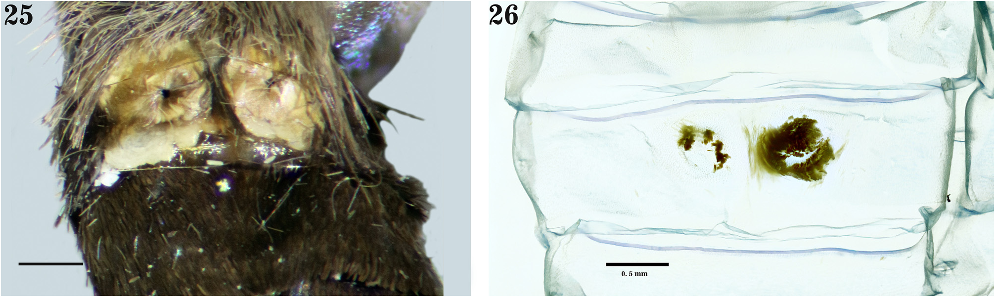
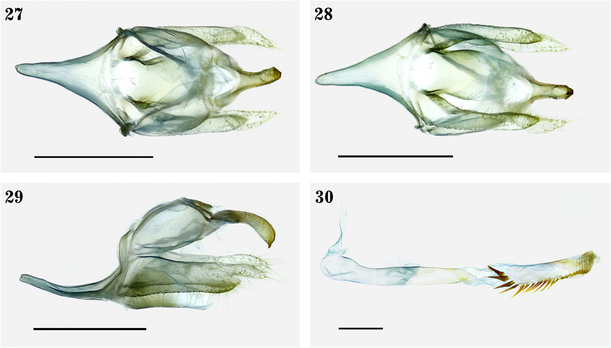
In recent works by Pinheiro & Araujo (2017) and Cerda (2017), the genitalic morphology of “ E. venata ” was described based on specimens from Venezuela and French Guiana, respectively. We reidentify those specimens as E. diversa sp. nov. for the following reasons: the type specimen of E. venata , is a female collected at the Jutahi River [= Jutaí] (Amazonas, Brazil) by J.W.H. Trail ( Fig. 19–20 View FIGURES 19–24 ), a Scottish botanist who explored the tributaries of the Amazon River (northwestern Brazil) for two years with the Amazon Steam Navigation Company, as well as Tefé, Óbidos, the Purús, Juruá, and Trombetas rivers among others ( Berkeley & Cook 1877; Anonymous 1920). In the present study, male and female specimens from places near the type locality of the species (Bretaña, Loreto) ( Figs. 21–24 View FIGURES 19–24 ) and other places in the Amazon of Peru were dissected ( Figs. 27–30 View FIGURES 27–30 ) (Genitalia # JGA-633, MUSM).
The differences in external morphology between both populations are minimal; the forewing in E. venata (near the type locality) is somewhat longer and in the dorsal side the brown spot on the tornus is smaller, while in the Venezuelan and French Guiana specimens, the forewing is somewhat wider and the brown spot in the dorsal side is larger. Moreover, the male of E. venata presents two androconial submedial patches circular in shape as an invagination on the IV abdominal tergite invagination ( Figs. 25–26 View FIGURES 25–26 ), while they are absent in the Venezuelan specimens ( Pinheiro & Araujo 2017). Regarding the male genitalia, specimens collected near the type locality of E. venata have an elongated saccus, a ventral valva process somewhat short and sclerotized, while the Venezuelan and French Guiana specimens present a short saccus and a ventral process of the valve sclerotized with a sharp distal end ( Pinheiro & Araujo 2017; Cerda 2017). Based on the aforementioned characters and the geographical distribution, we identified all specimens from Peru (adults and genital capsules) as E. venata ( Figs. 21–30 View FIGURES 19–24 View FIGURES 25–26 View FIGURES 27–30 ).
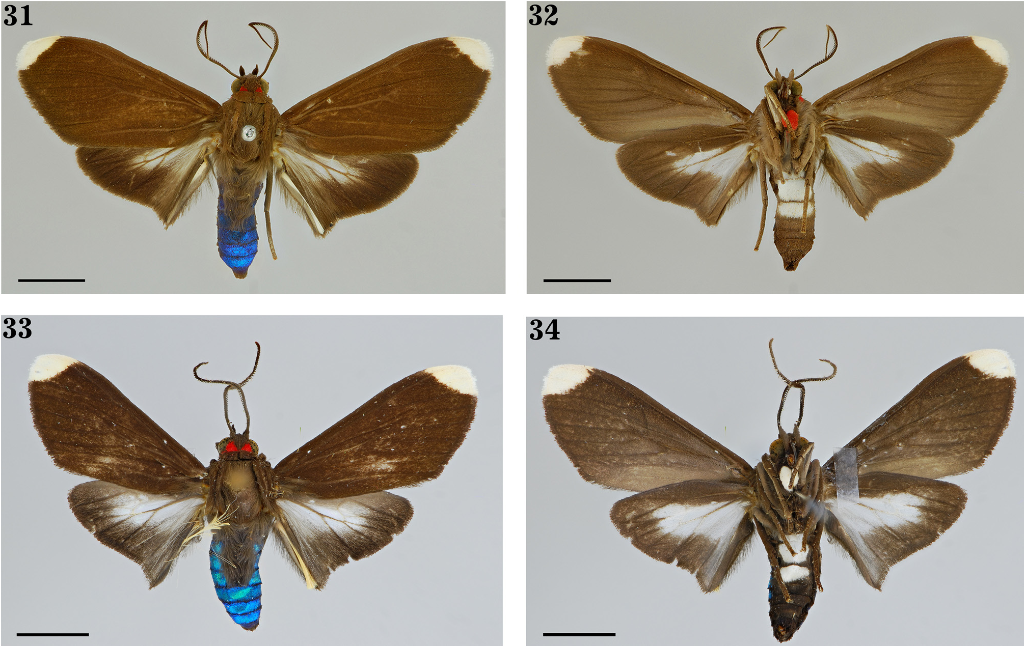
Episcepsis insularis sp. nov.
( Figs. 31–32 View FIGURES 31–34. 31–32 , 35–38 View FIGURES 35–38 )
Holotype male ( Figs. 31–32 View FIGURES 31–34. 31–32 ). Cuba, Santiago , Collection Wm Schaus ( USNM). 1 male paratype. Cuba, Santiago, Collection Wm Schaus (Slide USNM 115 About USNM , 961 About USNM ) ( USNM).
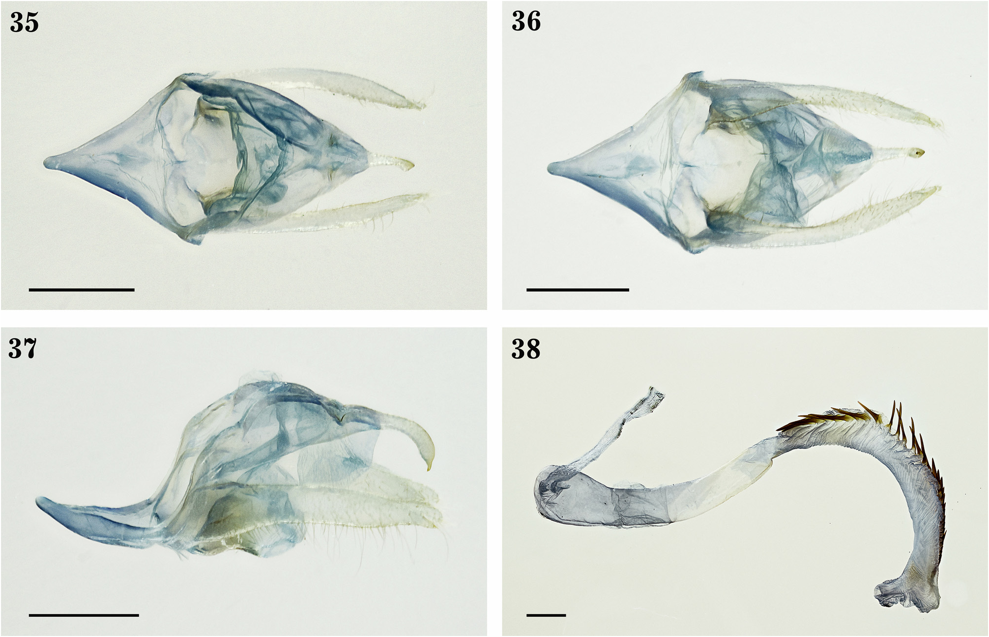
Diagnosis: Similar to Episcepsis thetis ( Linnaeus, 1771) ( Figs. 33–34 View FIGURES 31–34. 31–32 , 39–42 View FIGURES 39–42 ) (Slide USNM 115,972) and distinguished from it by having a more elongated and slenderer white spot on the apex of the forewing. Foreleg coxae red anteriorly in E. insularis sp. nov. while white in E. thetis . Valva with dorsal and ventral processes similar in length in E. insularis sp. nov. whereas ventral process longer than the dorsal one in E. thetis .
Description. Male ( Figs. 31–32 View FIGURES 31–34. 31–32 ). Head. Brown. Labial palps curved and reaching the vertex. First palpomere ventrally white. Second and third with anterior portion lighter than the posterior. Third, almost one third of the length of the second. Frontoclypeus with few white scales on the latero-superior surfaces. Occiput with two red spots on lateral surfaces. Antenna bipectinate brown; scapus with few white scales on the anteroproximal portion. Red spot on each side of the lateral surface of cervical region. Thorax. Brown. Tymbal organ present. Legs. Brown. Foreleg coxae red anteriorly. Trochanters with one white spot. Femora, whitish posterodistally, more evident in the second pair, and white scales on external sides, including part of the posterior side. Tibiae, with white scales on external sides, less notorious on the third pair. Tarsus with one white spot, less notorious on the third pair. Forewing. Length (16 mm) (n=2). Brown. Dorsal side, veins slightly lighter. Apex with one white spot from the costal margin including R 3 -R 4, R 4 -R 5 to M 1 -M 2. Ventral side, lighter than the dorsal one; light greyish brown from 1A+2A to the anal cell; white spot on the apex similar as in the dorsal side. Hindwing. Brown. Dorsal side, whitish area, with few brown scales; veins with dark brown scales. The whitish area including the posterior portion of the discal cell, proximal portion of M 2 -M 3, M 3 -Cu 1, Cu 1 -Cu 2 and half of Cu 1 -CuP decreasing towards the CuP. 1A+2A-3A modified in an enlarged invagination, deeper on the proximal portion; white hair-pencils originating from the basal portion of the cell. Ventral side, similar to the dorsal one, with white scales on the proximal area, except for the presence of a small white spot on the posterobasal portion of M 1 -M 2 and few white scales distally on CuP-1A+2A. Abdomen. Brown. Tergites with an iridescent blue hue; four anteriormost with piliform scales. Sternites of II to IV with a white spot in almost all its area. Male genitalia ( Figs. 35–38 View FIGURES 35–38 ) (Slide USNM 115, 961). Anterior portion of the tegumen V-shaped; posteromedial portion cup-shape. Uncus and tegumen united by a membrane. Uncus with a bifurcated base; lateral view, distal portion slightly globose and hook-like with a sharp apex. Saccus well-developed and Vshaped. Juxta cylindric, wider than long, with a concave anterior margin. Valva straight; numerous setae on the ventral portion and few on the distodorsal one; dorsal process membranous with setae and oval apex; ventral process sclerotized with sharpened apex; dorsal and ventral processes similar in length; in ventral view, sacculus slightly wide. Coecum slightly rounded. Aedeagus enlarged, somewhat sinusoid; distal portion slightly curved towards the right side; two distinct sclerotized areas distally: from right to left with a somewhat oval apex; length of the left sclerotized area 1/3 longer than the right one. Vesica everted dorsally, almost as long as the aedeagus, curved and directed towards the left side; Cornuti distributed along the vesica on the right side; decreasing in size distally.
Female: Unknown.
Etymology. insularis is a feminine adjective in the nominative singular that refers to the species locality ( Cuba).
Distribution. Presumably endemic to Cuba.
Remarks. The species E. thetis was described by Linnaeus (1771) based on the description and illustration of Drury (1770: Vol I, p. 57, pl.XXVI, fig. 4), with Jamaica as type locality and no mention of sex. The information provided by Drury (1770) corresponds to a male specimen that is presumably deposited at the Macleay Museum, Sydney University ( Australia). This specimen is the holotype by monotypy.
Episcepsis insularis sp. nov. and E. thetis ( Figs. 33–34 View FIGURES 31–34. 31–32 , 39–42 View FIGURES 39–42 ) are recorded from the Antilles and can be distinguished by external morphology and characters from the male genitalia. The whitish area of the hindwings (dorsal side) in E. insularis sp. nov. is more reduced than in E. thetis ; a small white spot present on the ventral side of M 1 -M 2 in E. insularis sp. nov., while proximally situated in E. thetis . Foreleg coxae red anteriorly in E. insularis sp. nov., while white in E. thetis . In relation to genitalia characters, the posterior portion of the valva of E. insularis sp. nov. is wider than in E. thetis , the dorsal and ventral processes of E. insularis sp. nov. are similar in length, while in E. thetis the ventral process is two times longer than the dorsal one; valva of E. insularis sp. nov. surpasss lightly the uncus apex while corresponding to one third of the uncus length in E. thetis ; cornuti more numerous in E. insularis sp. nov. than in E. thetis .
On the other hand, other similar species described from the Caribbean are: E. pseudothetis Fleming, 1959 from Trinidad and E. dominicensis Rothschild, 1911 from the Dominican Republic. Differences in the male genitalia were observed as well as in the external morphology of those species such as the presence of darker veins on the forewing of E. insularis sp. nov. (it is lighter in E. pseudothetis ), a white spot on the ventral side of hindwing posterobasal in the M 1 -M 2 of E. insularis sp. nov. (there is a spot extending proximally in E. pseudothetis ); foreleg coxae red anteriorly in E. insularis sp. nov. (white in E. pseudothetis ). Episcepsis dominicens is distinguishes from E. insularis sp. nov. by the presence of yellow scales on the lateral portions of the occiput and foreleg coxae anteriorly (red scales in E. insularis sp. nov.) and the whitish area of the hindwing (ventral side) is more reduced and less notorious in E. dominicensis .
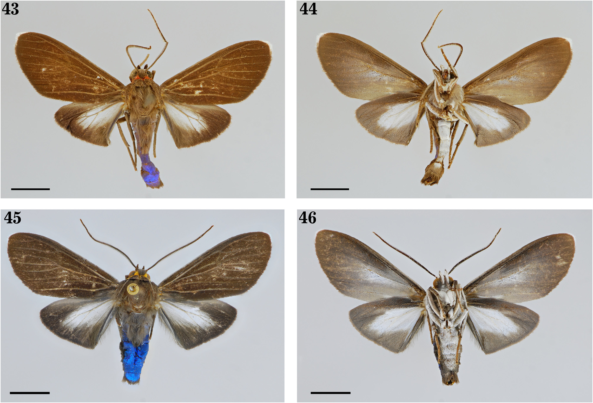
Episcepsis travassosi sp. nov.
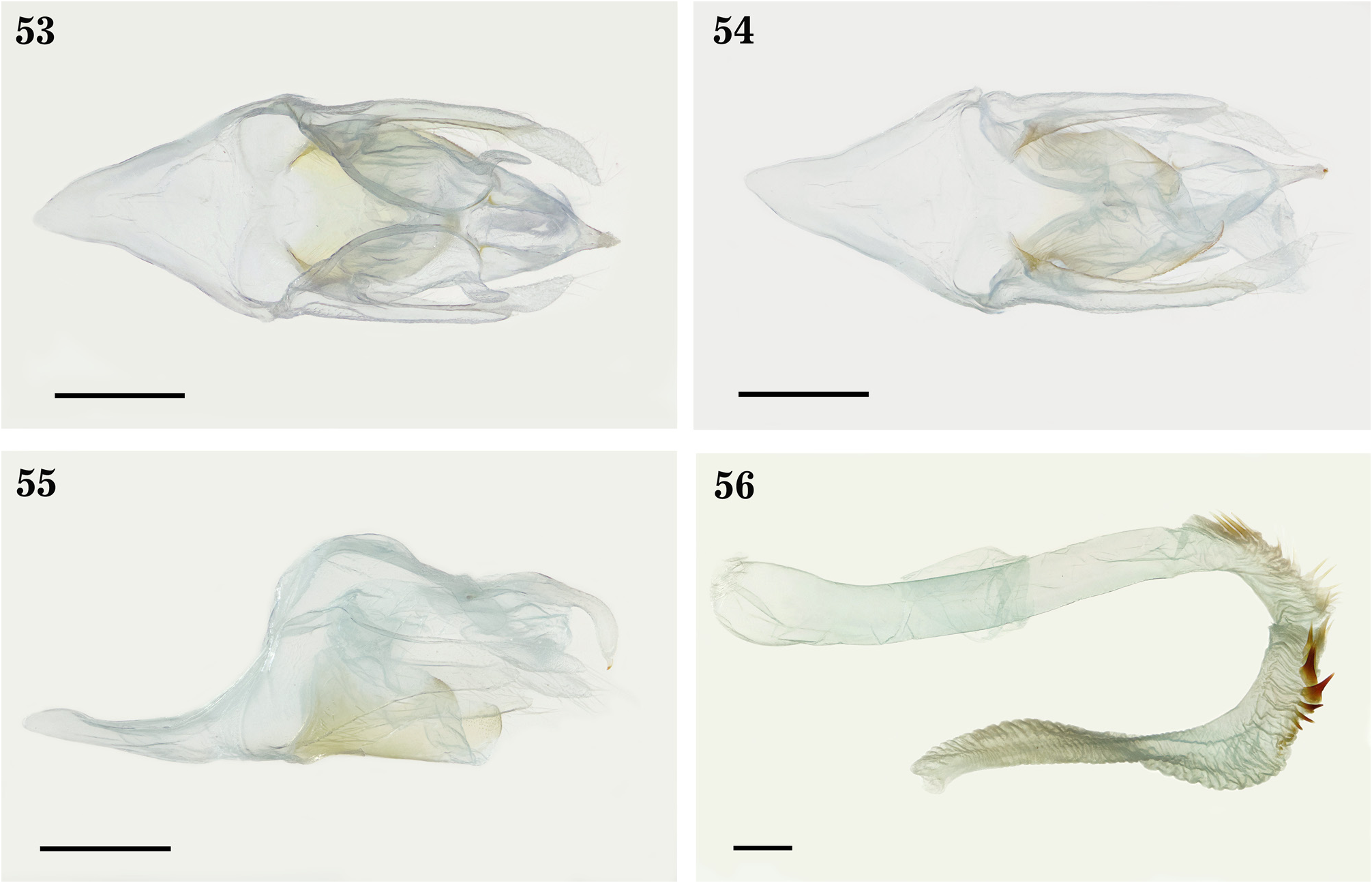
( Figs. 43–46 View FIGURES 43–46 , 49–52 View FIGURES 49–52 )
Holotype male ( Figs. 43–44 View FIGURES 43–46 ). Brazil, [Rio de Janeiro], [Angra] dos Reis, Jussaral, 29-9-935 [1935], TRAV. & OITICICA F. ( CEIOC). 11 paratypes. Brazil, 1 male, [Rio de Janeiro], Corcovado-Rio, X. 932, L. T.-COL. ( CEIOC); 1 male, [Rio de Janeiro], Angra [dos Reis], Jussaral , 8-934 [1934], TRAV. & ALMEIDA. ( CEIOC); 2 males, idem except, 9-934, TRAV. & OITICICA F. ( CEIOC); 1 male, idem except, 5-4-935 [1935]( CEIOC); 1 male, idem except, 20-9-935[1935] ( CEIOC); 1 male, idem except, 22-9-935[1935]( CEIOC); 1 male, idem except, 29-9-935[1935] ( CEIOC); 1 male, idem except, 27.x.936 [1936], TRAV. ET S. LOPES ( CEIOC); 1 male, [Brasil, Espírito Santo, Linhares] Brasil: ES Linhares, 40m, 20-29.ii.1992, V. O. Becker Col. / Col. BECKER 81282 ( USNM); 1 male, [ Brazil, Bahía, Camacan] Brazil: BA Camacan, 400-700m, 21-30.ix.1991, V. O. Becker Col. / Col. BECKER 83406 / Slide USNM 115,986 About USNM ( USNM).
Diagnosis. Similar to Episcepsis nigropunctata Cerda, 2017 . It is different because the two black androconial submedial patches on the IV abdominal tergite are smaller. Proximoventral portion of the valva with a triangular internal projection in E. travassosi sp. nov. whereas absent in E. nigropunctata . Ventral process of the valva of E. travassosi sp. nov. presents a flattened apex dorsoventrally while sharpened in E. nigropunctata .
Description. Male ( Figs. 43–44 View FIGURES 43–46 ). Head. Brown. Labial palps curved and reaching the vertex. First palpomere white ventrally; second, white anteriorly with a white horizontal line posteriorly; third, almost one third of the length of the second. Frontoclypeus with white latero-superior surfaces. Occiput with two red spots on lateral surfaces. Postgena predominantly reddish. Antenna bipectinate brown; scapus with white anterointernal portions; pedicel anteriorly white. A red spot on the lateral surfaces of the cervical region. Thorax. Brown. Patagia with few red scales on external portions and anterior margins. Tegulae, with a greyish line along the median portion. Pleura with white scales. Tymbal organ present. Legs. Brown. Foreleg coxae white anteriorly. Trochanters with a white spot. Femora with white scales on lateral portions, including part of the external and internal sides. Tibiae with white scales on external sides. Tarsus, with a white line, less evident on the second and third pair of legs. Forewing. Length (14–16 mm) (n = 12). Brown, with white apex margin. Dorsal side, with light brown veins. Ventral side, slightly greyish; with whitish scales on the proximal part of Cu 1 -Cu 2, on the anterior portion of Cu 1 -CuP and the anteroproximal portion of the anal cell. Hindwing. Brown. Dorsal side, whitish area very prominent, with few brown scales; dark brown veins. Whitish area comprising the posterior portion of the discal cell, proximal portion of M 2 -M 3 y M 3 -Cu 1, up to almost half of Cu 1 -Cu 2, more than half of Cu 2 -CuP decreasing in area towards the 1A+2A. 1A+2A-3A modified, with an enlarged invagination, deeper on the proximal portion; white hair-pencils originating from the basal part of the cell. Ventral side, similar to the dorsal one, except for the presence of white scales in part of the anterior side of the discal cell and the proximal portion of CuP-1A+2A; along the 1A+2A a line of white scales towards the distal portion. Abdomen. Brown. Tergites with an iridescent blue hue. Four anteriormost tergites with piliform scales. Tergite IV with two black androconial submedial patches covered by piliform scales of the tergite III. Sternites of the II to VII with white scales almost all their area. Male genitalia ( Figs. 49–52 View FIGURES 49–52 ) (CEIOC). Anterior portion of the tegumen V-shaped; median portion with a rectangular sclerite; posterolateral portions with digitiform projections. Uncus and tegumen united by a membrane. Uncus, with a bifurcated base and setae on the distodorsal portion; lateral side, uncus enlarged with distal portion slightly globose and a sharp apex. Saccus welldeveloped and V-shaped. Juxta subtriangular, somewhat rectangular posteriorly and anterolateral portion projected towards the dorsal portion; spicules on the lateromedial portion. Valva lightly curved to the centre; few setae on the ventral portion; lateral view with a broad proximal portion almost two times the width of its distal portion; dorsal process membranous with numerous setae; ventral process sclerotized, distal portion flattened dorsoventrally, rounded apex in dorsal view; dorsal process almost twice the length than the ventral one; proximoventral portion of the valva with a projection towards the internal portion of the base, almost triangular with numerous setae on its margin. Coecum slightly rounded. Aedeagus elongate, somewhat sinusoid, distally curved towards the right side; two sclerotized areas enlarged, wide and somewhat triangular distally; basal portion of the right side with a small rounded protuberance. Vesica everted dorsally, almost as wide as the aedeagus, slightly curved to the left. Cornuti on the right side along the vesica, decreasing in size distally; left side of the vesica with two enlarged sclerites.
Female: Unknown.
Etymology. travassosi is a latinized noun in the genitive singular dedicated to Lauro Pereira Travassos Filho (1918–1989), who contributed immensely to the study of Brazilian Arctiinae.
Distribution: The tropical forests of the Mata Atlântica in Bahia (Camacan), Espírito Santo (Linhares) and Rio de Janeiro (Angra dos Reis) states.
Remarks: The paratypes from Espírito Santo (Linhares) and Bahia (Camacan) differ in coloration ( Figs. 45– 46 View FIGURES 43–46 ) in comparison with the holotype ( Figs. 43–44 View FIGURES 43–46 ). Those specimens present yellow scales on the lateral portions of the occiput and cervical regions, and the lateral surface of the postgena; few yellow scales on the lateral portions and anterior margins of the patagia. Based on the morphological characters of the genitalia, both populations are conspecific.
The species E. nigropunctata was described from French Guiana, based on male specimens and the holotype is deposited at the Natural History Museum of Lyon (MHNL, France) ( Cerda 2017). The examination of external and internal morphology of specimens of Peru has shown that E. nigropunctata also occurs in Peru (MUSM), mainly in the Amazonian region of Ucayali, Huánuco, Cuzco and Madre de Dios, reaching up to 587 m elevation. E. travassosi sp. nov. distinguished from this species by having two small dark androconial patches on the submedial portions of the IV abdominal tergite whereas larger and evident in E. nigropunctata . The proximoventral portion of the valve of E. travassosi sp. nov. presents a triangular projection towards the internal portion, while absent in E. nigropunctata ; the ventral process of the valve of E. travassosi sp. nov. presents a flattened apex dorsoventrally vs. sharpened in E. nigropunctata ; the juxta of E. travassossi sp. nov. presents spicules on the lateromedial portions which are lacking in E. nigropunctata . The genitalia of E. nigropunctata was published by Cerda (2017).
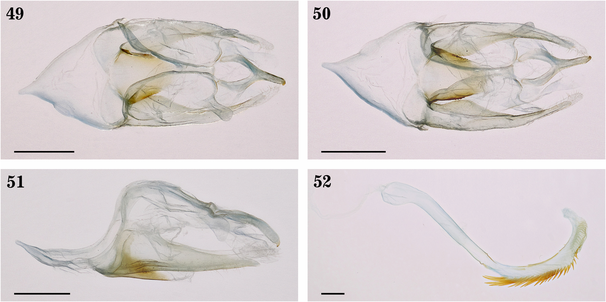
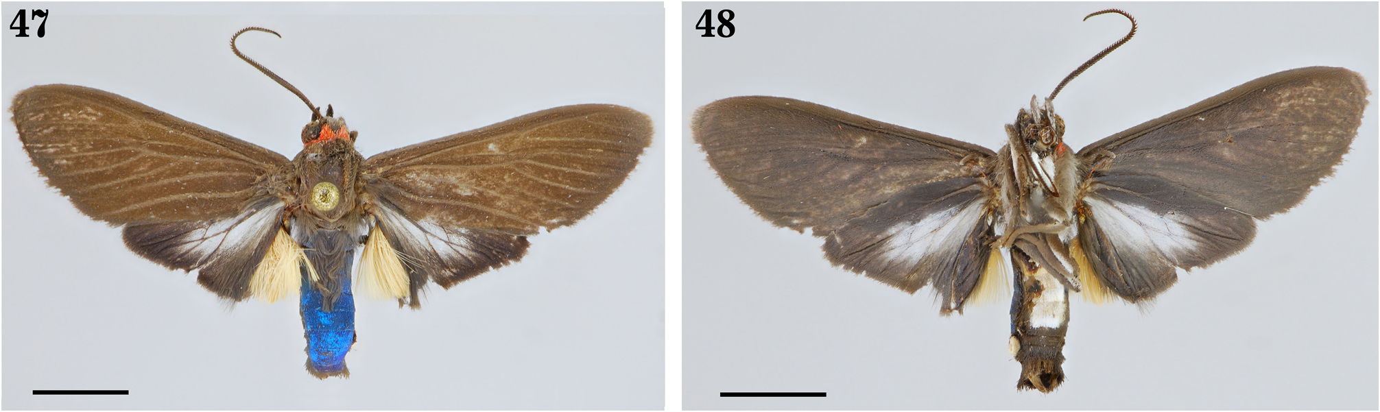
Another species similar to E. travassosi sp. nov. is E. gnoma ( Butler, 1877) , described from a male specimen from Padauiry River, Amazonas ( Brazil) collected by J.W.H Trail and deposited at the NHMUK. Also E. gnoma occurred in the northern Amazon of Peru (Loreto) ( Figs. 47–48 View FIGURES 47–48 , 53–56 View FIGURES 53–56 ) (Genitalia # JGA, 571-MUSM). E. travassosi sp. nov. distinguished from it by the presence of whitish area of the hindwing whereas wider than in E. gnoma ; E. travassosi sp. nov. presents two small dark androconial patches on the submedial portions of the IV abdominal tergite while absent in E. gnoma . Related to the male genitalia, the valva of E. travassosi sp. nov. is narrower than in E. gnoma ; ventral process of E. travassosi sp. nov. is flattened and enlarged dorsoventrally vs. short and almost one-fourth of the length than the dorsal one in E. gnoma ; the juxta of E. travassossi sp. nov. presents spicules on the lateromedial portions which are lacking in E. gnoma .. Both E. nigropunctata and E. gnoma present an allopatric distribution in relation to E. travassosi sp. nov.
No known copyright restrictions apply. See Agosti, D., Egloff, W., 2009. Taxonomic information exchange and copyright: the Plazi approach. BMC Research Notes 2009, 2:53 for further explanation.
|
Kingdom |
|
|
Phylum |
|
|
Class |
|
|
Order |
|
|
Family |
|
|
Genus |