
Nemertopsis mitellicola, Kajihara, Hiroshi, 2007
|
publication ID |
https://doi.org/10.5281/zenodo.176154 |
|
DOI |
https://doi.org/10.5281/zenodo.6246625 |
|
persistent identifier |
https://treatment.plazi.org/id/03E32426-FFDE-8E27-FF45-5644AE0EE005 |
|
treatment provided by |
Plazi |
|
scientific name |
Nemertopsis mitellicola |
| status |
sp. nov. |
Nemertopsis mitellicola sp. nov. ( Figs 7–11 View FIGURE 7 View FIGURE 8 View FIGURE 9 )
Etymology: The specific name indicates the host organism of the new species, the gooseneck barnacle, Capitulum mitella ( Linnaeus, 1767) (Cirripedia: Thoracica: Lepadomorpha).
Material examined: Three specimens, collected among barnacles along with the above described specimens of Nemertopsis quadripunctata , 8 July 1999. Holotype, ZIHU-3204, serial transverse sections of the complete body, total 52 slides: 6 µm, anterior end of body ( 1 cm long), 12 slides; 8 µm, rest of the body, 40 slides. Paratypes: ZIHU-3205, serial transverse sections of head ( 1.5 cm long), 8 µm, 15 slides; ZIHU-3206, serial longitudinal sections, 12 µm, 12 slides.
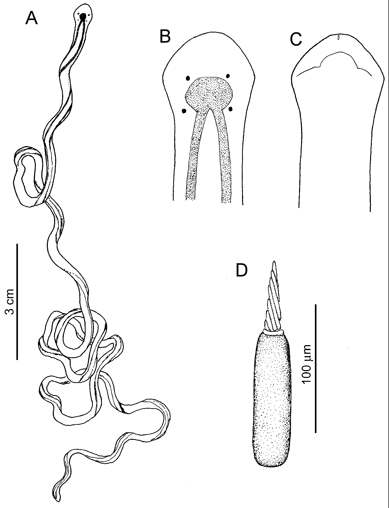
External features: Body slender ( Fig. 7 View FIGURE 7 A), up to 15 cm long, 0.4 mm wide. Head round diamond shape, wider than succeeding body. Four eyes arranged in corners of trapezium ( Fig. 7 View FIGURE 7 B). Pair of cephalic furrows on ventral surface of head below anterior eyes, running medially from edge of head, then suddenly curving forward to meet each other near tip of head ( Fig. 7 View FIGURE 7 C). Body colour cream-white. Dark brown cephalic pigment patch, from which two dorsal stripes running posteriorly; these stripes usually faint in posterior portion of body.
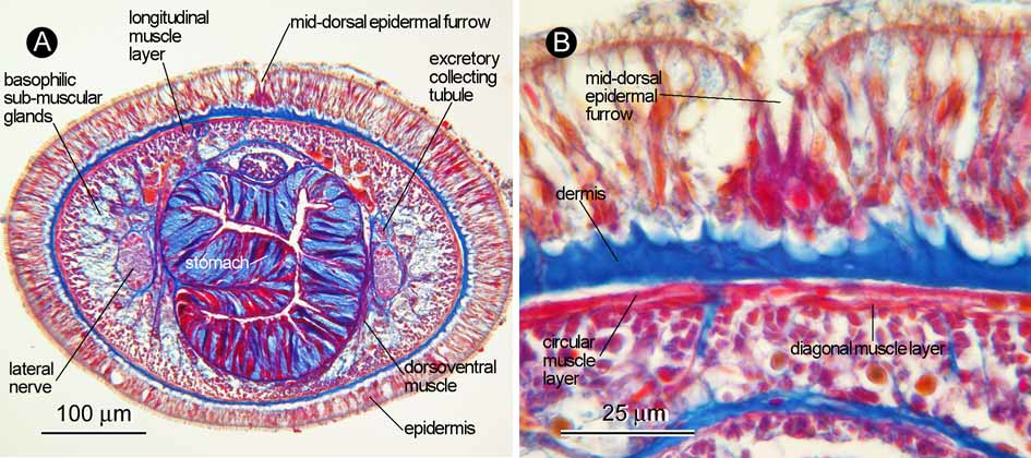
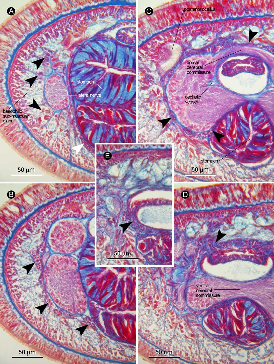
Body wall, musculature, and parenchyma: Ciliated epidermis ( Fig. 8 View FIGURE 8 A), mostly 40–50 µm thick in brain region. Dark staining glandular cells in proximal zone absent. Mid-dorsal epidermal furrow extending from tip of head, becoming shallow and inconspicuous in intestinal region; furrow usually containing acidophilic cells in its basal portion ( Fig. 8 View FIGURE 8 B). Dermis well developed, mostly 3–10 µm thick, its distal surface forming cup-like structures ( Fig. 8 View FIGURE 8 B). Body-wall musculature composed of outer circular and inner longitudinal muscle layers; diagonal muscle layer of lattice type present between outer circular and inner longitudinal muscle layers ( Fig. 8 View FIGURE 8 B). In foregut region outer circular layer up to 2–3 µm thick, inner longitudinal layer 10–20 µm thick. Dorsoventral muscles present in foregut and intestinal regions. In anterior foregut region, lateral portions of body-wall longitudinal muscle layer medio-anteriorly sending fibres traversing through basophilic sub-muscular glands to run adjacent to dorsal, lateral, and ventral sides of lateral nerve cord ( Fig. 9 View FIGURE 9 A); in same region, or in its slightly anterior region, dorsal and ventral portions of body-wall longitudinal muscle layer becoming divided by inner and outer portions by posterior extension of basophilic cephalic glands, medially sending fibre bundles ( Fig. 9 View FIGURE 9 A); these medial fibre bundles uniting to form incomplete inner longitudinal muscle layer covering rhynchocoel-brain-foregut complex ( Fig. 9 View FIGURE 9 B); in brain region, ventral portion of inner longitudinal layer penetrated by alimentary canal to situate between ventral commissure and alimentary canal ( Fig. 9 View FIGURE 9 C, D); most fibres of inner longitudinal muscle layer contributing to proboscis insertion ( Fig. 9 View FIGURE 9 D, E); small portion of inner longitudinal muscle layer appearing to lead farther anteriorly beyond proboscis insertion running around rhynchodaeum and oesophagus. Parenchymatous connective tissues sparingly developed throughout body.
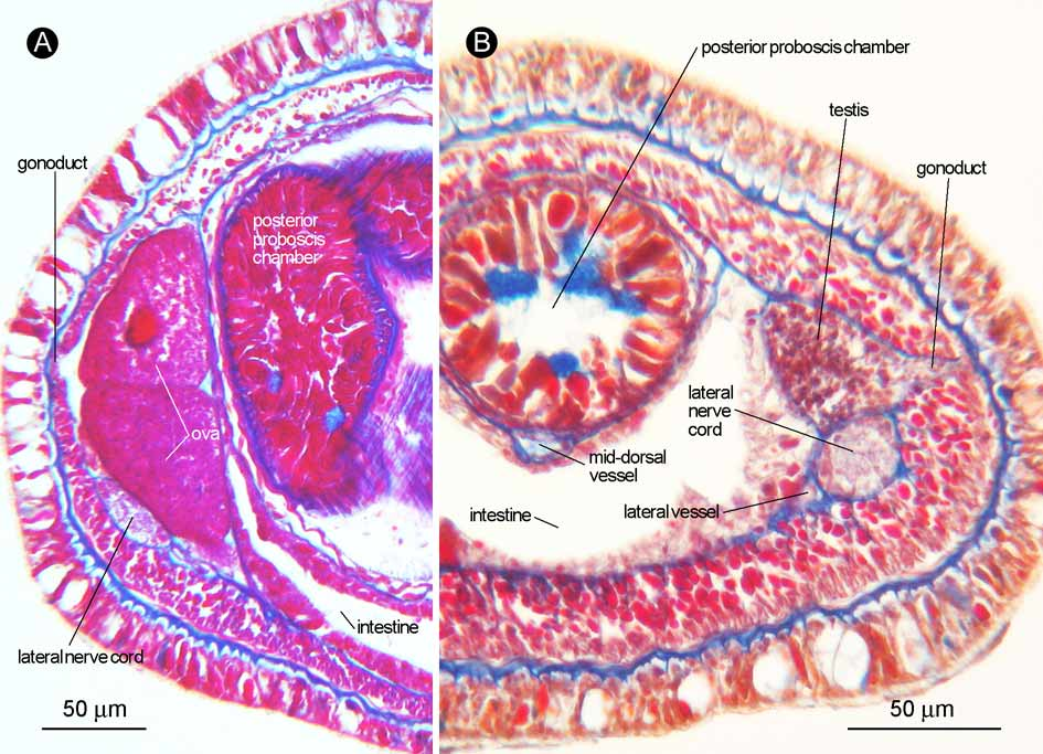
Proboscis apparatus: Proboscis pore opening subterminally ( Fig. 10 A) and leading into rhynchodaeum ( Fig. 10 B); rhynchodaeal epithelium about 3–8 µm thick; rhynchodaeal sphincter not found. Rhynchocoel extending for about 15% of body length, its wall composed of outer circular and inner longitudinal muscle layers. Proboscis short; its anterior chamber attaining up to 53% of body diameter and composed of three muscle layers (inner and outer circular, middle longitudinal); middle longitudinal layer containing 8 proboscis nerves ( Fig. 10 C). Anterior portion of anterior proboscis chamber possessing no circular muscles and glandular cells ( Fig. 10 E). Stylet basis cylindrical, 100 µm long; central stylet spirally sculptured ( Fig. 7 View FIGURE 7 D), up to 57 µm long; stylet/basis ratio 0.57; two accessory stylet pouches, each containing two to three accessory stylets. Posterior proboscis chamber possessing acidophilic glandular epithelium, inner longitudinal, and outer circular muscle layers ( Fig. 11 View FIGURE 11 A, B).
Alimentary system: Oesophagus opening to ventral wall of rhynchodaeum just in front of proboscis insertion, distally invested with longitudinal muscle fibres ( Fig. 10 B); oesophageal epithelium up to 8 µm thick, lacking cilia and glandular cells. Stomach histologically divisible into two regions: short anterior region possessing epithelium dominated by acidophilic glandular cells ( Fig. 9 View FIGURE 9 C); main posterior region possessing epithelium mainly containing basophilic glandular cells ( Fig. 9 View FIGURE 9 A). Stomach epithelium mostly 20–40 µm thick. Intestinal caecum extending forward to reach under posterior region of stomach, anteriorly not bifurcated, laterally possessing several pairs of small and shallow diverticula ( Fig. 10 D). Main intestinal canal possessing normal construction, bearing throughout its length shallow lateral lobes.
Blood vascular system: Pair of cephalic vessels meeting anteriorly above rhynchodaeum, posteriorly passing through cerebral ring and leading to lateral vessels; mid-dorsal vessel branching off from either side of lateral vessel (left in holotype, ZIHU-3204; right in paratype, ZIHU-3205), entering rhynchocoel just posterior to dorsal ganglia to form vascular plug, about 23 µm in diameter ( Fig. 10 E), soon coming down to run between rhynchocoel and alimentary canal. No transverse metameric connectives between mid-dorsal and lateral vessels.
Nervous system: Brain covered by outer neurilemma, with neither inner neurilemma nor neurochord cells. Dorsal and ventral commissures about 13 µm and 20 µm thick, respectively ( Fig. 9 View FIGURE 9 C, D). Lateral nerve cords containing single neuropile; single myofibril running along upper part of neuropile ( Fig. 10 F).
Apical organ and cephalic glands: Single apical organ, 20–30 µm wide, opening at tip of head just above proboscis pore ( Fig. 10 A). Basophilic cephalic glands ( Fig. 10 B) extending post-cerebrally beneath body-wall longitudinal muscle layer on lateral sides of body ( Fig. 10 F) back to pyloric region ( Fig. 10 D). Small and spherical Orange-G-staining acidophilic glandular cells, 2–3 µm diameter, distributed throughout body length just below body-wall longitudinal muscle layer ( Fig. 10 F). In foregut region, large acidophilic glandular cells, up to 20 µm long, aggregated on dorsolateral sides of body among basophilic glands ( Fig. 10 E).
Sense organs: Pigment cup ocelli ( Fig. 10 B) about 50–70 µm in diameter; anterior pair situated near tip of head, posterior pair just in front of proboscis insertion. Cerebral sense organs about 50 µm wide and 70 µm tall ( Fig. 10 B), situated in front of brain, opening ventrally to exterior at cephalic furrow on each side.
Excretory system: Excretory system consisting of thin-walled collecting tubules, up to 20 µm in diameter, running along lateral nerve cords extending from brain region to anterior intestinal region. Pair of efferent canals passing ventrolaterally to open at nephridiopore in posterior foregut region ( Fig. 10 F).
Reproductive system: Sexes separate. Gonads arranged laterally to intestine in row on each side; gonoduct passing above lateral nerve cord. Ovaries up to 170 µm tall, each containing two to three eggs, 80–90 µm in diameter ( Fig. 11 View FIGURE 11 A). Testes up to 50 µm in diameter ( Fig. 11 View FIGURE 11 B).
Remarks: The anatomy of the present species, in particular the short rhynchocoel, the intestinal caecum without anterior bifurcation, the small and simple cerebral sensory organs located some distance in front of the brain, the four eyes, the lateral nerve cords with no accessory nerves, and the mid-dorsal epidermal furrow, closely resembles that described for Nemertopsis quadripunctata , and the two species clearly belong to the same genus. The mid-dorsal epidermal furrow is less developed than that in N. quadripunctata ; in a paratype (ZIHU-3205) the furrow is not apparent in the pre-cerebral region. Nemertopsis mitellicola sp. nov. can be distinguished from all the congeners in having a spirally sculptured central stylet. The new species resembles N. tetraclitophila Gibson, 1990 in possessing a cephalic pigment patch and a symbiotic relationship between cirripedes, but differs from the latter in the following characteristics (those in brackets referring to the condition found in N. tetraclitophila ): body size 15 cm long, 0.4 mm wide [ 0.4–3.5 cm long, 2 mm wide]; dorsal stripes continuous to the cephalic patch [not continuous]; and the central stylet/basis length is 57µm/100µm [30µm/120µm].
No known copyright restrictions apply. See Agosti, D., Egloff, W., 2009. Taxonomic information exchange and copyright: the Plazi approach. BMC Research Notes 2009, 2:53 for further explanation.
|
Kingdom |
|
|
Phylum |
|
|
Class |
|
|
Order |
|
|
Family |
|
|
Genus |