
Enghophyllum, Lazányi & Vagalinski, 2013
|
publication ID |
https://doi.org/ 10.5852/ejt.2013.70 |
|
DOI |
https://doi.org/10.5281/zenodo.3846681 |
|
persistent identifier |
https://treatment.plazi.org/id/03E5878D-0477-054C-FD9E-47935ECEB362 |
|
treatment provided by |
Carolina |
|
scientific name |
Enghophyllum |
| status |
gen. nov. |
Genus Enghophyllum View in CoL gen. nov.
urn:lsid:zoobank.org:act:15008961-CD77-4325-B694-13BA1F7E514E
Type species
Brachyiulus (Chromatoiulus) naxius Verhoeff, 1901 View in CoL
Diagnosis
A genus of Brachyiulini , differing from contribal genera by the following combination of characters: promere broad, shield-like, in situ protruding mostly posteriad, completely covering opisthomere and gonopodal sinus; transverse muscles and coxal apodemes of the promere fully reduced. Opisthomere with 3 well-differentiated processes: lateral (lp), basal posterior (bpp) and apical posterior (app). Solenomere rather simple, tubular.
Etymology
This genus is named in honor of Prof. Henrik Enghoff from the ZMUC, not only for his vast contribution to our knowledge of diplopods, but also for his continuous encouragement and help during our work with Brachyiulini .
Description
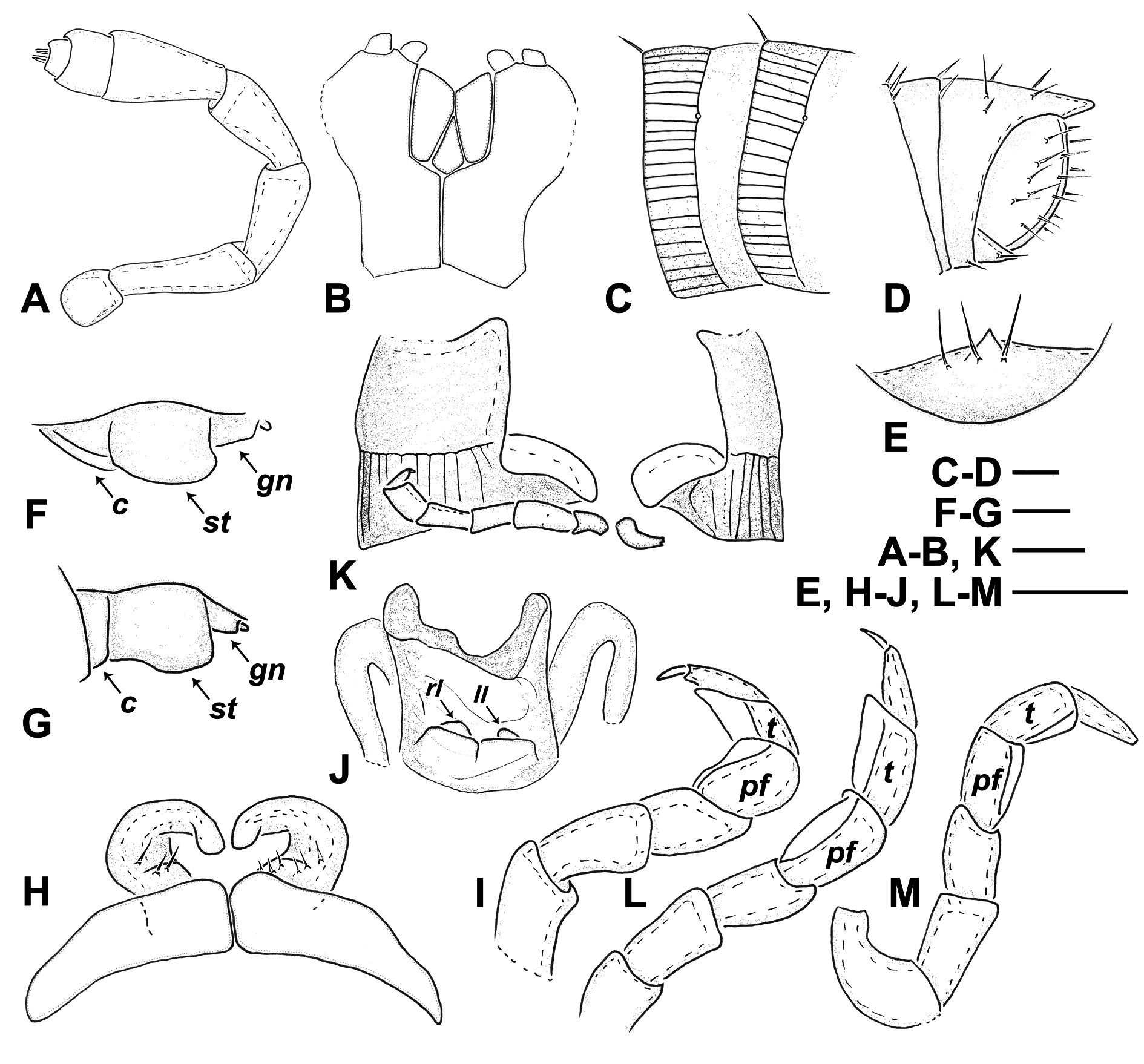
Two frontal, four supralabral and 14-18 labral setae. Antennomeres 4 and 5 subequal in length ( Fig. 1A View Fig ). Gnathochilarium as on Fig. 1B View Fig . Metazona sparsely striated, as emphasised by Verhoeff (1901); ozopores right on the suture ( Fig. 1C View Fig ). Preanal process straight, anal valves sparsely pilose ( Fig. 1D View Fig ); subanal scale triangular, with protruding tip ( Fig. 1E View Fig ).
Males
Mandibular stipes slightly protruding ( Fig. 1 View Fig F-G). First leg-pair like simple, rounded, somewhat converging hooks; the two hooks converging in an obtuse angle ( Fig. 1H View Fig ). Second leg-pair ( Fig. 1I View Fig ) with two ventral pads: on postfemur (pf) and tibia (t). Penis ( Fig. 1J View Fig ) significantly small, deeply hidden in penis sac, with two very short lobes and two rounded small lamellae running parallel, i.e., not diverging (rl and ll for right and left lamella). Pleurotergum of the 7 th body ring protruding like a simple shovel (broken in the unique holotype of E. sifnium gen. et sp. nov., thus investigated only in E. naxium , see Fig. 1K View Fig ). Walking legs ( Fig. 1 View Fig L-M) with two ventral pads: on postfemur (pf) and tibia (t). Tarsus of midbody legs slightly longer than tibia: tarsus / tibia = (1.05-1.1) / 1.
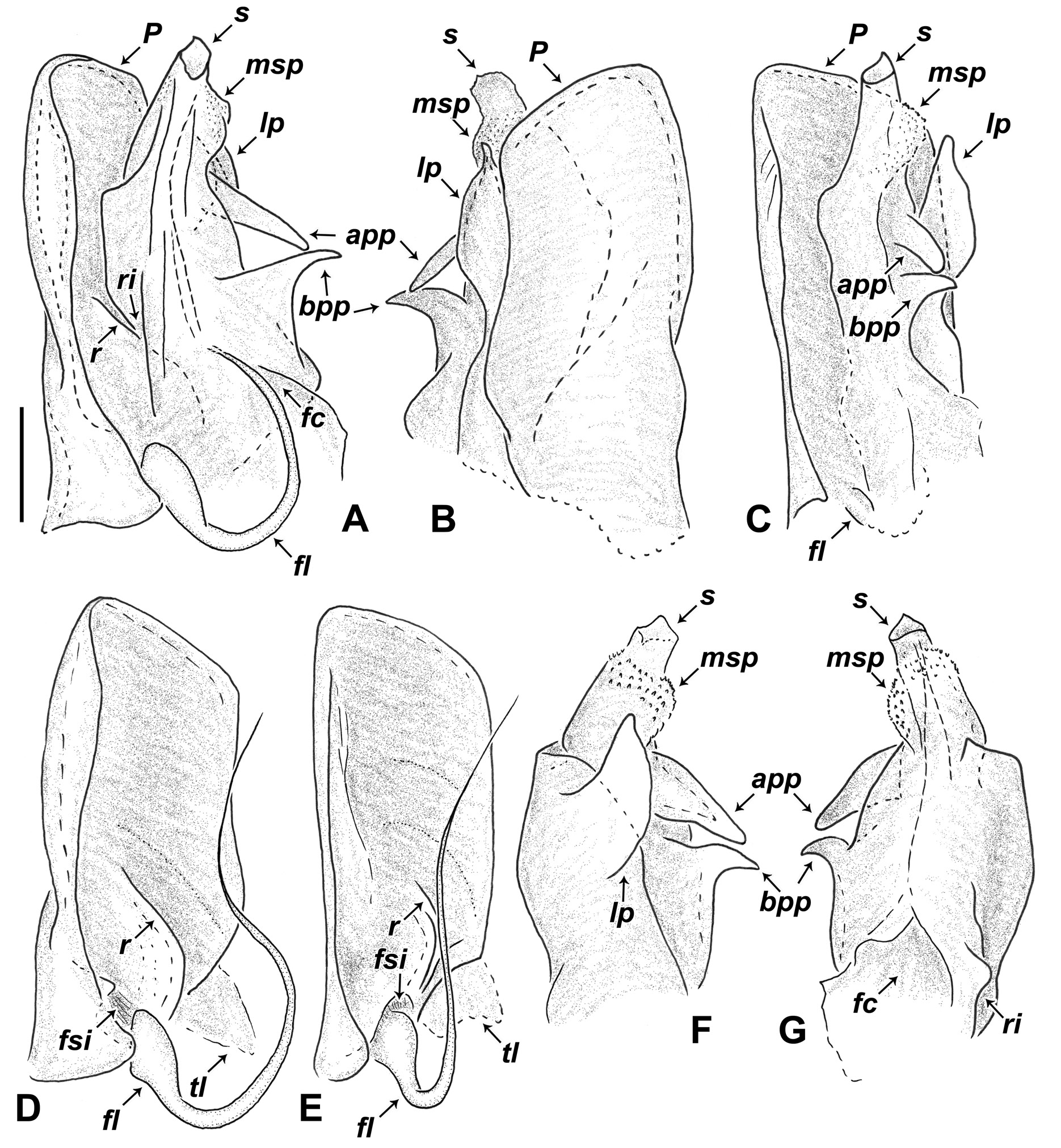
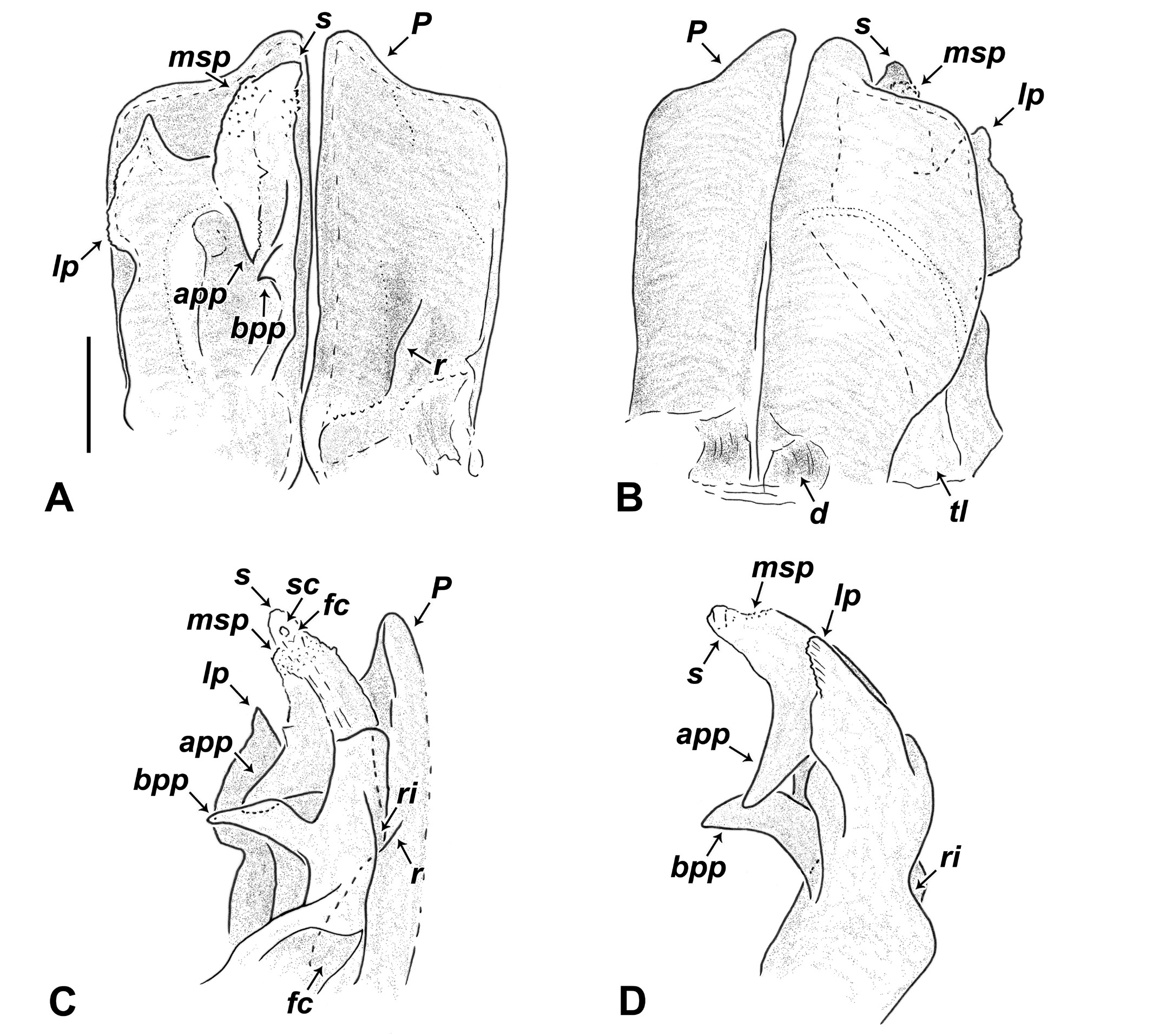
Gonopods noticeably big compared to body size, directed mostly posteriad. Promere bevelled in laterobasal corner to fit into the inner curve of the 7 th body ring; latero-basal corner of promere only with thin triangular lamella (tl) ( Fig. 2 View Fig D-E). Promeres attached to each other in their meso-basal corner with a chitinous bridge. A short, oblique ridge (r) on posterior surface of promere ( Figs 2A View Fig , D-E, 4A, C). Flagellum emerging from a well-developed sinus (fsi) ( Fig. 2 View Fig D-E). At the level of this sinus a deepening on anterior surface of the promere (d) ( Fig. 4B View Fig ). Opisthomere entirely concealed under the promere, protruding postero-ventrad, the two promeres tightly closing the ventral opening on 7 th ring like a pair of shutters. Opisthomere not embedded in the groove formed by the oblique ridge on the promere’s posterior surface (unlike in Megaphyllum ), but “riding” on the ridge ( Figs 2A View Fig , 4C View Fig ): posterior basis of opisthomere with a characteristic riffle (ri) formed by chitinous lamellae, where the ridge of the promere is positioned ( Figs 2A, G View Fig , 4 View Fig C-D). Opisthomere with a well-developed lateral process (lp) and an apical and a basal posterior process (app and bpp). Solenomere (s) simple, with no processes but basally with a micro-spinose pillow (msp) ( Figs 2 View Fig A-C, F-G, 4A-D). Flagellum channel and sperm canal (fc and sc) each with an apical opening ( Fig. 4C View Fig ).
Females
The only available female of E. sifnium gen. et sp. nov. is in stadium IX (with 8 rows of ocelli) and proved to be subadult, but females of E. naxium in stadia VIII-IX (with 7-8 rows of ocelli) had fully developed vulvae, which we used here for the description of female sexual characters. First two legpairs slightly swollen.
Vulva ( Fig. 3 View Fig ): Subcylindric in shape, the mesal half shorter, the opening large, oval, apical. Operculum shorter than bursa, with around 20-25 setae. Mesal and lateral sclerites with 2-3 setae each. Apodematic tube not opening apically into a longitudinal median cleft as in, e.g., Megaphyllum species, but into a wide sinus (si). Around this sinus a thin wall, formed by the apical part of the bursa. These walls with small dot-like pores; most setae (around 20/side) emerging from this apical region. Apodematic tube (at) ending in two receptaculi seminis or ampullae. Central ampulla (ca) drop-like. Distal, globular ampulla (da) joining the apodematic tube through a twisted connecting tube (ct). Connecting tube very long, thus the distal ampulla hanging out of the bursa; easily broken off during preparation.
Remarks
The everted penes of the adult E. naxium male from Antiparos was quite soft, amorphous and huge, contrary to the stout, minute penes, deeply hidden under the 2 nd leg-pair coxae, observed in the E. naxium male from Mavri Islet and the E. sifnium gen. et sp. nov. male from Sifnos. It is possible that penes vary in size between copulating and non-copulating periods.

No known copyright restrictions apply. See Agosti, D., Egloff, W., 2009. Taxonomic information exchange and copyright: the Plazi approach. BMC Research Notes 2009, 2:53 for further explanation.
|
Kingdom |
|
|
Phylum |
|
|
Class |
|
|
Order |
|
|
Family |
|
|
Tribe |
Brachyiulini |