
Micridium halidaii ( Matthews, A. 1868 )
|
publication ID |
https://doi.org/10.11646/zootaxa.4242.2.2 |
|
publication LSID |
lsid:zoobank.org:pub:0DD1868D-4EF5-4FB7-AEE9-372644800A79 |
|
DOI |
https://doi.org/10.5281/zenodo.6045201 |
|
persistent identifier |
https://treatment.plazi.org/id/03E64B08-E322-FFFF-8585-5E65FC17EB90 |
|
treatment provided by |
Plazi |
|
scientific name |
Micridium halidaii ( Matthews, A. 1868 ) |
| status |
|
Micridium halidaii ( Matthews, A. 1868) View in CoL as Ptilium halidaii
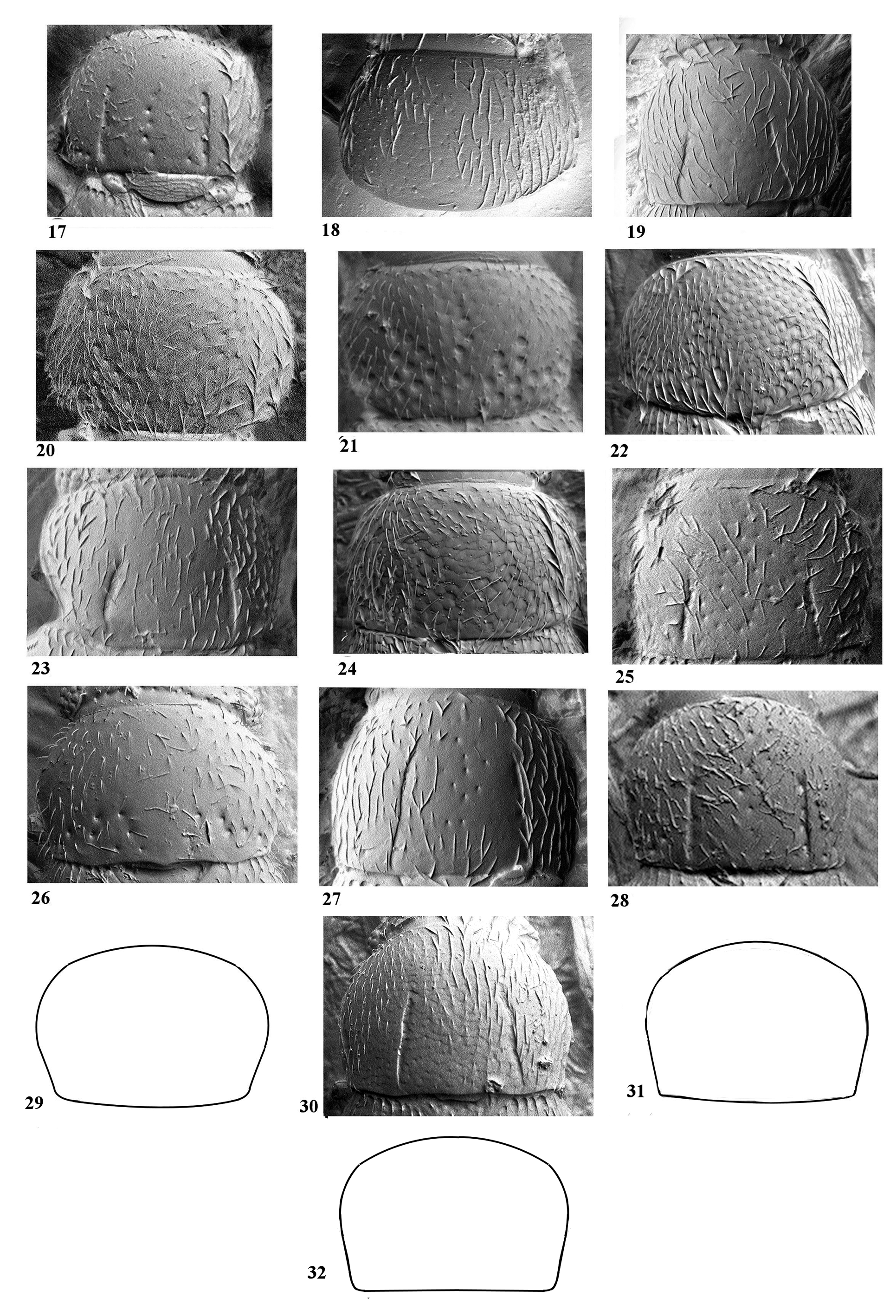
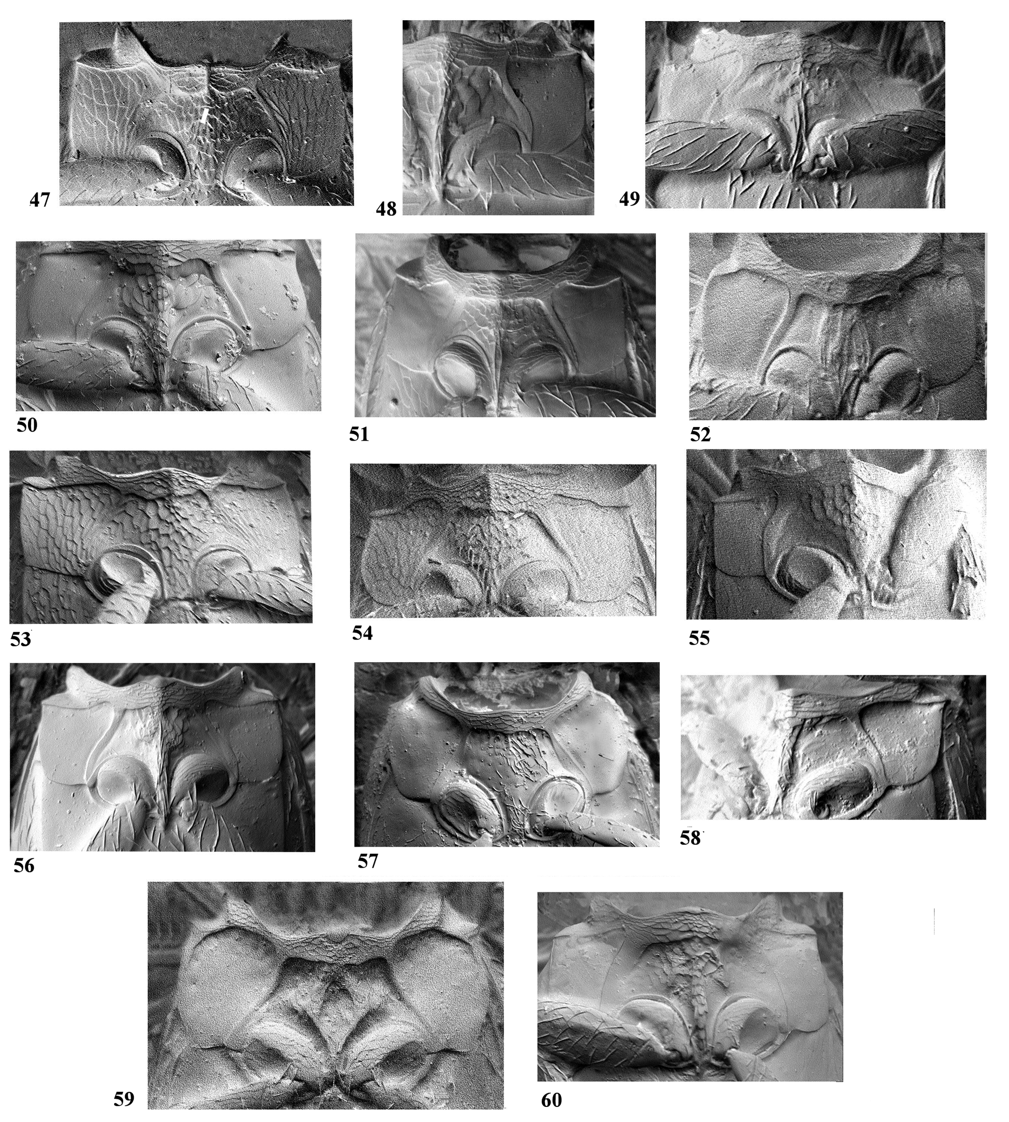
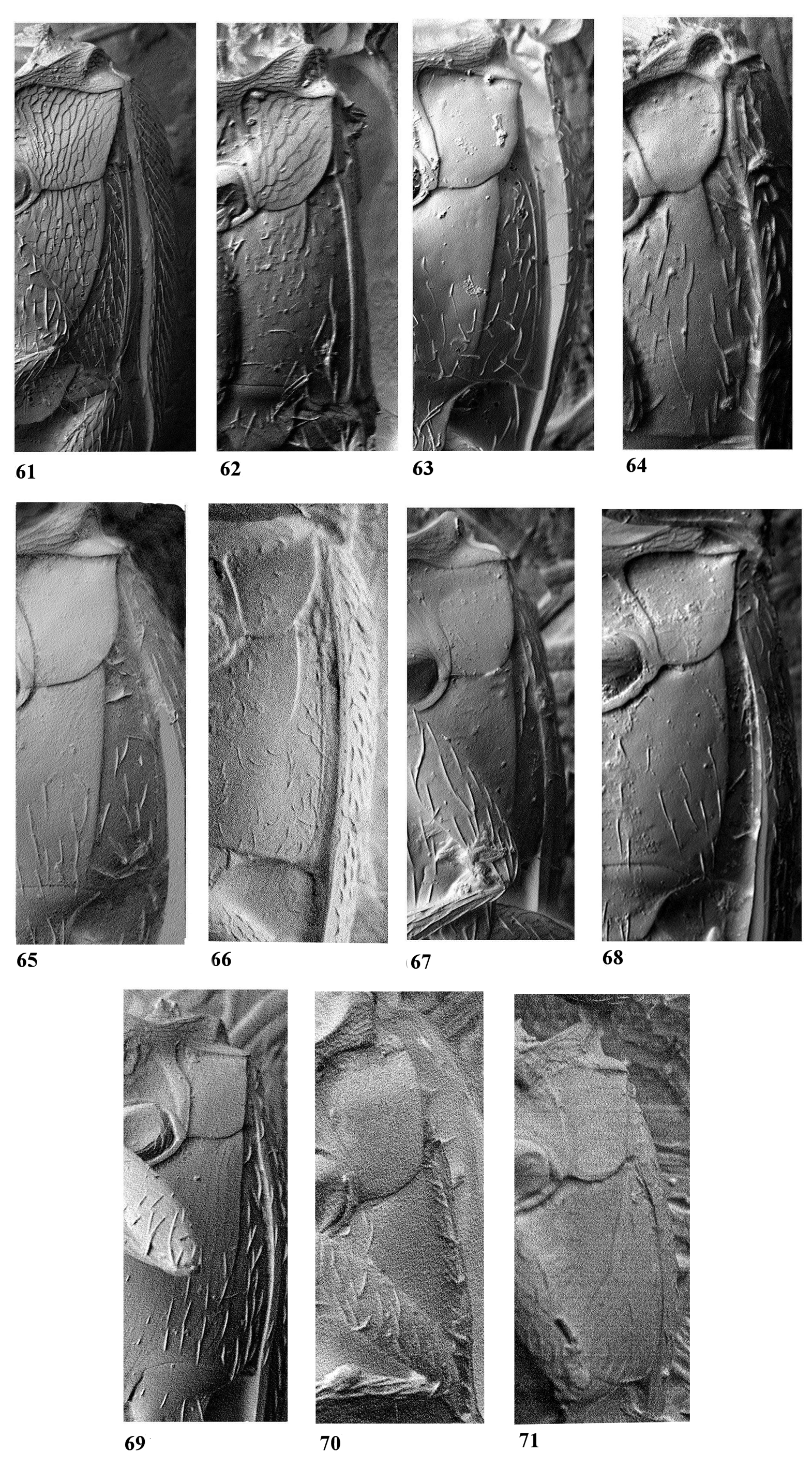
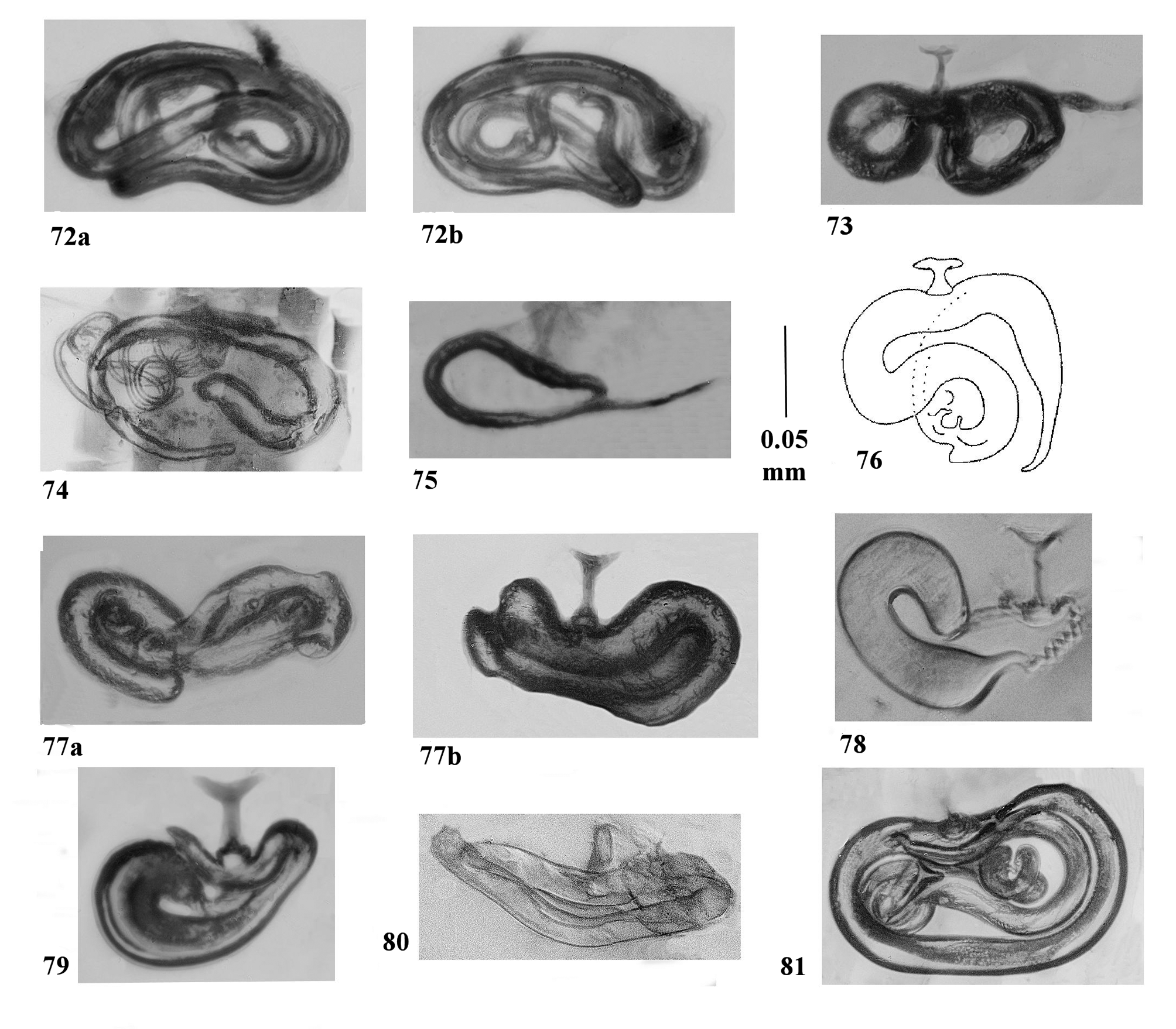
( Figs. 2 View FIGURES 1 – 14 , 25 View FIGURES 17 – 32 , 55 View FIGURES 47 – 60 , 69 View FIGURES 61 – 71 , 76 View FIGURES 72 – 81 , 84, 96, 101)
Habitus Fig. 2 View FIGURES 1 – 14 . Length 0.65 mm (as mounted with abdomen extruded 0.76 mm). Colour yellow brown, antennae and legs light yellow. Antonnomeres 3–11 length 0.19 mm. Width across eyes 0.18 mm. Mentum: narrowed towards base, sides slightly sinuate. Pronotum: punctured and pubescent, medial punctures larger than those at the sides; with two distinct, almost parallel, linear depressions reaching from the posterior margin to the centre, widest in the anterior half, lateral margins sinuate and hind angles clearly denoted, length 0.13 mm. width 0.22 mm, Fig. 25 View FIGURES 17 – 32 . Scutellum with four depressions at base Fig. 101 View FIGURES 100 – 101 . Elytra length 0.40, width 0.26 mm, punctured and pubescent, the punctures smaller than those on the pronotum; sides evenly rounded, wider just in front of centre, humeri with a row of concave depressions. Mesoventrum: keel narrow between the mesocoxae, not reaching metaventrum, Fig. 55 View FIGURES 47 – 60 . Mesepiventra without clear reticulation, straight before slight hind angles Fig. 55 View FIGURES 47 – 60 . Metaventrum: metepiventral sutures reaching +/– 3/4 of the distance from the mesoventrum to the hind coxae, Fig. 69 View FIGURES 61 – 71 .
Male: posterior intercoxal process of metaventrum with a fringe of flattened setae, Fig. 96. Aedeagus with a tapering point, Fig. 84.
Female: spermatheca Fig. 76 View FIGURES 72 – 81 .
Remarks. The habitus figure and measurements are taken from the holotype in BMNH. The description is based on the holotype and on a second, slightly smaller and darker specimen, in the Museum collected by J.A.Owen in Richmond Park, Surrey, UK on 7 January 1984 and determined by C. Johnson, this specimen was also used for the SEMs. A third specimen in a private collection mounted as a slide, also from Richmond, was borrowed to make the spermatheca drawing and figure of the male metaventral intercoxal process.
The holotype is mounted between two sheets of mica (?) attached to a card with the rear corners cut off. Labels are present as follows: 'halidayi Ih[?] July 1867 ' (pinned immediately below the specimen, apparently in Matthews' hand, rear corners also cut off); 'Type AM [monogram]' (in Matthews' hand on red card); 'haldaii' (in Matthews' hand on blue strip); ' Ptilium halidaii Mat. H. Britten 1931 '; ' Ptilium halidayi, Mat'; '2796' (Matthews/Mason collection number); ' Micridium halidaii Matth. C. Johnson det.'; 'in Matthews. Coll.'; 'Matthews coll 1904 - 120' (BMNH accession number) and 'Syntype' (added by BMNH). ( Fig. 2 View FIGURES 1 – 14 ).


No known copyright restrictions apply. See Agosti, D., Egloff, W., 2009. Taxonomic information exchange and copyright: the Plazi approach. BMC Research Notes 2009, 2:53 for further explanation.
|
Kingdom |
|
|
Phylum |
|
|
Class |
|
|
Order |
|
|
Family |
|
|
Genus |